Biol 226 (A&P II) Lab Exercise 9 - Respiratory System, Volumes, Rates, and Capacities
1/84
There's no tags or description
Looks like no tags are added yet.
Name | Mastery | Learn | Test | Matching | Spaced | Call with Kai |
|---|
No analytics yet
Send a link to your students to track their progress
85 Terms
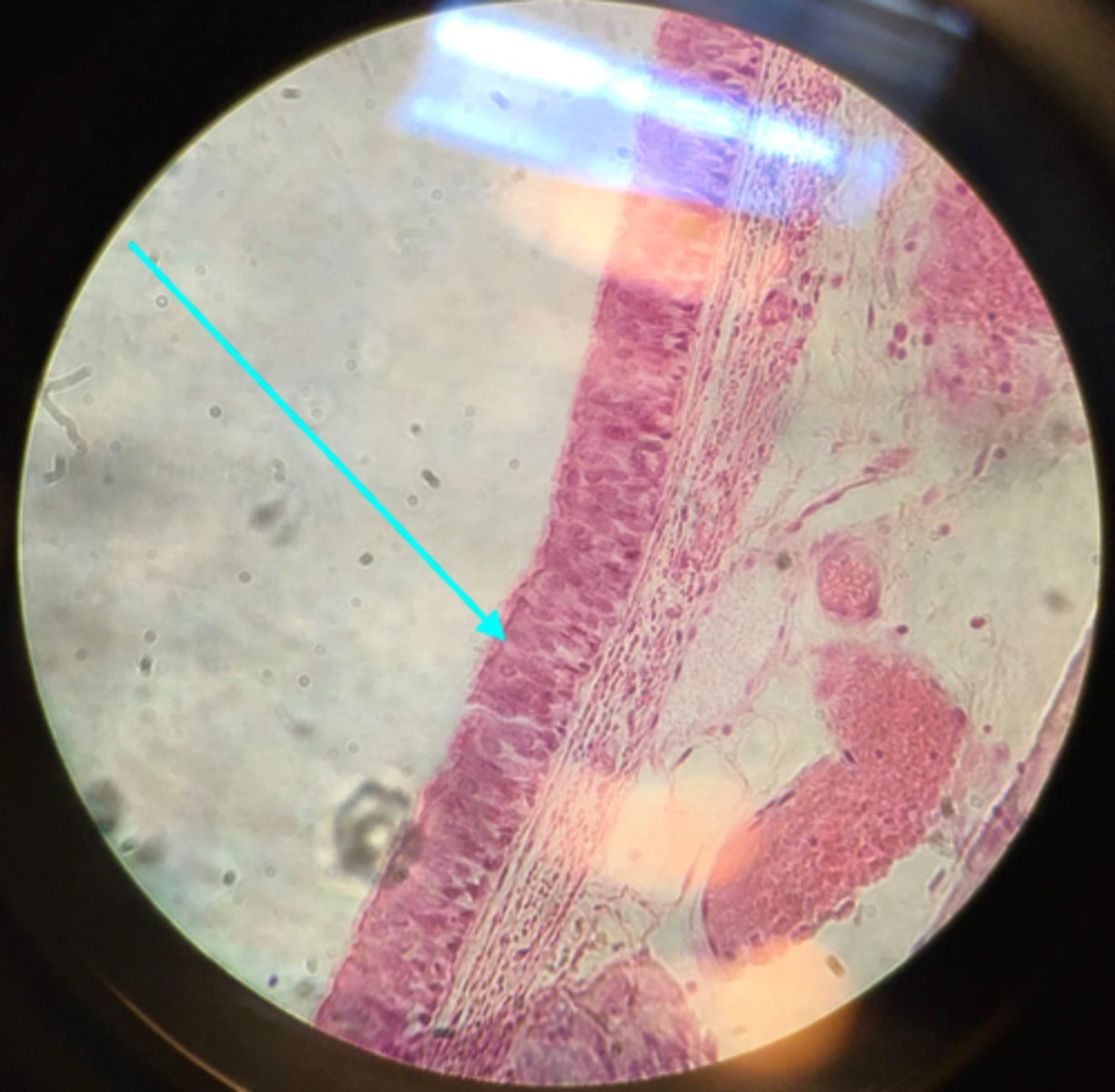
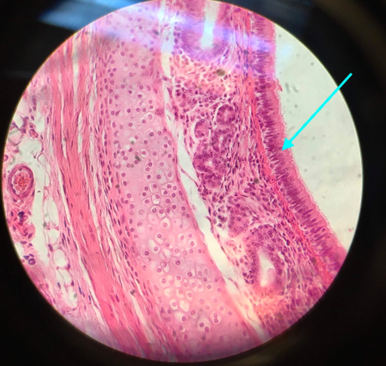
Ciliated pseudostratified columnar epithelium
What type of tissue is the blue arrow pointing at on this slide?

Basement membrane
Ciliated pseudostratified columnar epithelium is shown on this slide. What is the blue arrow pointing at?

Cilia
Ciliated pseudostratified columnar epithelium is shown on this slide. What is the blue arrow pointing at? These hair-like structures project out into the lumen

Goblet cells
Ciliated pseudostratified columnar epithelium is shown on this slide. What is the blue arrow pointing at? This cell secretes mucus.
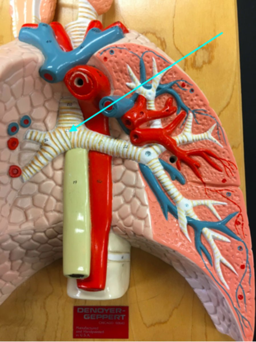
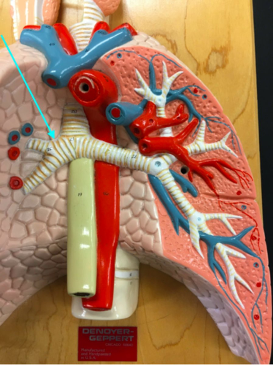
Bifurcation of the trachea
What structure of the heart/lung model is the blue arrow pointing at?
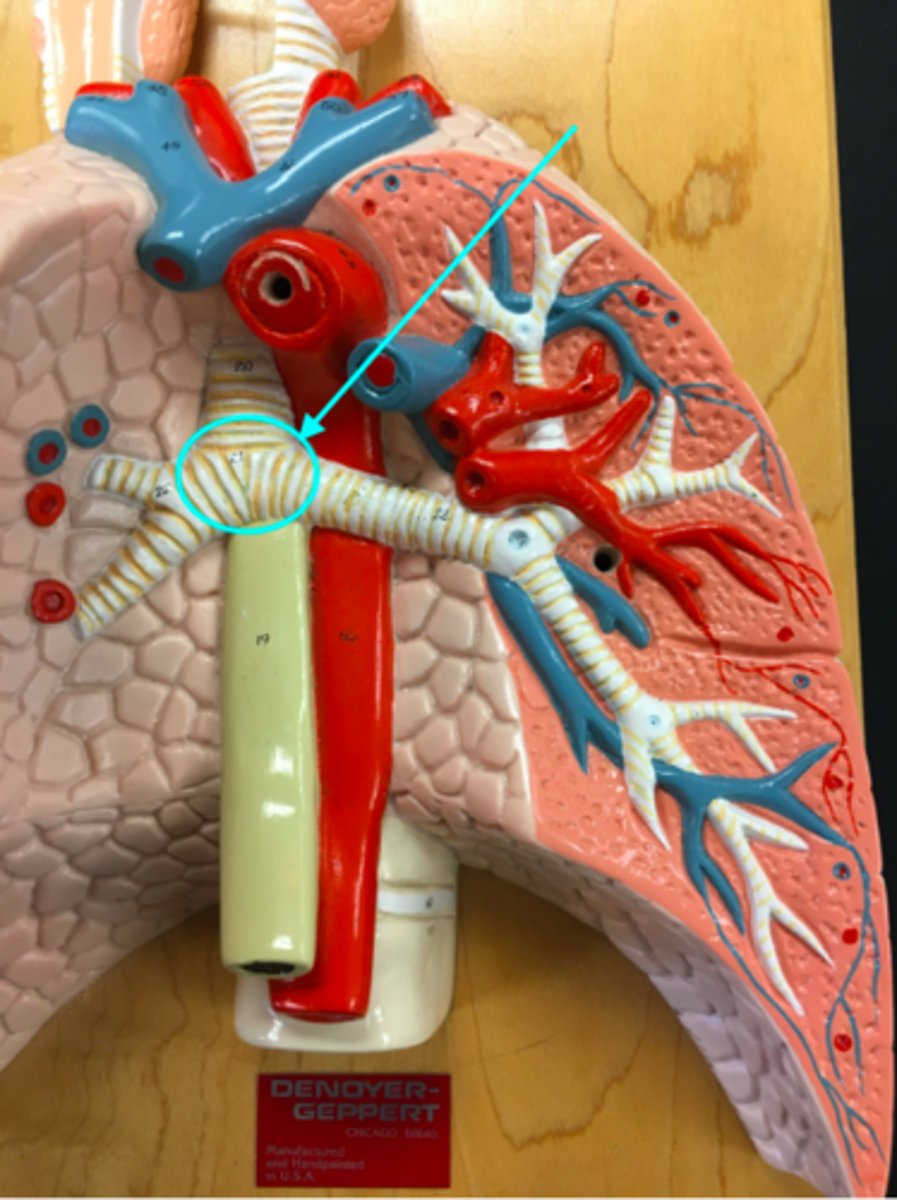
Carina
What structure of the heart/lung model is the blue arrow pointing at?
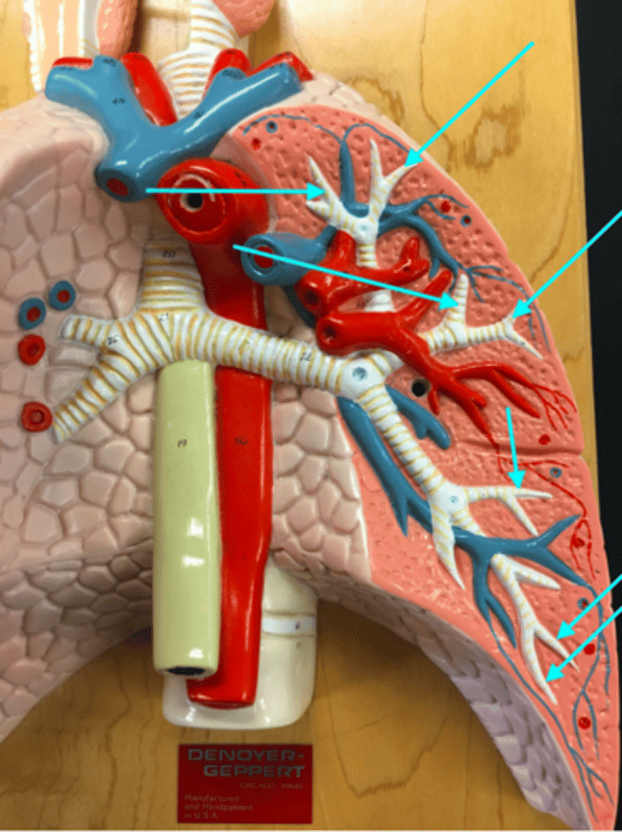
Bronchioles
What structures of the heart/lung model are the blue arrows pointing at?
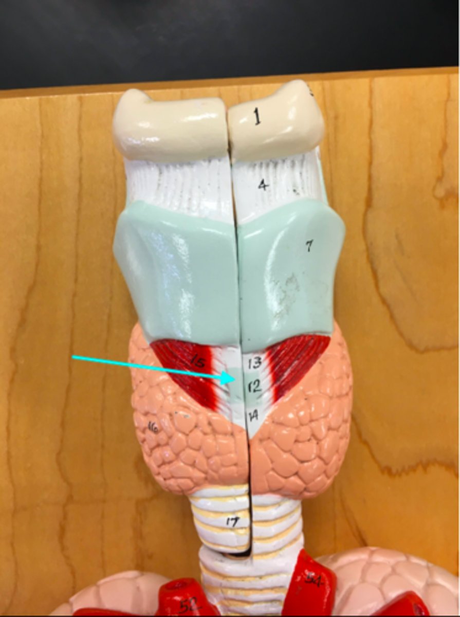
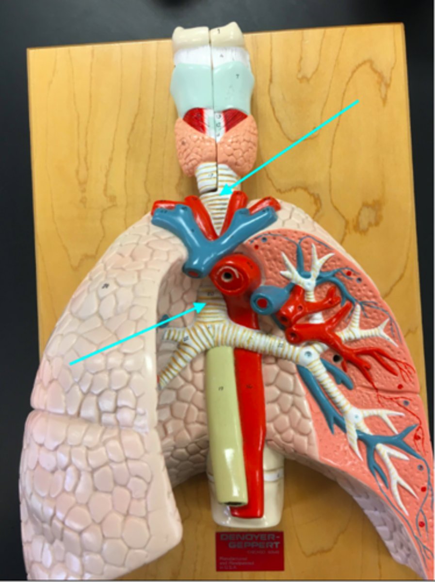
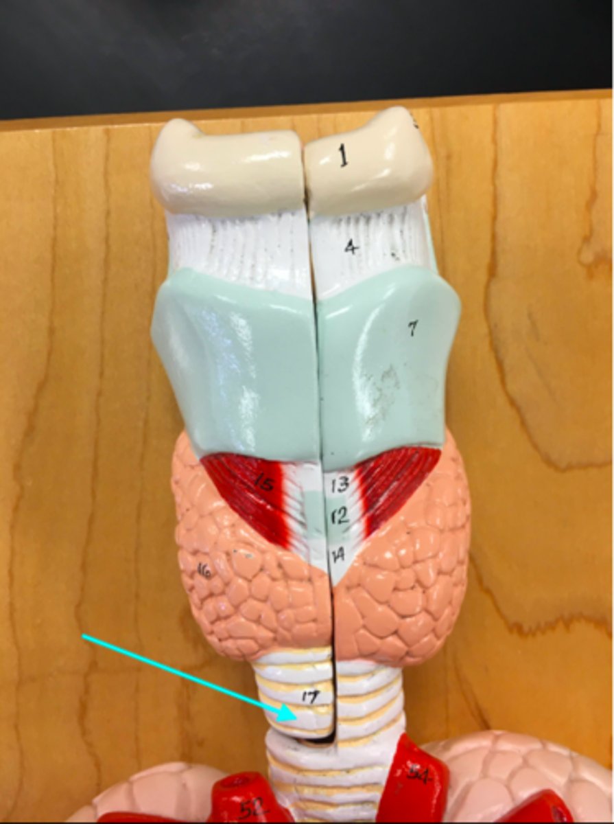
Cricoid cartilage
What structure of the heart/lung model is the blue arrow pointing at?
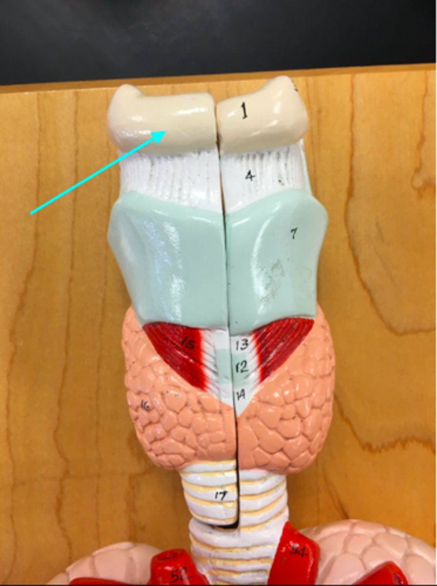
Epiglottis
What structure of the heart/lung model is the blue arrow pointing at?
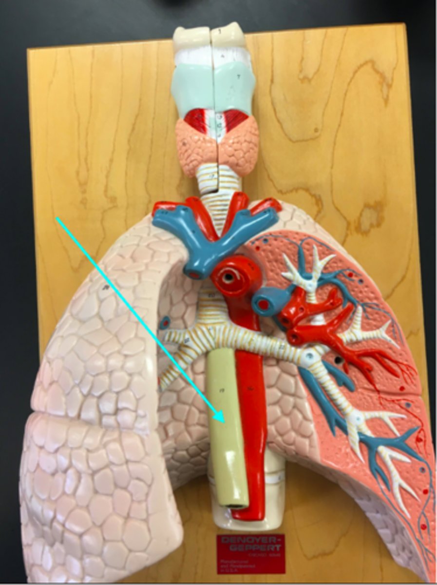
Esophagus
What structure of the heart/lung model is the blue arrow pointing at?
Hyoid bone
What structure of the heart/lung model is the blue arrow pointing at?
Lungs
What structure of the heart/lung model is the blue arrow pointing at?

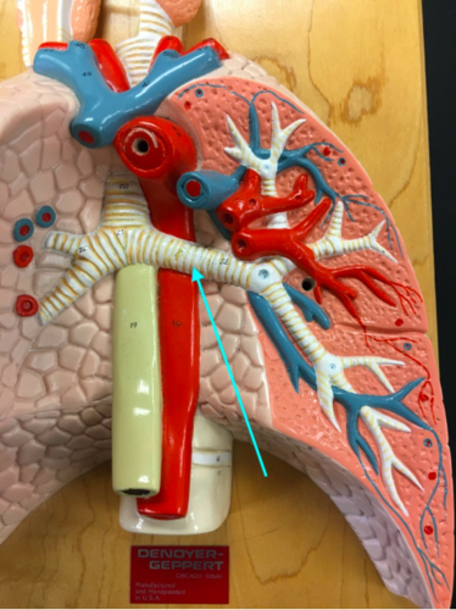
Right primary bronchi
What structure of the heart/lung model is the blue arrow pointing at?
Left primary bronchi
What structure of the heart/lung model is the blue arrow pointing at?
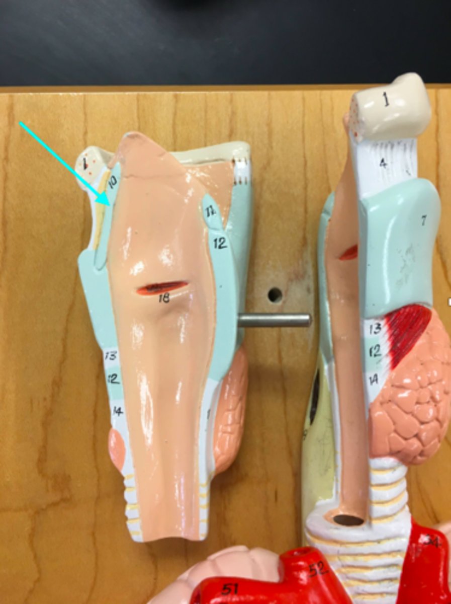
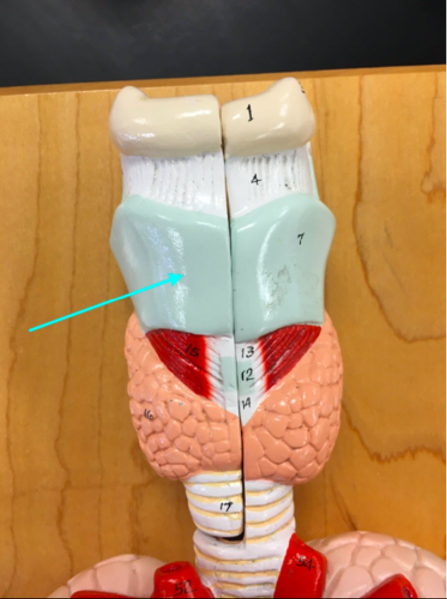
Thyroid cartilage
What structure of the heart/lung model is the blue arrow pointing at?
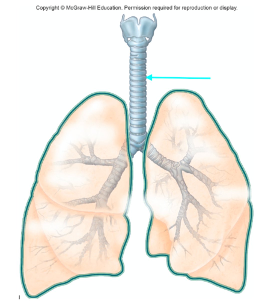
Trachea
What structure of the heart/lung model are the blue arrows pointing at?
Tracheal cartilage
What structure of the heart/lung model is the blue arrow pointing at? (The partial ring structure)
Carina
What structure of the lung diagram is the blue arrow pointing at?

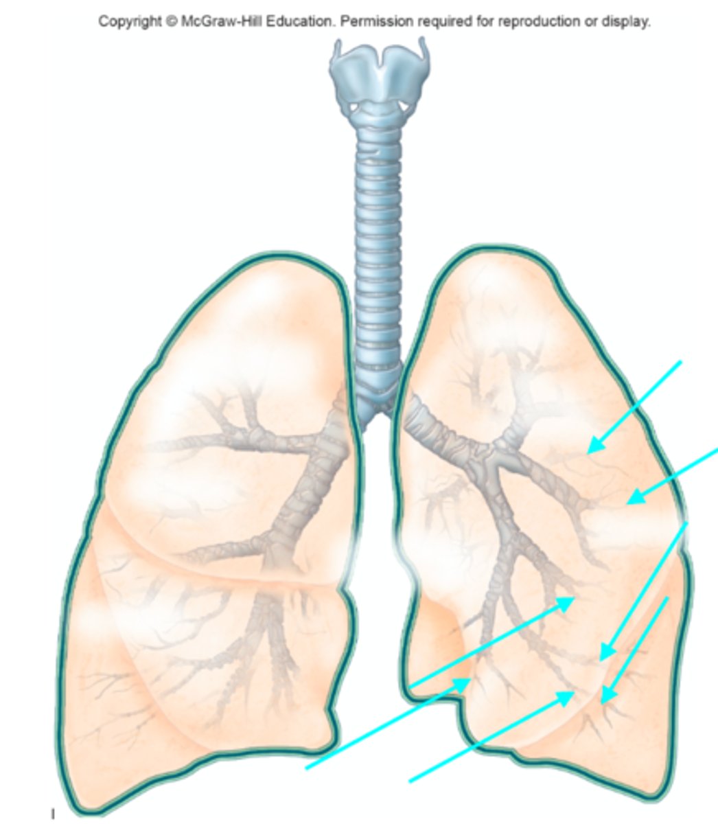
Bronchioles
What structures of the lung diagram are the blue arrows pointing at?
Cricoid cartilage
What structure of the lung diagram is the blue arrow pointing at?
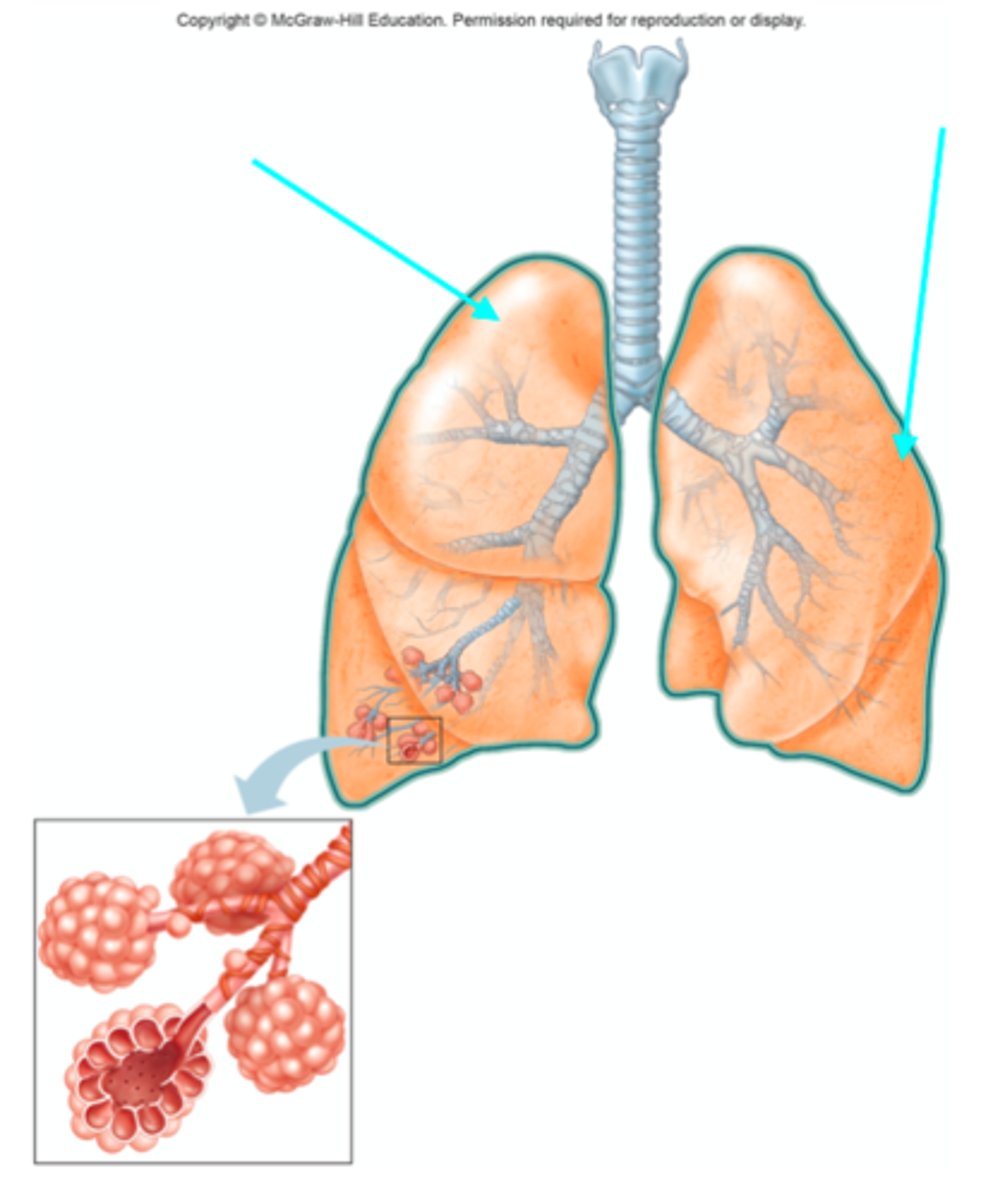
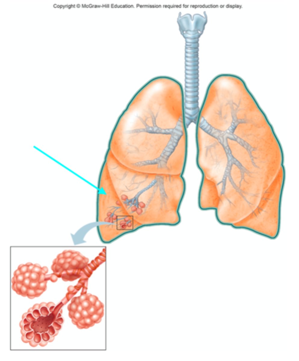
Lungs
What organ are the blue arrows pointing at?
Right superior (upper) lobe
Which lobe of the lungs is the blue arrow pointing at?

Right middle lobe
Which lobe of the lungs is the blue arrow pointing at?
Right inferior (lower) lobe
Which lobe of the lungs is the blue arrow pointing at?
Left superior (upper) lobe
Which lobe of the lungs is the blue arrow pointing at?

Left inferior (lower) lobe
Which lobe of the lungs is the blue arrow pointing at?

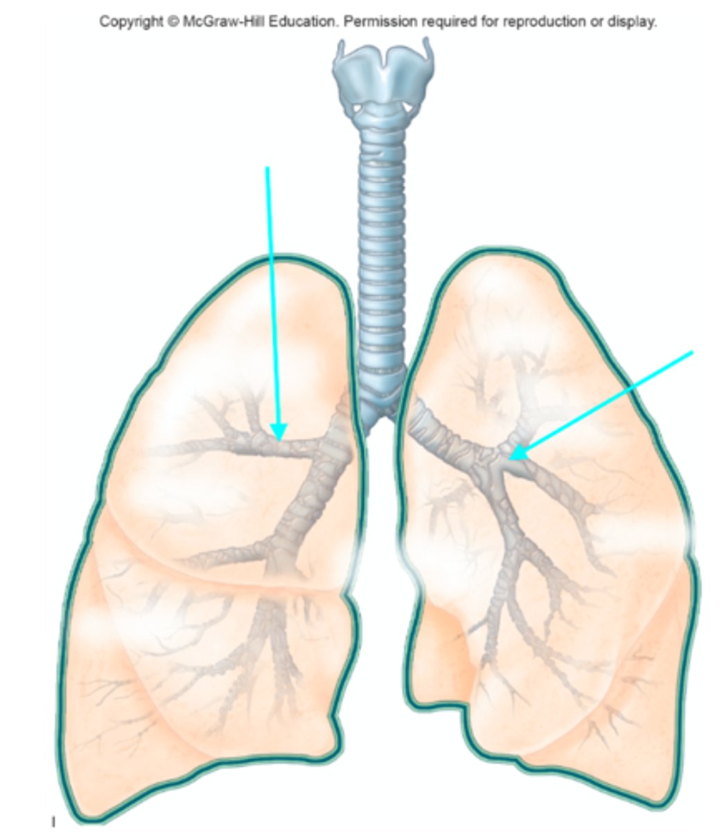
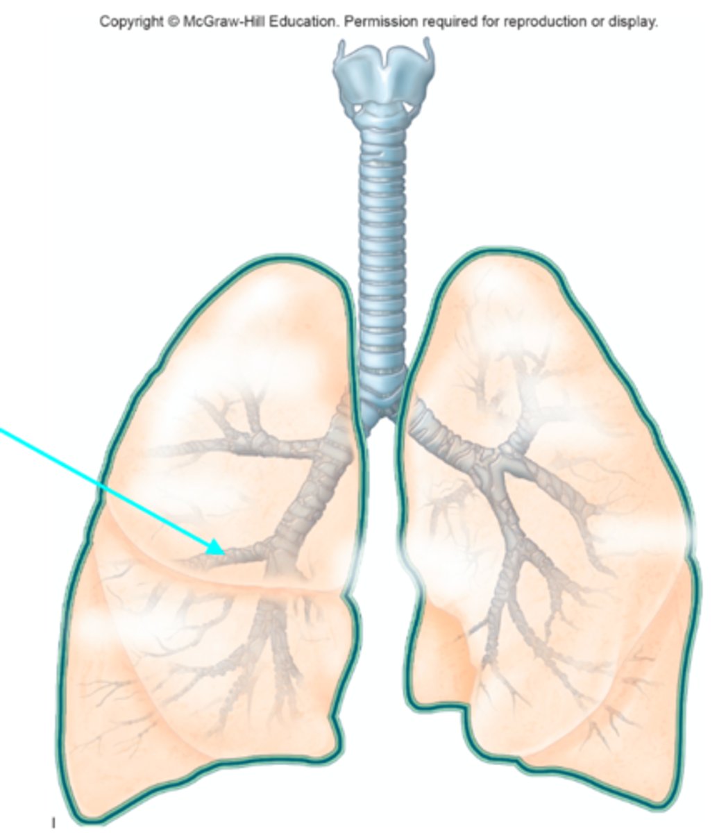
Right main (primary) bronchus
Which structure of the lung diagram is the blue arrow pointing at?

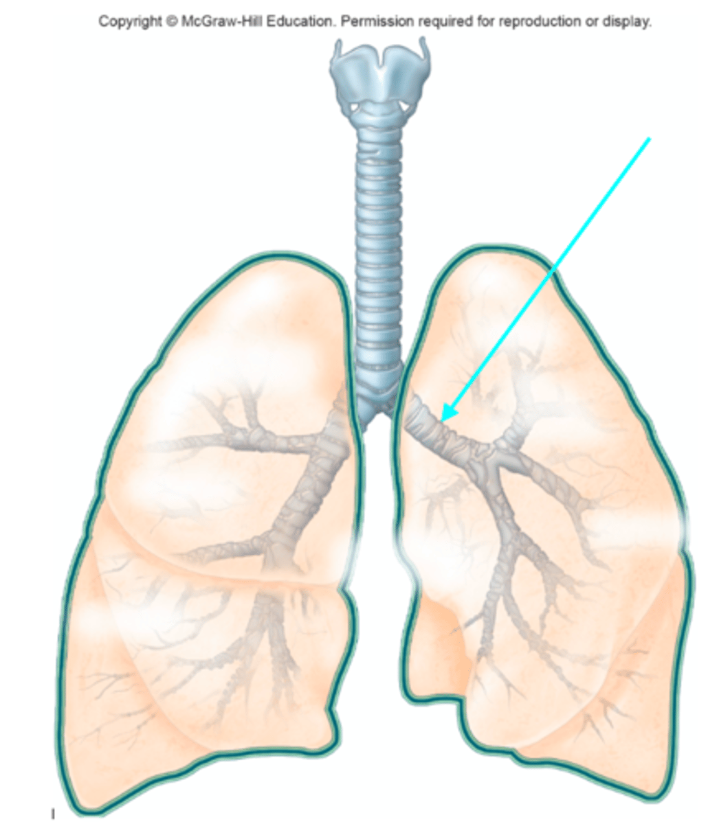
Right main (primary) bronchus
Which structure of the lung diagram is the blue arrow pointing at?

Left main (primary) bronchus
Which structure of the lung diagram is the blue arrow pointing at?
Lobar (secondary) bronchus
Which structure of the lung diagram is the blue arrow pointing at?

Segmental (tertiary) bronchus
Which structure of the lung diagram is the blue arrow pointing at?
Superior (upper) lobar bronchi
Which structures of the lung diagram are the blue arrows pointing at?
Inferior (lower) lobar bronchi
Which structures of the lung diagram are the blue arrows pointing at?

Middle lobar bronchus
Which structure of the lung diagram is the blue arrow pointing at?
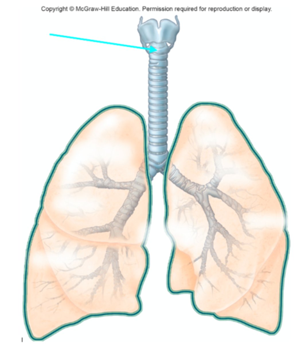
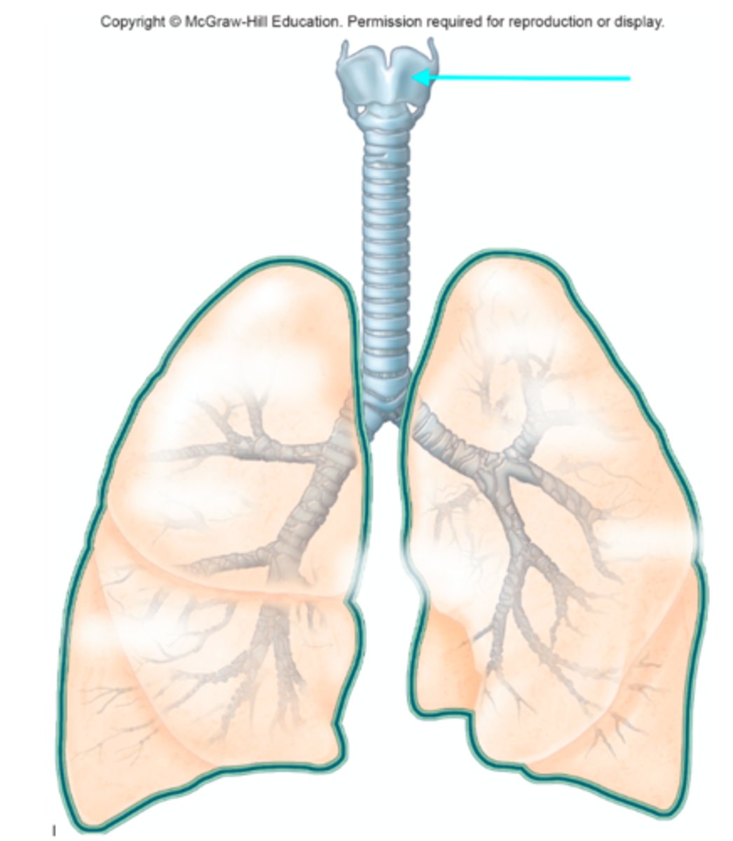
Thyroid cartilage
Which structure of the lung diagram is the blue arrow pointing at?
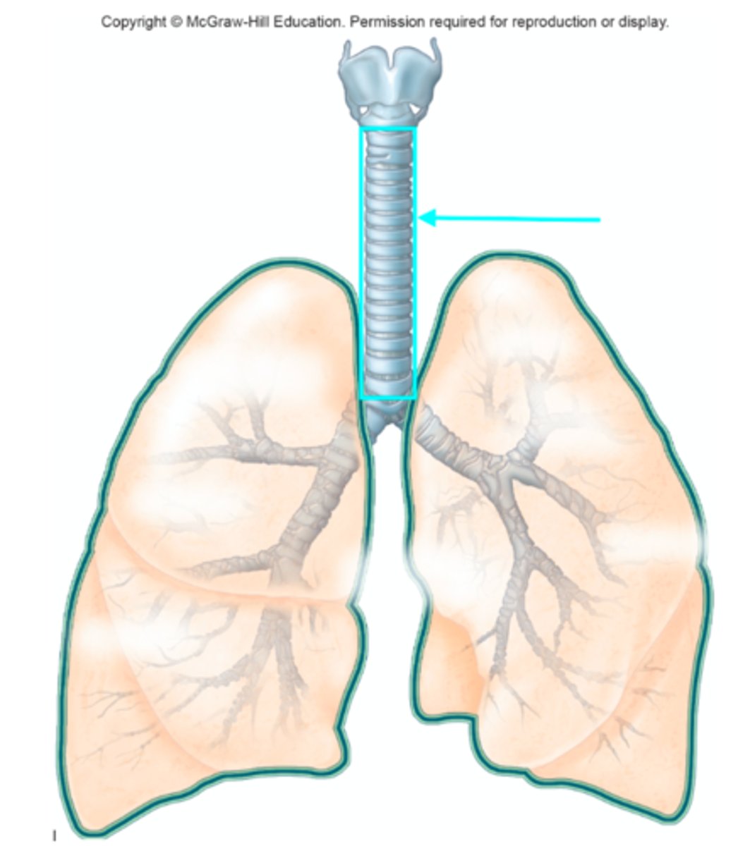
Trachea
Which structure of the lung diagram is the blue arrow pointing at?
Tracheal cartilage - cartilaginous ring
Which individual structure of the lung diagram is the blue arrow pointing at?
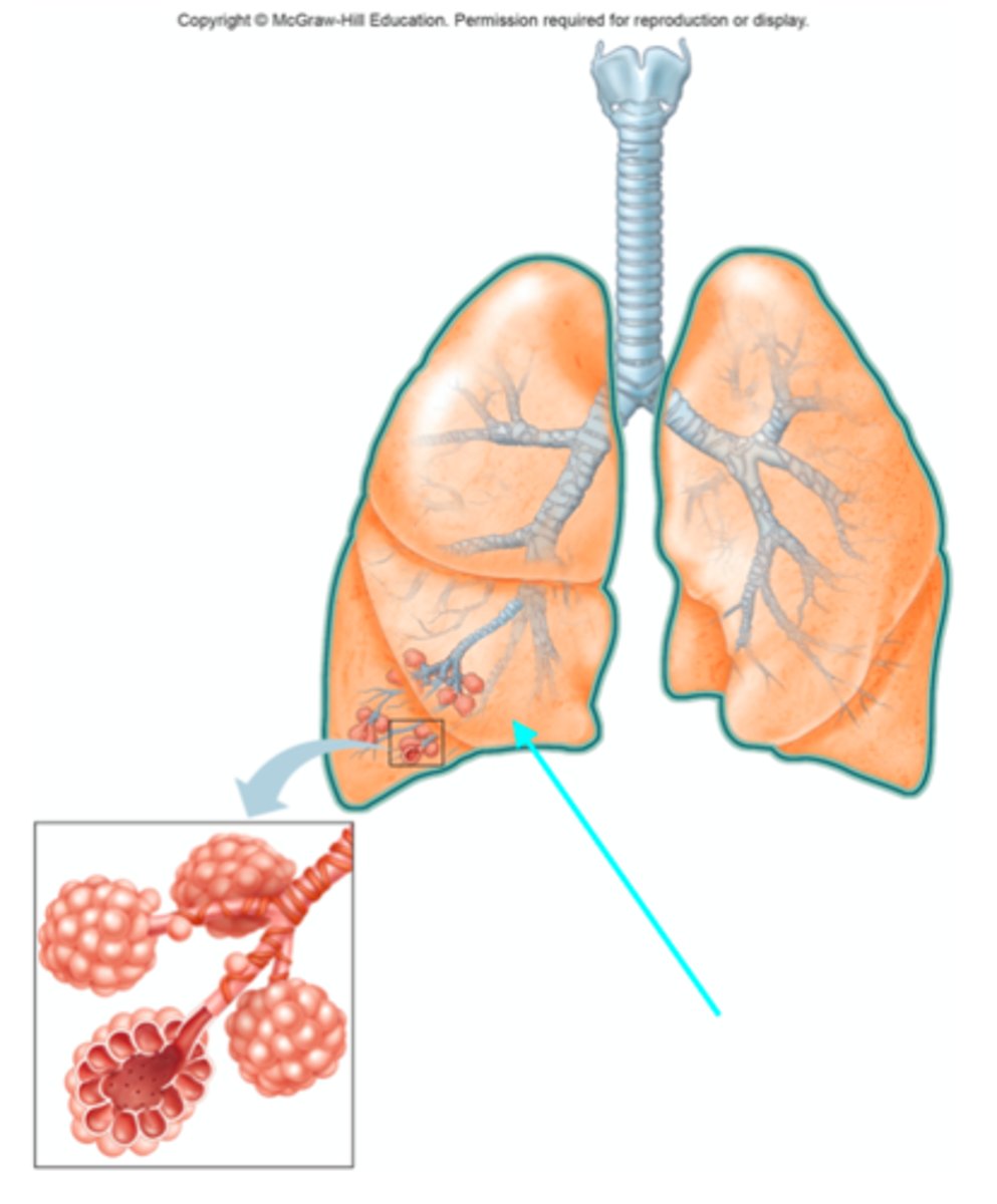
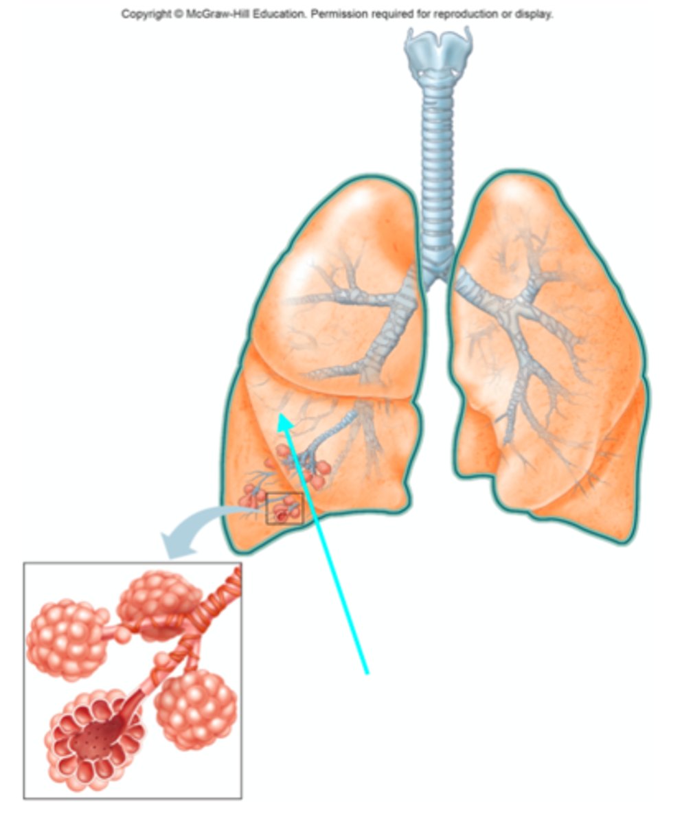
Terminal bronchiole
Which structure of the lung diagram is the blue arrow pointing at?
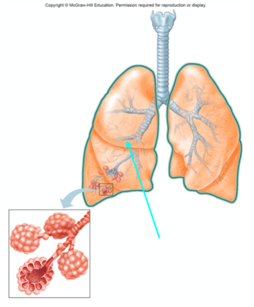
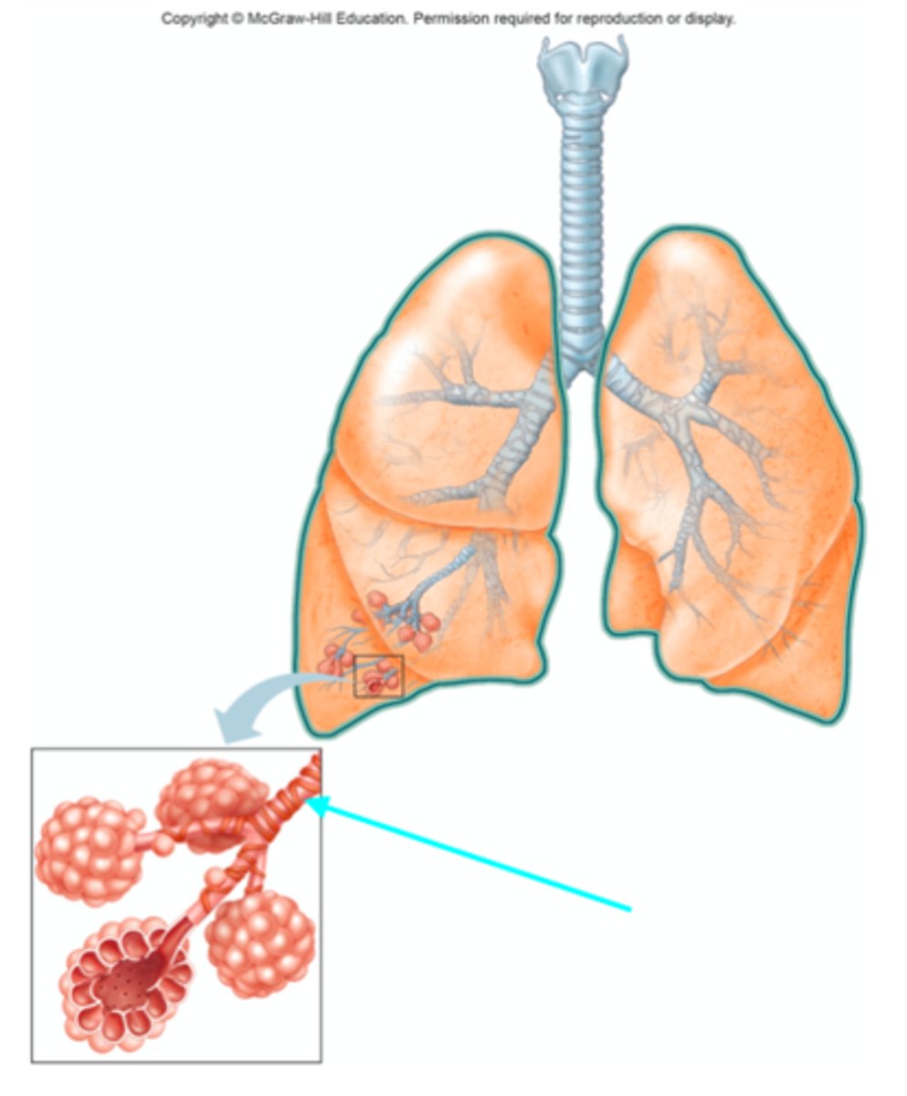
Respiratory bronchiole
Which structure of the lung diagram is the blue arrow pointing at?
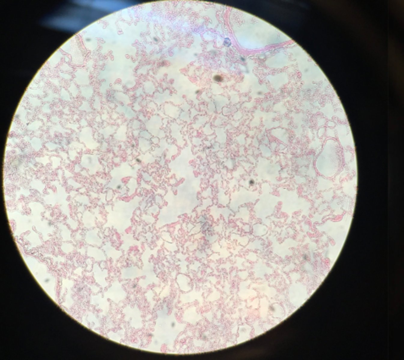
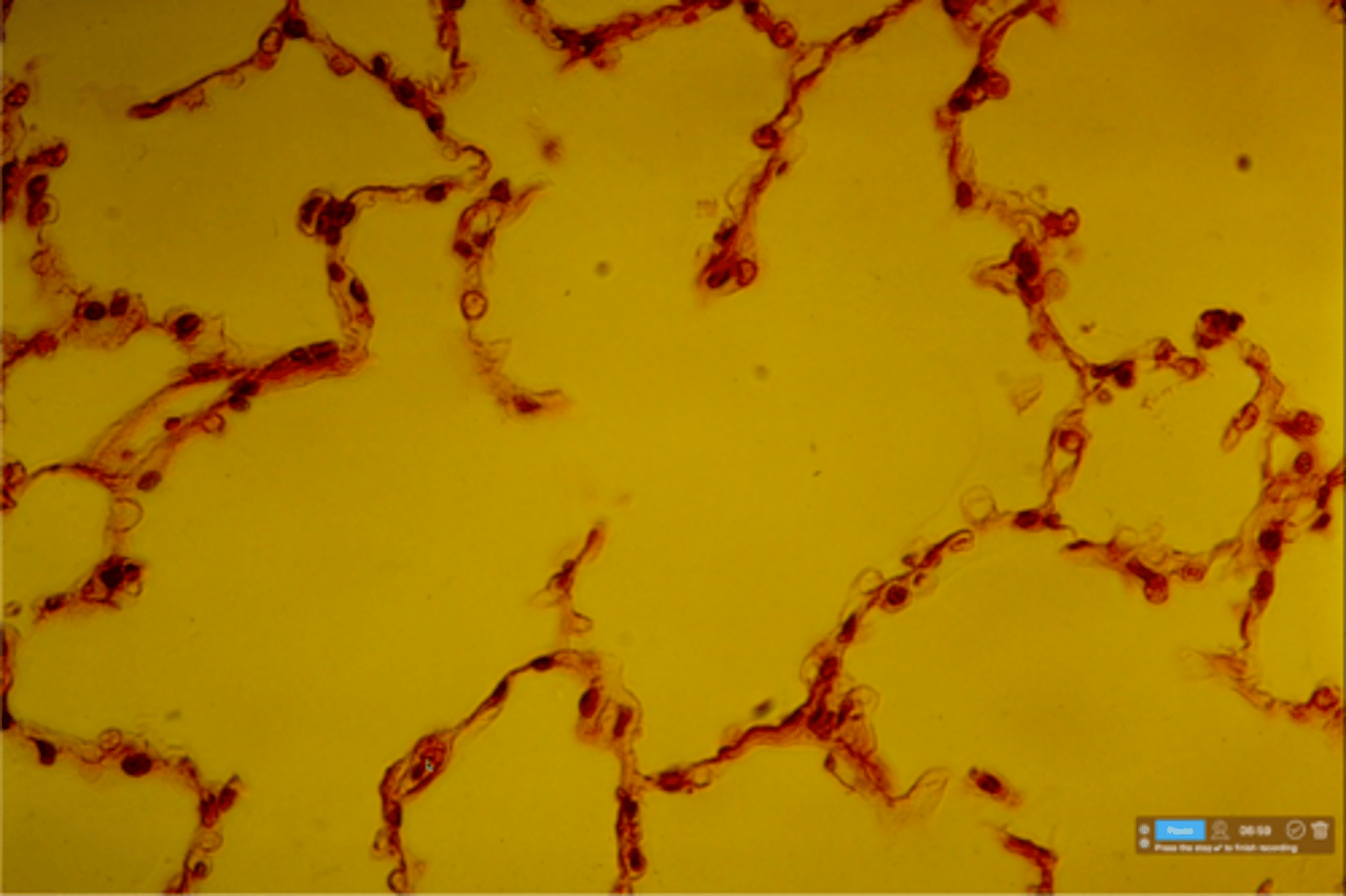
Normal lung tissue
What type of tissue is depicted on this microscope slide?
Alveolus
What structure is the blue arrow pointing at?

Simple Squamous epithelium
Which tissue is depicted on this slide? This makes up the walls of an alveolus.
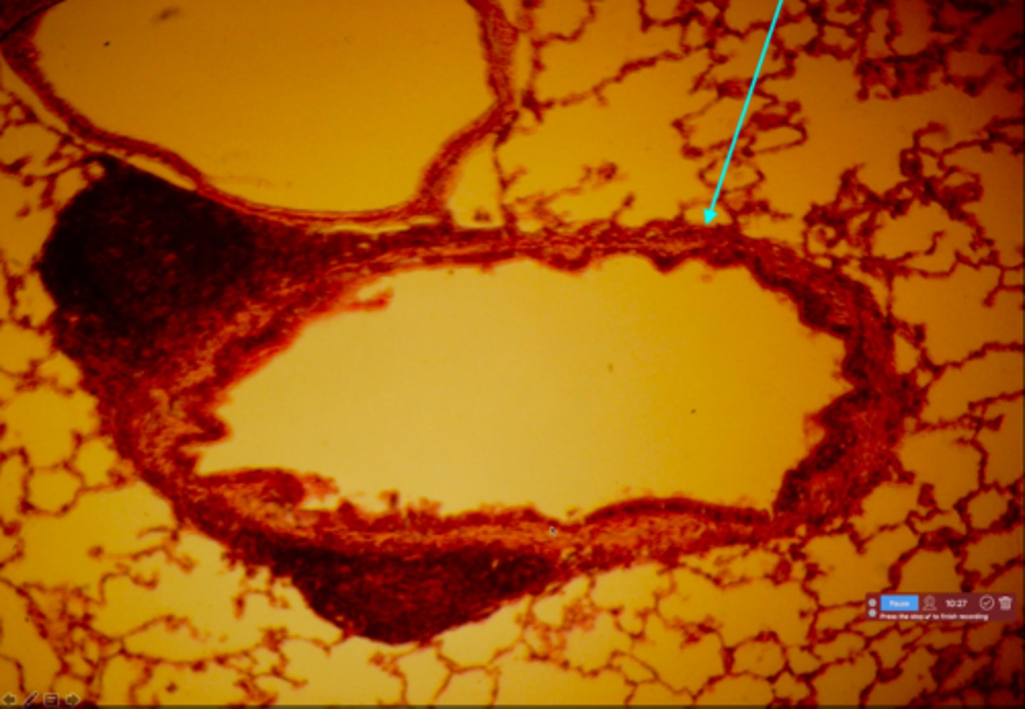
Intralobular bronchiole
What structure is the blue arrow pointing at?

Ciliated pseudostratified columnar epithelium
This is a slide showing an interlobular bronchiole. What type of cells lines this bronchiole?
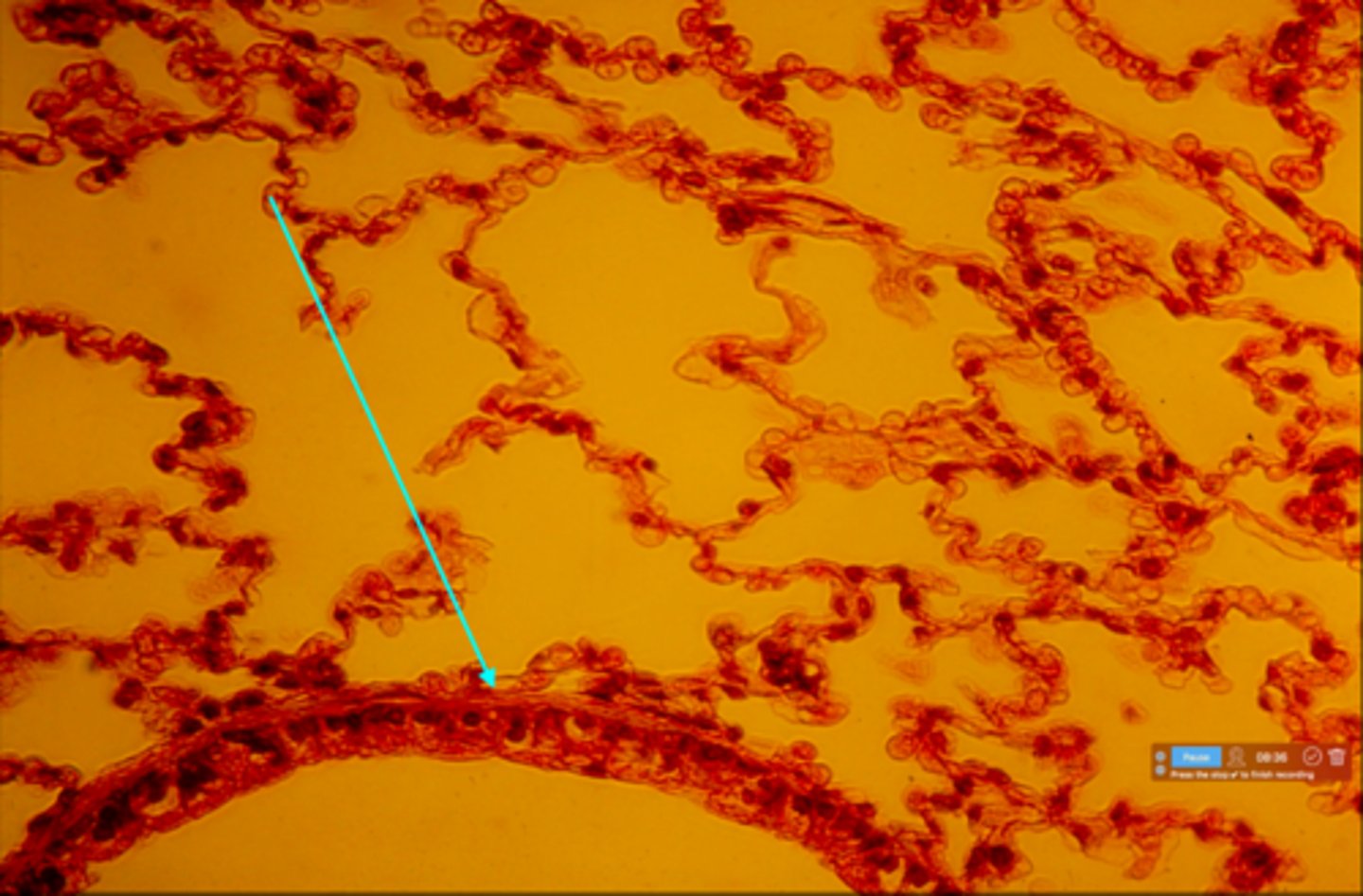
Respiratory bronchiole
What structure is the blue arrow pointing at?

Simple cuboidal epithelium
This is a slide showing a respiratory bronchiole. What type of cells lines this bronchiole?
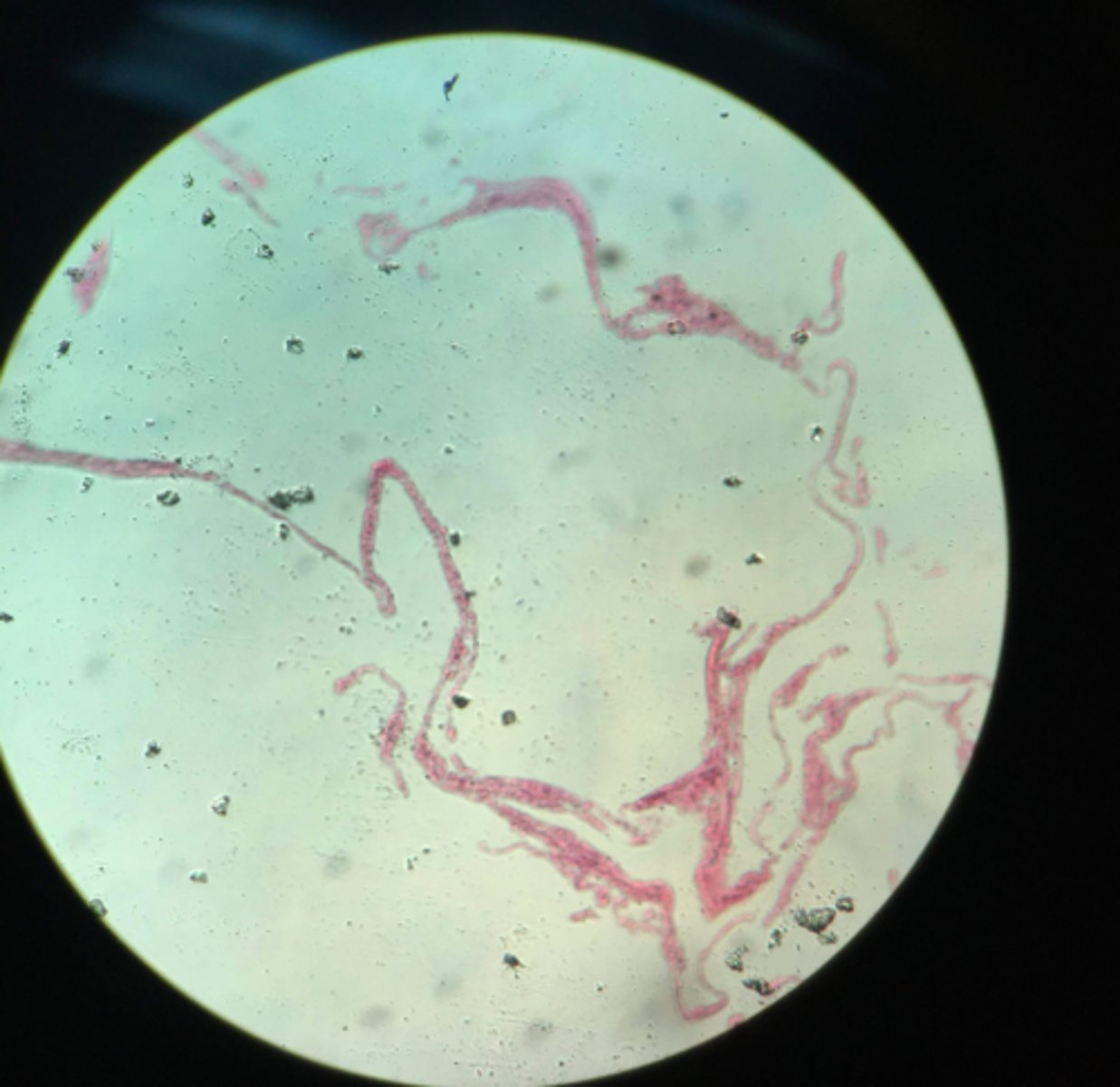
Lung tissue from a patient with emphysema
What type of tissue depicted on this slide? What condition did this person have?
Trachea
What organ is shown on this microscope slide?

Ciliated pseudostratified columnar epithelium
This is a slide depicting the trachea. What layer of cells is the blue arrow pointing at?
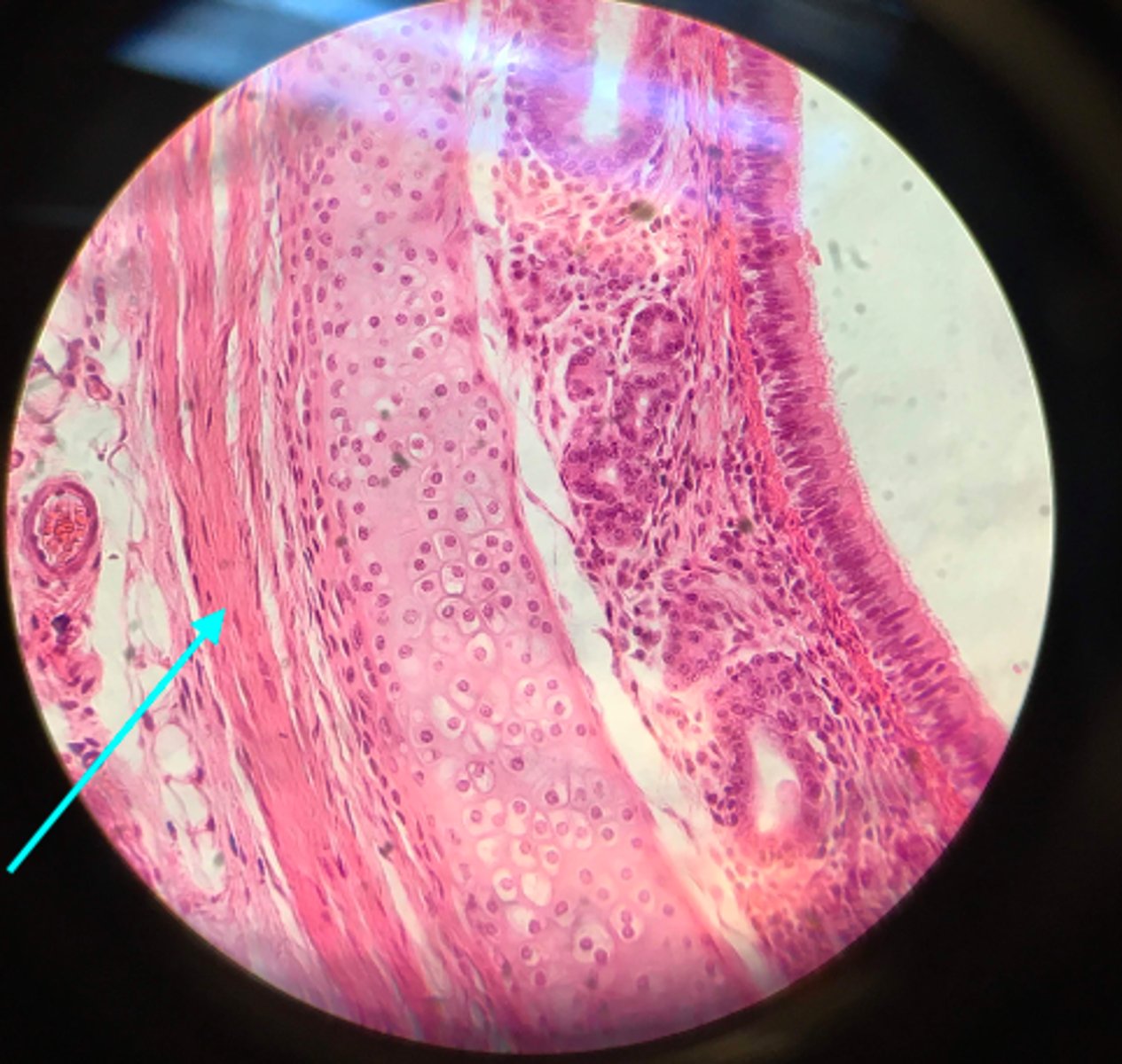
Hyaline cartilage
This is a slide depicting the trachea. What type of tissue is the blue arrow pointing at?

Smooth muscle tissue
This is a slide depicting the trachea. What type of tissue is the blue arrow pointing at?
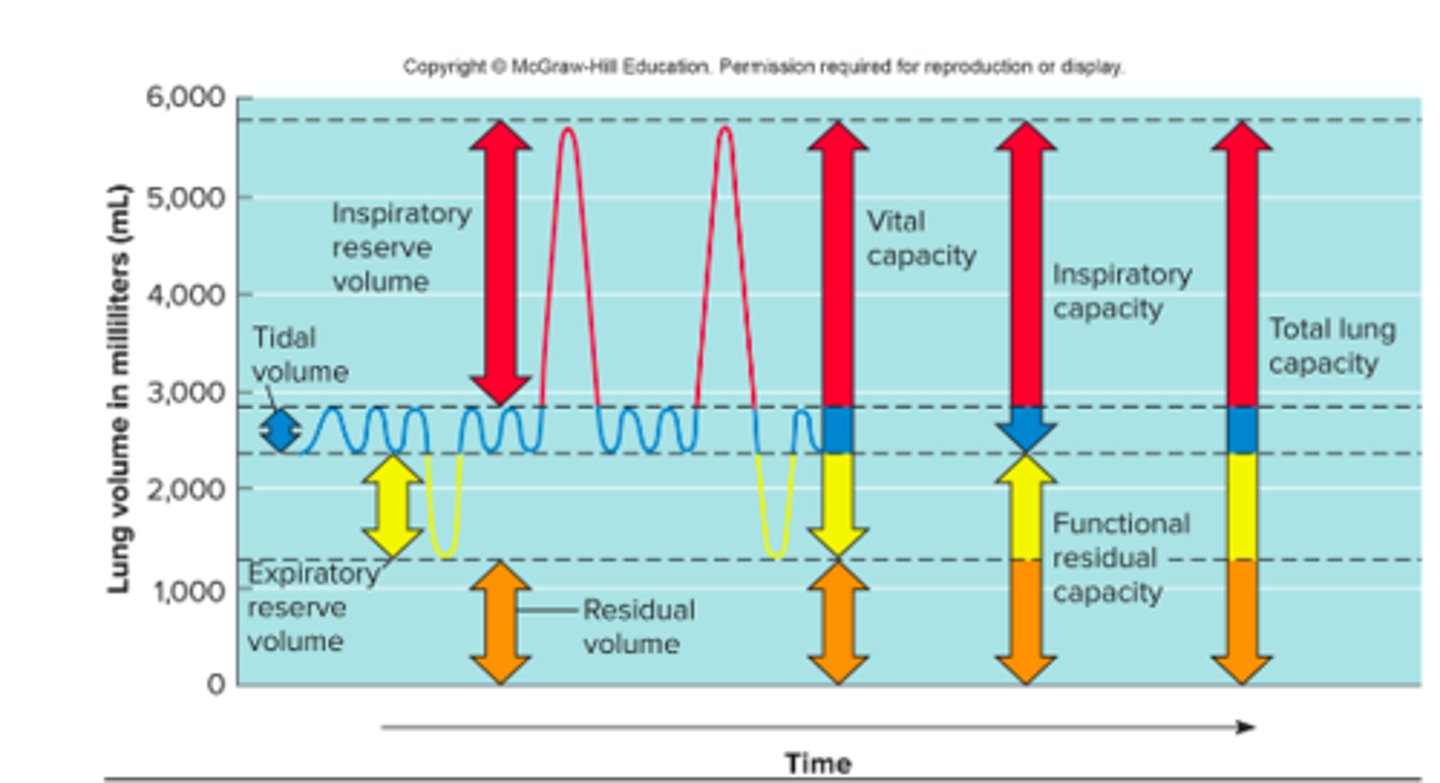
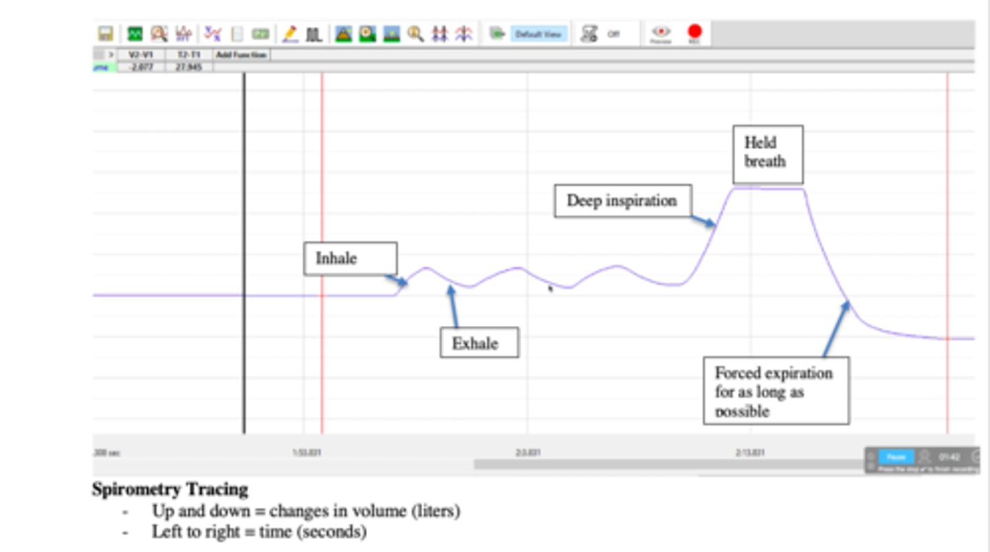
Tidal volume (TV)
What volume is shown on this spirometer tracing?
Inspiratory reserve volume (IRV)
What volume is shown on this spirometer tracing?
Expiratory reserve volume (ERV)
What volume is shown on this spirometer tracing?
Vital capacity (VC)
What capacity is shown on this spirometer tracing?
Inspiratory capacity (IC)
What capacity is shown on this spirometer tracing?
Functional residual capacity (FRC)
This capacity of the lungs can be calculated by combining residual volume and the expiratory reserve volume
Total lung capacity (TLC)
This is the maximum amount of air contained in lungs after a maximum inspiratory effort. This is the maximum amount of air the lungs can hold. This capacity can be calculated by combining Inspiratory reserve volume, tidal volume, expiratory reserve volume, and residual volume.
Tidal volume
This volume is the difference between the peak of normal inhalation and the trough of normal exhalation. This is normal breathing and is the volume of air from one normal exhalation
Inspiratory reserve volume
This is the volume of air one can inhale beyond normal inhalation.
Expiratory reserve volume
This is the volume of air one can exhale after normal exhalation.
Vital capacity
This capacity is the maximum amount of air that can be exhaled from the lungs. This goes from peak inhalation to the plateau of maximum exhalation. This can be calculated by adding Inspiratory reserve volume, tidal volume, and expiratory reserve volume
Inspiratory capacity
This capacity is the maximum amount of air that can be inhaled. This goes from the end of normal exhalation to the peak of forced inhalation. This can be calculated by adding inspiratory reserve volume and tidal volume.
Residual volume
This is the volume of air that one cannot exhale; this is always present in the lungs.
Inspiratory capacity (IC)
Inspiratory reserve volume (IRV) + Tidal volume (TV) = ?
or
Total lung capacity (TLC) - Functional residual capacity (FRC) = ?
or
Vital capacity (VC) - Expiratory reserve volume (ERV) = ?
Functional residual capacity (FRC)
Expiratory reserve volume (ERV) + Residual volume (RV = ?
or
Total lung capacity (TLC) - Inspiratory capacity (IC) = ?
Vital capacity (VC)
Inspiratory reserve volume (IRV) + Tidal volume (TV) + Expiratory reserve volume (ERV) = ?
or
Inspiratory capacity (IC) + Expiratory reserve volume (ERV) = ?
or
Total lung capacity (TLC) - Residual volume (RV) = ?
Total lung capacity (TLC)
Inspiratory reserve volume (IRV) + Tidal volume (TV) + Expiratory reserve volume (ERV) + Residual volume (RV) = ?
or
Vital capacity (VC) + Residual volume (RV) = ?
or
Inspiratory capacity (IC) + Functional residual capacity (FRC) = ?
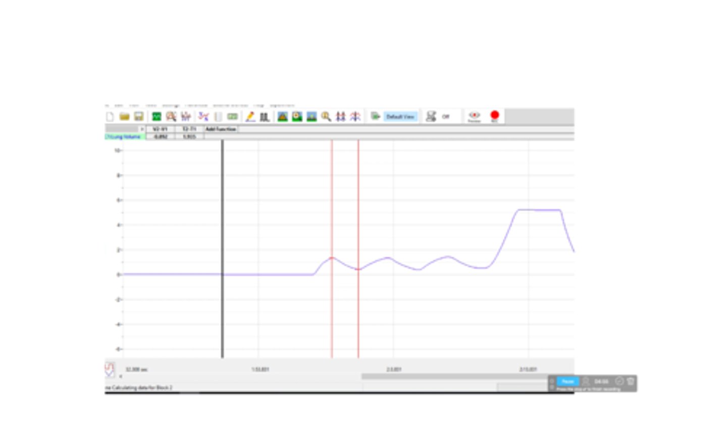
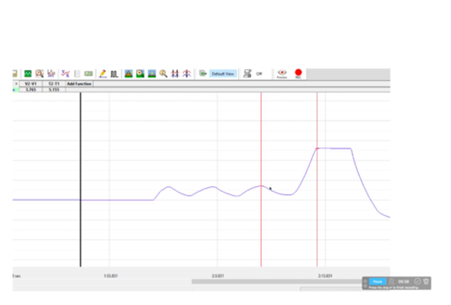
Duration of one breath
What is depicted on the spirometry tracing?
RR = Breaths/minute
- First calculate how long the respiratory cycle duration goes on
- Go from one peak of the tidal volume to the next peak = 1 breath/x seconds
- Convert 1 breath/x seconds to breaths/minute
How does one calculate respiratory rate (RR)?
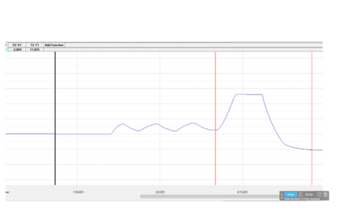
Respiratory rate (RR)
This is the rate determining how many breaths one breathes in a minute
Minute ventilation
This is the volume of air moved through all respiratory passages in one minute
MV = RR x TV
Respiratory rate * Tidal volume = Minute ventilation in mL/min
How does one calculate minute ventilation (MV)?
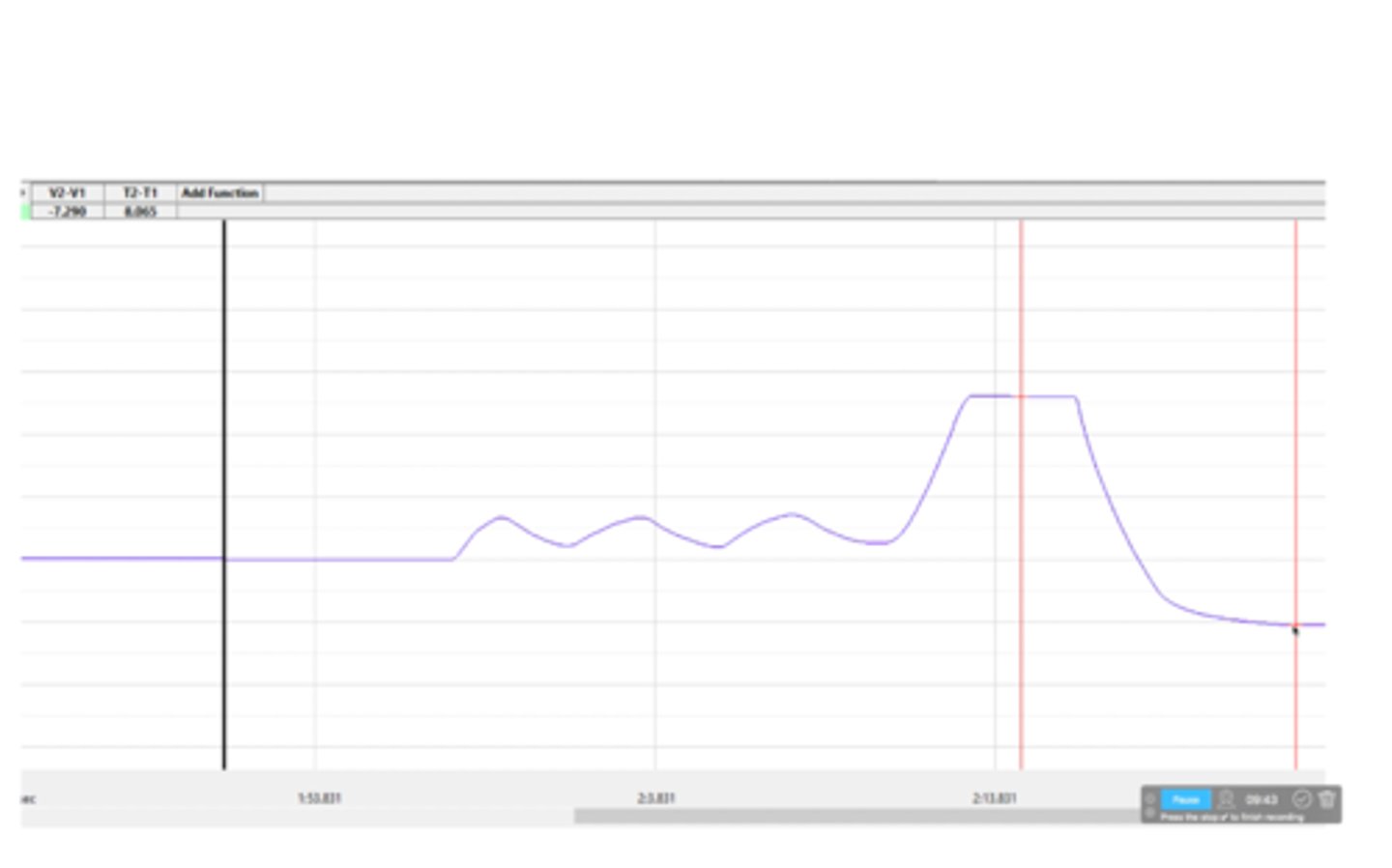
Dead space (DS)
This is the volume of air in each breath that does not participate in gas exchange. This number is given to you for calculations. The volume consists of air in the trachea, major bronchi, and many bronchioles where exchange does not take place
AVR = RR x (TV-DS)
Respiratory rate * (Tidal volume - Dead space) = Alveolar ventilation rate in mL/minute
How does one calculate alveolar ventilation rate (AVR)?
Alveolar ventilation rate (AVR)
This is the volume of air that participates in gas exchange in one minute - must subtract volume of dead space (volume not involved in gas exchange)
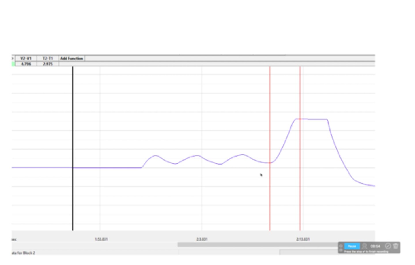
3,259 mL
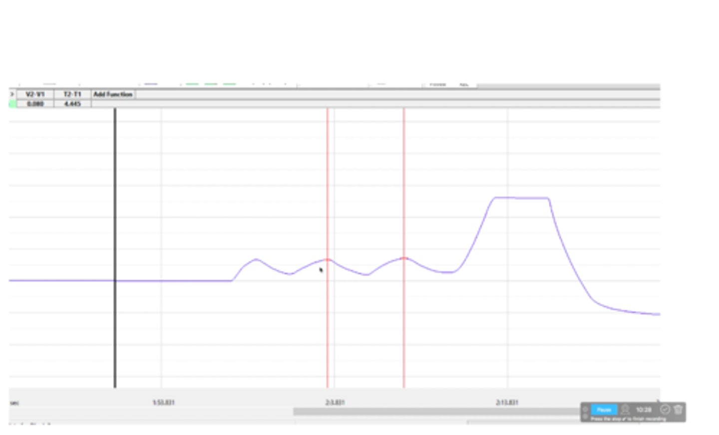
What is the forced expiratory volume after 1 second (FEV1)? This occurs from the start of forced exhalation to one second after

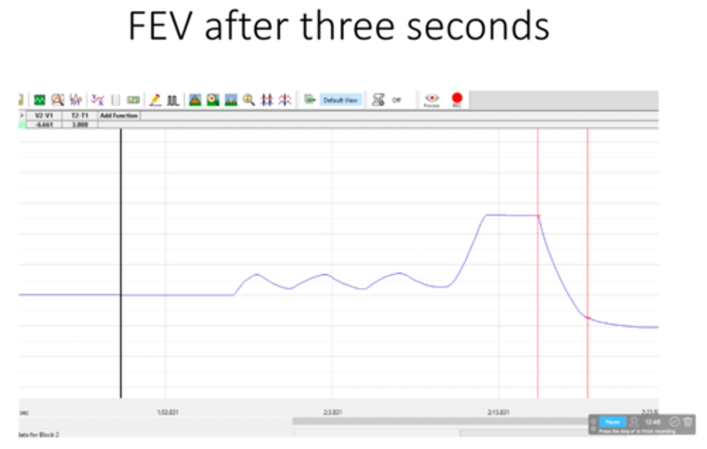
6,661 mL
What is the forced expiratory volume after 3 seconds (FEV3)? This Occurs from the start of forced exhalation until 3 seconds after
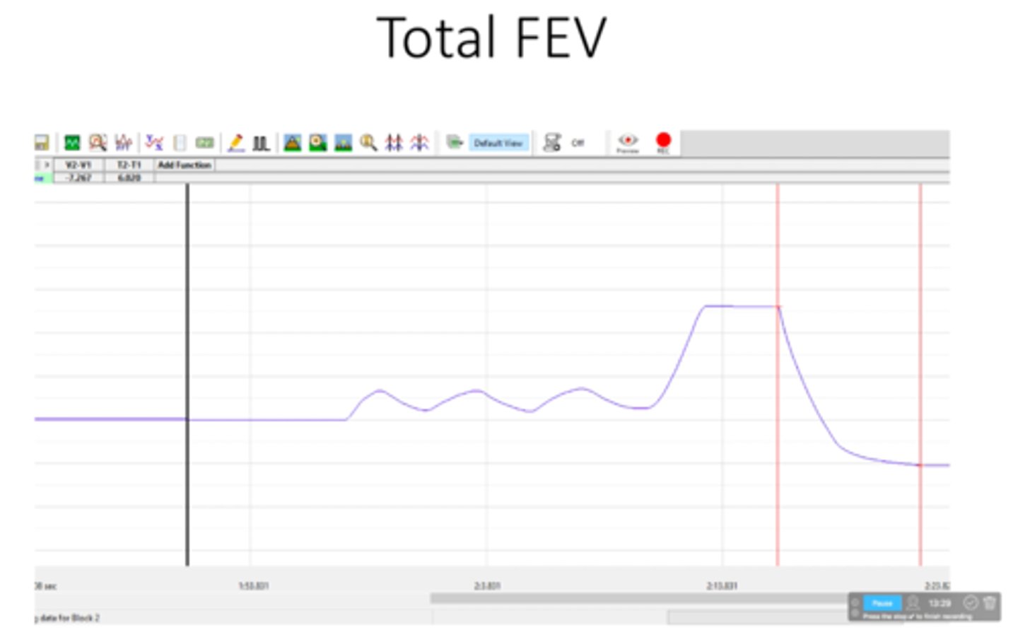
7,267 mL
What is the total forced expiratory volume (FEVT)? This occurs from start of forced exhalation until it plateaus and no more can be exhaled
FEV1/FEVT = 3,259 mL / 7,267 mL x 100% = 45%
Forced expiratory volume, 1 second (FEV1) = 3,259 mL ; Forced expiratory volume, total (FEVT) = 7,267
Calculate Forced expiratory volume, % total after 1 second (%FEVT1)
FEV3/FEVT = 6,661 mL / 7,267 mL x 100% = 92%
Forced expiratory volume, 3 seconds (FEV3) = 6,661 mL ; Forced expiratory volume, total (FEVT) = 7,267 mL
Calculate forced expiratory volume, % total after 3 seconds (%FEVT3)
A normal individual should be able to exhale ~75% of the total volume in the first second and ~95% of the total volume within the first 3 seconds.
%FEVT1 = ~75%
%FEVT3 = ~95%
What are the "normal" values for %FEVT1 and %FEVT3?
Patients with emphysema or forms of COPD may not be able to forcefully exhale the volume of air in their lungs as quickly as someone without those conditions. Damage to the alveoli and structures of the lungs make it difficult for the lungs to recoil and release their contents. Inhalation may not be the problem, but upon forcing exhalation, it would not be possible to do so efficiently. This would decrease the Forced expiratory volume % totals after 1 and 3 seconds
What is a condition that would affect the values for %FEVT1 and %FEVT3? (Forced expiratory volume, % total after 1 and 3 seconds)