functional anatomy midterm
1/138
There's no tags or description
Looks like no tags are added yet.
Name | Mastery | Learn | Test | Matching | Spaced | Call with Kai |
|---|
No analytics yet
Send a link to your students to track their progress
139 Terms
what 3 bones make up the pelvis
ilium, ischium, pubis (innominate bones)
are the bones that make up the pelvis innominate or nominate?
innominate
what is an innominate bone?
fused bone
where is the acetabulum?
where innominate bones meet
what are the key differences between the male and female pelvis?
male = less curved sacrum & coccyx, less wide, smaller pubic arch angle
the head of the humerus is on the ________ side
medial
the greater trochanter is on the _______ side of the femur
lateral
the linear aspera is on the ______ aspect of the femur
posterior
the ___________ joins the right and left pubic bones together
pubic symphysis
what is the function of the acetabular labrum?
deepens socket of joint
acts as a suction cup
what type of joint is the hip?
ball and socket
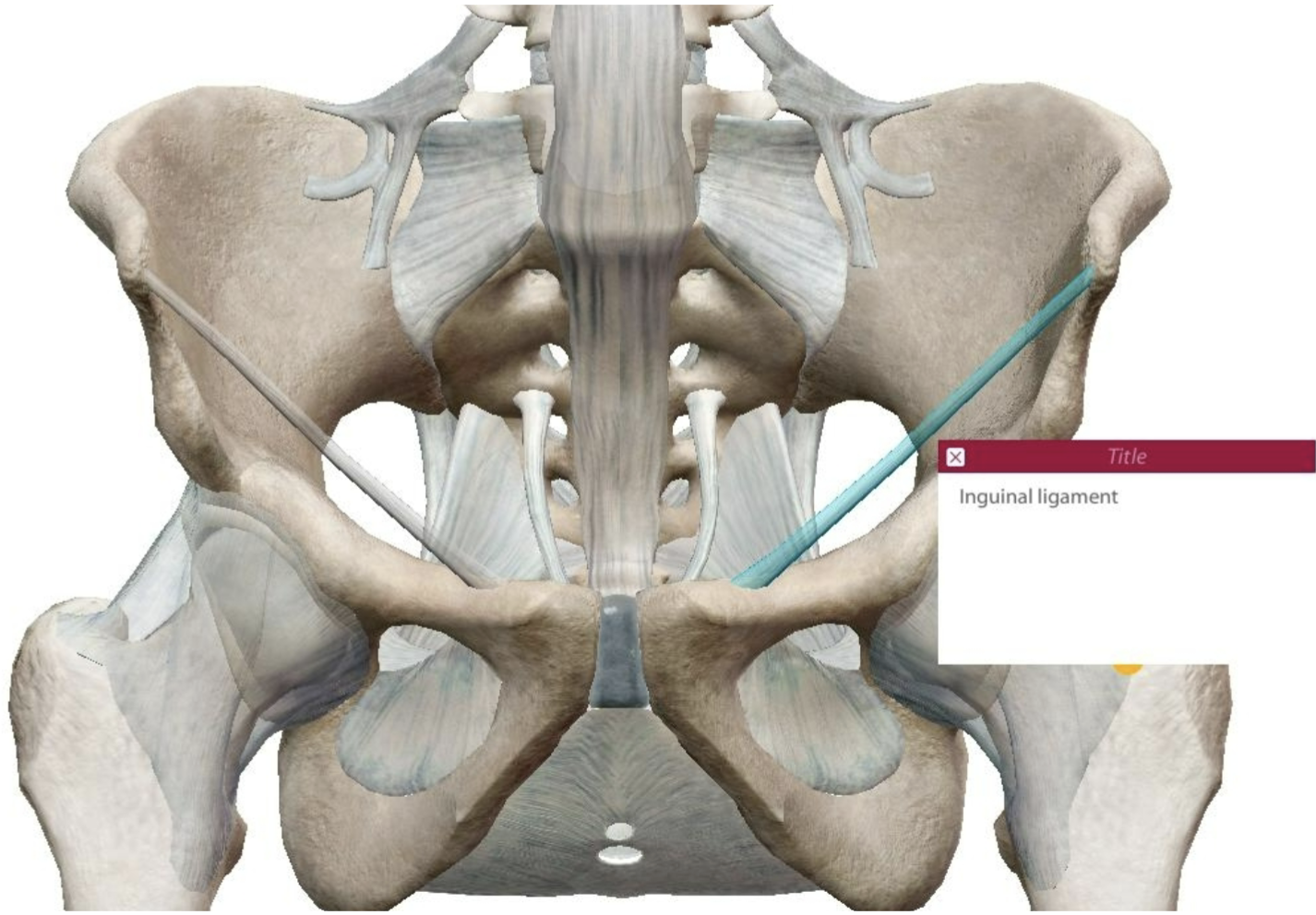
where is the inguinal ligament?
ASIS → pubic bone
the __________ ligament is also called the “Y ligament”
illiofemoral
the ligamentum teres is unique in that it has _________ that passes through the ligament
blood supply
what motion does the pubofemoral ligament resist?
hyper abduction
the iliacus, sartorius, and rectus femoris are all hip flexor muscles that are innervated by what nerve?
femoral nerve
the _____ head of the biceps femoris originates on the ischial tuberosity
long
all of the hamstring muscles EXCEPT the _________ are innervated by the tibial nerve
short head of biceps femoris
what are the actions of the gluteus medius muscle?
hip abduction
external rotation (posterior fibers)
internal rotation (anterior fibers)
list the muscles with insertions on the greater trochanter
gluteal group (max, med, min)
external rotators (piriformis, superior & inferior gemellus, obturator internus, quadratus femoris)
list the muscle with origins on the ischial tuberosity
long head biceps femoris
semitendinosus
semimembranosus
all of the adductor muscles are innervated by the ______ nerve except the pectineus which is innervated by the femoral nerve
obturator
all of the adductor muscles are innervated by the obturator nerve except the pectineus which is innervated by the _______ nerve
femoral
what comprises the femoral triangle and why is it an important region of the hip?
inguinal ligament, sartorius, adductor longus
it is the area where the neurovascular structures pass through anteriorly and can be easily accessed
describe the general location of the following nerve paths: femoral, obturator, tibial, fibular
femoral- anterior, lateral aspect of leg
obturator- anterior medial aspect of leg
tibial- posterior, medial aspect of leg
fibular- posterior, lateral aspect of leg
what muscles are in the anterior compartment?
quadriceps & sartorius
what nerve runs through the medial compartment?
obturator
what muscle group is in the posterior compartment?
hamstrings
the ___________ is viewed in the frontal plane and is the angle between the head and neck to the shaft of the femur
angle of inclination
the angle of inclination is viewed in the frontal plane and is the angle between the head and neck to the ______ of the femur
shaft
angles greater than 125 degrees for the angle of inclination would indicate
coxa valga
the ________ is viewed in the transverse plane and is the angle between the head & neck to the femoral condyles
angle of torsion
the angle of torsion is viewed in the transverse plane and is the angle between the ___________ to the femoral condyles
head & neck
what is anteversion?
angle of torsion > 15 degrees
may observe toe in posture
hip flexors
iliacus
psoas major
sartorius
rectus femoris
iliacus
origin: iliac fossa
insertion: lesser trochanter
action: hip flexion, trunk flexion when seated
nerve innervation: femoral
psoas major
origin: anterior aspects of lumbar vertebra
insertion: lesser trochanter
action: hip flexion, trunk flexion when seated
nerve innervation: L1-L4 spinal nerves
sartorius
origin: ASIS (Anterior Superior Iliac Spine)
insertion: pes anserine
action:
hip- flexion, abduction, external rotation
knee- flexion, internal rotation
nerve innervation: femoral
rectus femoris
origin: AIIS (Anterior Inferior Iliac Spine)
insertion: tibial tuberosity
action:
hip- flexion
knee- extension
nerve innervation: femoral
hip extensors
gluteus maximus
biceps femoris
semitendinosus
semimembranosus
gluteus maximus
origin: sacrum superior gluteal line of ilium
insertion: IT (iliotibial) band gluteal tuberosity
action:
hip- extension, abduction, external rotation
nerve innervation: inferior gluteal
biceps femoris
origin:
long head- ischial tuberosity
short head- linea aspera
insertion: head of fibula, posterolateral tibia
action:
hip- extension
knee- flexion
nerve innervation:
long head- tibial
short head- common peroneal/fibular
semitendinosus
origin: ischial tuberosity
insertion: anterior medial tibial shaft pes anserine
action:
hip- extension
knee- flexion
nerve innervation: tibial
semimembranosus
origin: ischial tuberosity
insertion: posterior medial tibial condyle
action:
hip- extension
knee- flexion
nerve innervation: tibial
hip abductors
gluteus medius
gluteus minimus
tensor fascia latae
gluteus medius
origin: iliac crest gluteal surface
insertion: greater trochanter
action:
hip- abduction, internal rotation (anterior fibers), external rotation (posterior fibers)
nerve innervation: superior gluteal
gluteus minimus
origin: lateral surface of ilium
insertion: greater trochanter
action:
hip- abduction, internal rotation
nerve innervation: superior gluteal
tensor fascia latae
origin: ASIS (anterior superior iliac spine)
insertion: IT (iliotibial) band
action:
hip- abduction, flexion, internal rotation
nerve innervation: superior gluteal
hip adductors
adductor longus
adductor brevis
adductor magnus
gracilis
pectineus
adductor longus
origin: pubis
insertion: linea aspera
action:
hip- adduction, external rotation
nerve innervation: obturator
adductor brevis
origin: pubis
insertion: linea aspera
action:
hip- adduction, external rotation
nerve innervation: obturator
adductor magnus
origin: pubis & ischial tuberosity
insertion: adductor tubercle, linea aspera
action:
hip- adduction, external rotation
nerve innervation: obturator
gracilis
origin: pubic
insertion: anterior medial tibia, pes anserine
action:
hip- adduction
knee- flexion, internal rotation
nerve innervation: obturator
pectineus
origin: pubis
insertion: lesser trochanter, linea aspera
action:
hip- adduction, flexion, external rotation
nerve innervation: femoral
what bone is also known as the thigh?
femur
what bones are also known as the leg?
tibia & fibula
the pes anserine is located on the ______ side of the tibia
medial
what 3 muscles insert on the pes anserine?
sartorius, gracilis, semitendinosus
gerdy’s tubercle is located on the _____ side of the tibia
lateral
what are the 3 joints of the knee?
patellofemoral, tibiofemoral, proximal tibiofibular joint
true or false: the tibiofemoral joint is a traditional hinge joint
false; modified
the ________ femoral condyle is longer and the medial condyle is wider
lateral
the lateral femoral condyle is longer and the ________ condyle is wider
medial
because the femoral condyles differ in size and shape, in order to achieve full knee extensnion, the tibia needs to externally rotate during the last 20-30 degrees of extension. This is called the _______________
screw home mechanism
the patella is an extremely moveable structure. why is this potentially problematic?
prone to dislocation or sublxation
what type of joint is the proximal tibiofibular joint?
arthroidal
what is the purpose of the menisci?
dissipates pressure, reduce friction, provide support
what does cruciate mean?
cross
which ligaments are situated inside of the knee joint and cross over another?
anterior & posterior cruciate ligaments
what type of forces does the ACL resist?
anterior translation of tibia, internal rotation of tibia
where is the PCL located?
origin: lateral surface of medial femoral condyle
insertion: posterolateral tibial plateau
where are the collateral ligaments located?
on the sides (medial & lateral)
the MCL resists ___ forces
valgus
and the LCL resists _____ forces
varus
the patellar tendon is also known as the _______________
patellar ligament
what nerve innervates all of the quadriceps muscles?
femoral
all of the quadriceps muscles cause what action to occur?
knee extension
list all of the muscles that cause knee flexion to occur
biceps femoris, semimembranosus, semitendinosus, gastrocnemius, gracilis, sartorius
what is the purpose of the bursae?
lubrication at areas of higher friction
what does the q-angle measure?
angle between ASIS & tibial tubercle (axis is at patella)
what is a normal q-angle (degrees)?
~15 degrees
what does genu varum look like clinically?
bow legged
what does genu valgum look like clinically?
knock knees
what are the contents of the popliteal fossa?
sciatic nerve
popliteal artery
saphenous vein
knee extensors
rectus femoris
vastus medialis
vastus laterais
vastus intermedius
rectus femoris
origin: AIIS (anterior inferior iliac spine)
insertion: tibial tuberosity
action:
hip- flexion
knee- extension
nerve innervation: femoral
vastus medialis
origin: intertrochanteric line & medial linea aspera
insertion: tibial tuberosity
action:
knee- extension
nerve innervation: femoral
vastus lateralis
origin: greater trochanter, linea aspera
insertion: tibial tuberosity
action:
knee- extension
nerve innervation: femoral
vastus intermedius
origin: upper 2/3 of anterior surface of femur
insertion: tibial tuberosity
action:
knee- extension
nerve innervation: femoral
knee flexors
gastrocnemius
hamstring muscles
gracilis
sartorius
gastrocnemius
origin: medial & lateral femoral condyles
insertion: calcaneus via achilles tendon
action:
knee- flexion
ankle- plantar flexion
nerve innervation: tibial
what bones are included in the tarsal bones?
cuneiforms (3), cuboid, navicular, calcaneus, talus
the __________ bone is wedge shaped and located on the medial side of the foot
navicular
where is the cuboid bone located?
lateral side of foot
the heelbone is also known as the __________
calcaneus
the talus is _____ shaped on the superiorsurface where it articulates with the distal tibia and fibula
dome
the base of the metatarsal is located ______ and the head is located distally
proximally
the base of the metatarsal is located proximally and the _____ is located distally
head
what is unique/different about the first toe when considering the phalanges?
there’s no middle phalynx
true or false: all the toes have distal, middle, and proximal phalanges
false