Diagnostic Imaging Week 3- Arthritis of the Extremities
1/16
There's no tags or description
Looks like no tags are added yet.
Name | Mastery | Learn | Test | Matching | Spaced | Call with Kai |
|---|
No analytics yet
Send a link to your students to track their progress
17 Terms
What is the most common form of arthritis
DJD
What are the main symptoms of DJD
-Pain
-Stiffness
-Crepitus
-Deformity
-Swelling
-Normal lab studies
What are the 3 types of DJD
Primary
Secondary
Erosive osteoarthritis
What are the features of primary DJD (cause, age, sex, joint)
-unknown cause
-5th-6th decade
-females 10:1
-weight-bearing joints
What are the features of secondary DJD (cause, age, sex, joint)
-known cause
-2nd-6th decade
-equal sex distribution
-any joint
What is the pathology of DJD
-Begins focally, increases in size
-Loss of chondroitin sulphate leads to break down of articular cartilage and adjacent bone
-Synovial fluid escapes in subchondral bone, forming subchondral bone cysts
What are the main radiological features of DJD
-Osteophytes
-Subchondral sclerosis
-Non-uniform joint space narrowing
-Subchondral cysts
-Loose bodies
-Asymmetrical distribution
-Subluxation
What is the colour of the cortices in CT vs MRI scans?
CT: white
MRI: black
Rheumatoid arthritis demographic
-Onset typically 20-60 y/o
-Highest incidence 40-50 y/o
-Under 40 = Females 3:1
-Over 40 = Equal sex distribution
What are the radiological features of rheumatoid arthritis
-Uniform loss of joint space
-Marginial erosion
-Subchondral bone cysts
-Juxta-articular osteoporosis
-Ankylosis
-Boutenniere and swan neck deformity
-Subcutaneous soft tissue mass
-Enthesopathy
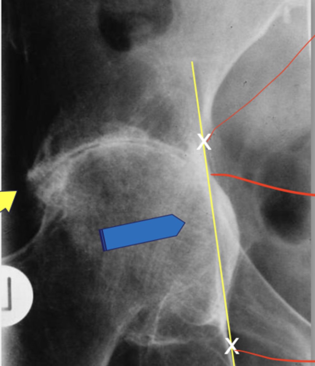
In what condition is protrusio acetabuli often seen? How do you detect it?
-Rheumatoid arthritis
-Align most lateral aspect of greater sciatic notch with obturator foramen
-Draw line done
-If femoral had crosses the line, it is positive
What are the main features of Erosive Osteoarthritis (EOA) (sex, age, type, location)
-Females
-40-50 y/o
-Inflammatory
-Seronegative
-DIPs and PIPs
What are the main radiographic features of EOA
-Non-uniform loss of joint space
-Sclerosis
-Osteophytes
-Gull wing erosions
-Periostitis
-Deformity
What's the main difference between psoriatic arthritis and erosive osteoarthritis
-PA: Mouse ears
-EOA: Gull wings
-PA maintains bone mineralisation adjacent to the involved joint, EOA does not
What are the main affected areas of PA
-DIPs
-Small joints of the feet
-SIJ
-Spine
-Occasionally knee, hip, shoulder
What are the main radiographic findings of PA
-Asymmetric distribution
-Normal bone mineralisation
-Mouse ear erosions (erosion+ fluffy periostitis)
-Narrow or widened joint space
-Pencil in cup deformity
-Bony ankylosis
-Acro osteolysis
-Os trigonum
-Enthesitis
-Ivory phlanax
-Sacrolitis
-Non- marginal syndesmophyte formation
What are the main features of PA (age, presentation)
-20-50 y/o
-Sausage digits
-Skin lesions
-Presence of nail changes
-DIPs
-Seronegative (ESR normal)