Toes, Foot, and Ankle Positioning
1/271
There's no tags or description
Looks like no tags are added yet.
Name | Mastery | Learn | Test | Matching | Spaced | Call with Kai |
|---|
No analytics yet
Send a link to your students to track their progress
272 Terms

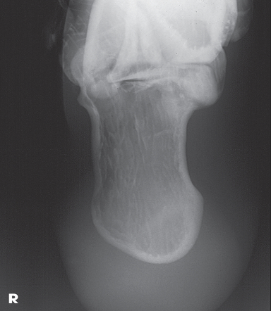
Name this projection.
Lateral calcaneus

What type of projection is it?
Mediolateral

Is the patient on the affected or unaffected side?
Affected

State specifically where the CR enters.
Perpendicular to the calcaneus // 1" distal to the medial malleolus
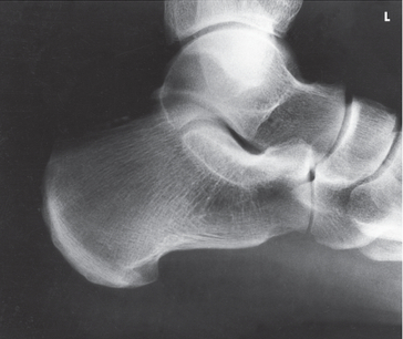
Which tarsal is in profile?
Calcaneus

Which radiographically significant landmark is open?
Sinus tarsi

Name this projection.
AP oblique toes

State the degree and type of rotation used.
Medial rotation 30-45 degrees

Which aspect of the foot rests on the IR?
Medial aspect

Where does the CR enter?
Perpendicular to 3rd MTP joint
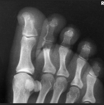
When radiographing the 1st-2nd toes, which oblique should be used?
Medial (rotate on medial/unaffected side)

When radiographing the 4th-5th toes, which oblique should be used?
Lateral (rotate on lateral/affected side)

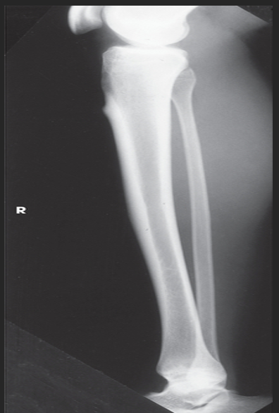
Name this projection.
AP leg (tib fib)

What SID is used?
48 inches
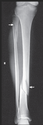
What anatomy is placed parallel to the IR?
Patella and femoral condyles

What position is the patient in?
Supine w/ foot dorsiflexed

What anatomy must be included on the radiograph?
Entire tibia and fibula plus adjacent joints

What should be done if the entire tib/fib cannot fit on the IR?
Either rotate the IR to be diagonal to the leg OR take two separate images (a bigger image of whichever joint is more in pain and a smaller image of the other joint)

How far beyond the adjacent joints should you see light?
1.5"

What part of the tib/fib should be free of superimposition?
Fibula midshaft

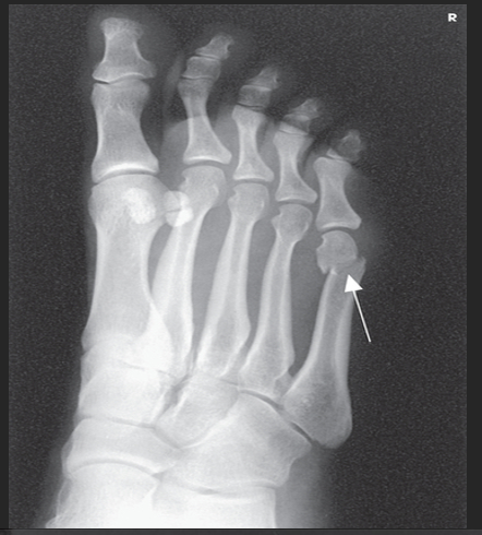
Name this projection.
Tangential sesamoids

What anatomy is of primary interest?
Sesamoid bones

State the method name if the patient is in the prone position.
Lewis

The Holly method is used when the patient is
supine

Where does the CR enter?
Perpendicular and tangential to 1st MTP joint
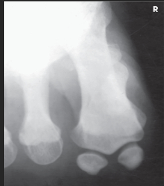
What anatomy is in profile?
Sesamoid bone and metatarsal head

Name this projection.
AP axial foot

What type of projection is it?
Dorsoplantar

Which surface of the foot should touch the IR?
Plantar surface

How is the CR directed (angle)?
10 degrees posteriorly

Where does the CR enter?
To the base of the 3rd metatarsal

Which tarsals should be seen on the radiograph?
The tarsals that are anterior to the talus (all cuneiforms, cuboid, and navicular)

Overlap of the ________ metatarsal bases should be seen.
2nd-5th
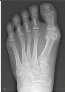
There should be equal space between _________ metatarsals.
2nd-4th

What joint spaces (if any) should be open?
IP, MTP, and TMT joints (+ the space between the 1st and 2nd cuneiforms)

Name this projection.
AP oblique ankle

State the degree and type of rotation used.
45 degrees medially

Where does the CR enter?
Perpendicular to the ankle; midway between the malleoli
Which aspect of the ankle is best visualized?
Lateral

Which joint space must be open?
Distal tibiofibular joint

Name this projection.
Lateral toes

Describe the patient position for the 2nd toe.
Seated or lateral recumbent: rolled onto the unaffected side with the foot resting on its medial aspect

Describe the patient position for the 4th toe.
Seated or lateral recumbent: rolled onto the affected side with the foot resting on its lateral aspect

What type of projection is used for the 1st toe?
Lateromedial

What type of projection is used for the 5th toe?
Mediolateral

Where will the CR enter for the 1st toe?
Perpendicular to the IP joint
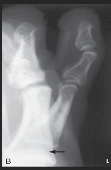
Where will the CR enter for the 3rd toe?
Perpendicular to the PIP joint

At a minimum, what anatomy must be seen on the radiograph?
Proximal phalanx

What anatomy is in profile?
Toenail

Name this projection.
Lateral leg (tib-fib)

What type of projection is it?
Mediolateral

What SID is used?
48"

Describe the position of the patient.
recumbent onto the affected side; keep the knee slightly flexed to obtain a true lateral

What anatomy must be ⟂ to the IR?
Femoral condyles and patella
Where does the CR enter?
Perpendicular to the midpoint of the leg

What must be seen on the radiograph?
Entire tibia and fibula with adjacent joints

Describe how to evaluate for a true lateral.
Distal fibula superimposed by posterior half of tibia; slight overlap between tibia and fibular head; separation of tibial and fibular bodies (not at ends)

Name this projection.
AP oblique foot

Describe the position of the patient.
Seated or supine with their knee flexed and lower leg/foot rotated medially 30 degrees
Where does the CR enter?
Base of the 3rd metatarsal

What is demonstrated?
The lateral side of the foot from the toes to heel, the sinus tarsi, and the tuberosity of 5th metatarsal

The ________ metatarsals should be free of superimposition.
3rd-5th

Which metatarsals should be superimposed?
1st and 2nd

What could be done to demonstrate the opposite aspect of the foot?
Rotate the foot out laterally 30 degrees

Name this projection.
AP axial toes

How is the CR directed?
15 degrees posteriorly
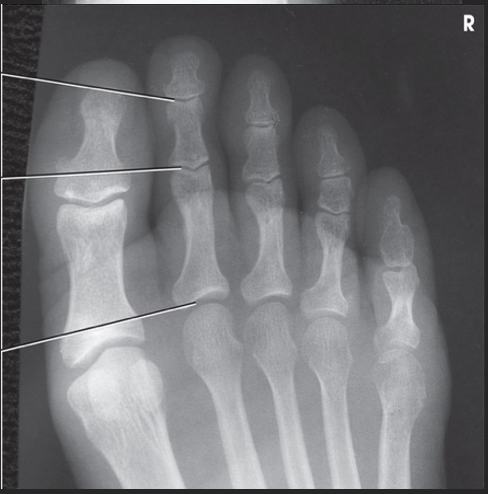
Where does the CR enter?
3rd MTP joint (or MTP joint of affected toe)

What structures must be seen?
All phalanges and distal metatarsal; open IP and MTP joints

Why is this projection preferred over the alternative?
It opens the IP joints better than a regular AP
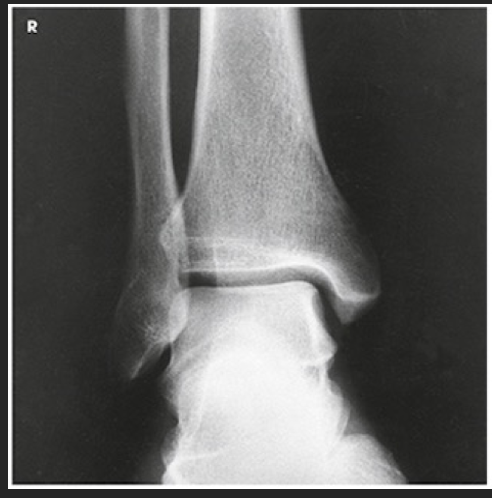
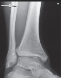
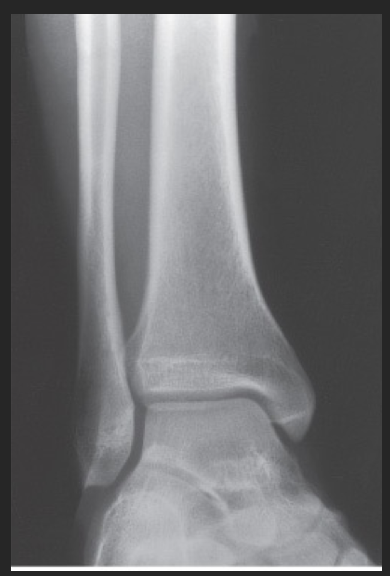
Name this projection.
AP ankle

Describe how the anatomy of interest is positioned.
Patient is seated, or supine, with the affected leg extended onto the IR; the ankle is in true anatomic position w/ dorsiflex

Where does the CR enter?
Perpendicular through the ankle joint (midway between malleoli)

Is there any rotation?
No

What structures are seen?
Ankle joint, distal tib/fib (medial and lateral malleoli), and the proximal talus

Which aspect of the ankle is best visualized?
Medial

What anatomy will overlap?
Tibiofibular joint and slight superimposing of the talus and distal fibula

Name this projection.
Lateral foot
What type of projection is it?
Mediolateral

On which side should the patient be positioned?
Affected side

What 2 things should be ⟂ to the IR?
Patella and plantar surface

Where does the CR enter?
Perpendicular to the base of the 3rd metatarsal

What is shown in profile?
The entire foot

How can you guarantee that the foot is in a true lateral position?
Place your hand perpendicular to the IR and line the foot up with your hand to obtain a true lateral; if you do not do this, the foot will be over-rotated, and the malleoli/metatarsals will not superimpose properly

How can you tell if the foot is in a true lateral?
Open sinus tarsi, metatarsal superimposition, fibula superimposes tibia on posterior half

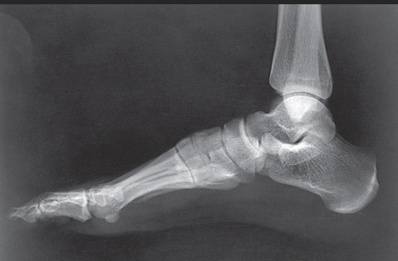
Name this projection.
Axial calcaneus

If the patient is supine, what type of projection is it?
Plantodorsal

How is the CR directed?
40 degrees cephalic (toward heel)

Where does the CR enter?
Plantar surface near 3rd metatarsal base

What anatomy is in profile?
Sustentaculum tali
What joints are seen?
Calcaneocuboid, talocalcaneal (subtalar)

What type of projection is used when the patient is prone?
Dorsoplantar

Name this projection.
Lateral ankle
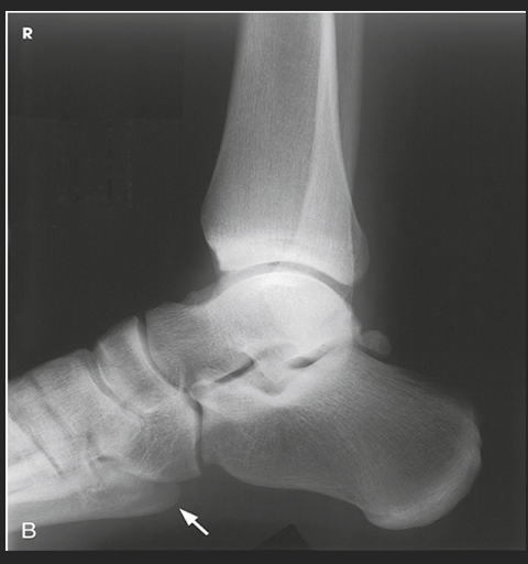
What type of projection is it?
Mediolateral

Which side is the patient turned toward?
Affected side

Where does the CR enter?
Perpendicular to the ankle joint, entering at the medial malleolus

How do you evaluate for a true lateral?
The tibiotalar joint will be well visualized, and the fibula will be over the posterior half of the tibia

How much of the distal tibia and fibula must be included?
1/3

Which joint is well visualized?
Tibiotalar

Name this projection.
AP oblique mortise joint
State the degree and type of rotation used.
15-20 medially