MICRO LAB MIDTERM
1/19
There's no tags or description
Looks like no tags are added yet.
Name | Mastery | Learn | Test | Matching | Spaced | Call with Kai |
|---|
No analytics yet
Send a link to your students to track their progress
20 Terms
Safety procedures and protocols
clean work area with disinfectant each period before and after lab
wash hands before and after lab and when you need to step out of lab
dont smoke, eat, chew gum, or drink in lab
keep hands away from face
do not apply lip balm
cloes toed shoes
lab coats worn. no baggy sleeves, floppy shirttails, neckties etc.
long hair tied back
fingernails short
follow teachers instructions on phone
spillage or breakage, inform lab instructor, quarantine area
notify instructor of any injury
know location of all safety equipment
labels include: initials, date, lab section number, medium/test, organism
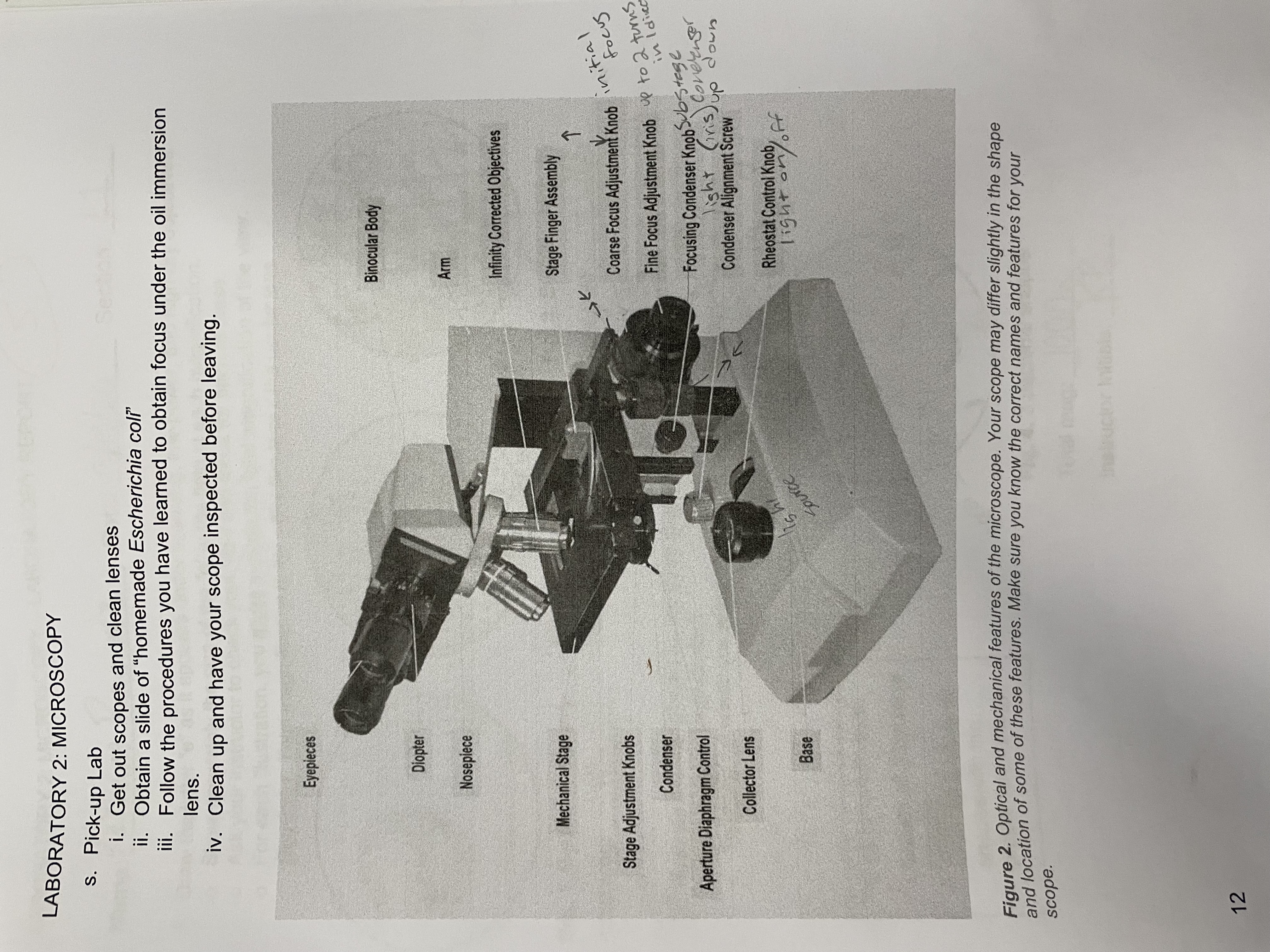
microscope parts
Microscope objective magnification
total magnification= power of ocular lens x power of objective lens
Lens | Magnification (Objective) | Magnification (Ocular) | Total Magnification |
|---|---|---|---|
Scanner (red) | 4× | 10× | 40× |
Low Power (yellow) | 10× | 10× | 100× |
High Power (blue) | 40× | 10× | 400× |
Oil Immersion (white) | 100× | 10× | 1000× |
Microscope usage
Scope should be stored like this
cord folded and secured. Placed atop the stage so the cord does not dangle
high power objective (blue) in viewing position
stage all the way down
rheostat on lowest setting
free of oil, stain, and dirt (clean with lens paper)
Things to remember
never use coarse adjustment when using high dry or oil immersion lenses
when using oil and cannot focus, do not drag high power lens through the oil. Rotate the nosepiece past the scanning lens to the low power lens
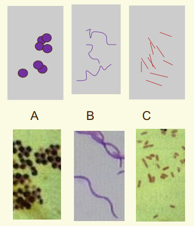
Bacterial cell morphology
Coccus- spheres
Bacillus- rods
Spirillum- squiggles
Categorize microbes into Kingdoms and subdivisions
Kingdom Plantae
Kingdom Animalia
Kingdom Protista
protozoa
amoeboid: (100x) single-celled heterotrophs; pseudopodia
ciliated: (100x) single-celled; cilia
flagellated: (1000x) flagellated protozoa
higher algae:(100x)
chlorophyta:
euglenophyta: single flagellum
volvox: thousands of cells linked together. easily be seen with naked eye.
Kingdom Fungi: composed of hyphae arranged in mycelium
yeast: (100-400x) multicellular do not bear hyphae
filamentous fungi:(100x) black bread mold; horizontal hyphae
lichen:
Kingdom Monera (1000x)
archaea:
bacteria:
cyanobacteria:
Aseptic techniques for smearing and culturing
types of stains and stain theory
🔹1. Simple Stains
Purpose: Show cell shape, size, and arrangement
Dye: One basic (positively charged) dye
Examples:
Methylene blue
Crystal violet
Safranin
What you see: Blue/purple/pink cells on a light background
Why it works: Basic dyes bind to negatively charged cell components (DNA, cell wall)
🔹 2. Differential Stains
Used to distinguish between groups of microorganisms based on structural differences.
A. Gram Stain
Separates Gram+ (purple) vs Gram– (pink)
Based on peptidoglycan thickness and outer membrane
B. Acid‑Fast Stain
Identifies Mycobacteria (TB, leprosy)
Acid‑fast = red
Non–acid‑fast = blue
Based on mycolic acids (waxy lipids)
C. Endospore Stain (Schaeffer–Fulton)
Spores = green
Vegetative cells = pink
Detects Bacillus and Clostridium
D. Capsule Stain
Capsules = clear halos
Background = dark
Cells = light
Uses negative staining (acidic dyes)
E. Flagella Stain
Coats flagella to make them thick enough to see
🔹 3. Special / Structural Stains
Highlight specific structures.
Negative Stain
Uses acidic dyes (eosin, nigrosin)
Background stains, cells remain clear
Great for capsules and morphology
Endospore Stain
(Already listed under differential)
Flagella Stain
(Already listed)
Metachromatic Granule Stain
Shows storage granules (e.g., Corynebacterium)
Lactophenol Cotton Blue
Used for fungi
🧬 II. Stain Theory (Why Stains Work)⭐ 1. Charge Interactions
Basic dyes (crystal violet, safranin, methylene blue)
→ Positively charged
→ Bind to negatively charged bacterial surfacesAcidic dyes (nigrosin, eosin)
→ Negatively charged
→ Repelled by bacteria → stain the background
⭐ 2. Cell Wall Chemistry
Different stains exploit structural differences:
Gram Stain Theory
Gram+ = thick peptidoglycan traps CV‑I complex
Gram– = outer membrane dissolves, thin PG loses dye
Acid‑Fast Theory
Mycolic acids are waxy, hydrophobic
Carbol fuchsin + heat penetrates
Acid‑alcohol cannot remove it
Endospore Theory
Spores are highly resistant
Malachite green + heat forces dye in
Safranin stains vegetative cells afterward
⭐ 3. Solubility & Permeability
Some dyes require heat to penetrate (acid‑fast, endospore)
Some stains rely on lipid solubility (carbol fuchsin)
⭐ 4. Contrast Creation
Stains increase contrast between:
Cell vs background
Cell types vs each other
Cell structures vs cell body
Microbiological stains fall into simple, differential, and special categories. All stains rely on charge interactions, cell wall chemistry, and dye solubility to make invisible cells visible and distinguishable.
gram stain
Type of Stain
A differential stain that separates bacteria into Gram‑positive and Gram‑negative based on cell wall structure.
Name of Methods
Gram Staining Method (developed by Hans Christian Gram)
Procedure:
Primary stain: Crystal violet
Mordant: Iodine (forms CV‑I complex)
Decolorizer: Alcohol or acetone‑alcohol
Counterstain: Safranin
Chemicals:
Crystal violet (primary stain)
Gram’s iodine (mordant)
Alcohol/acetone (decolorizer)
Safranin (counterstain)
Appearance and results:
Gram‑positive:
Purple
Thick peptidoglycan retains CV‑I complex
Gram‑negative:
Pink/red
Alcohol dissolves outer membrane → thin peptidoglycan loses CV‑I → takes up safranin
acid fast stain
Type of Stain:
Differential stain
Separates acid‑fast organisms (with mycolic acids) from non–acid‑fast organisms.
Name of Methods:
Ziehl–Neelsen Method (hot method)
Kinyoun Method (cold method)
Procedure:
1. Prepare smear
Air‑dry and heat‑fix.
2. Primary stain
Flood with carbol fuchsin.
Ziehl–Neelsen: heat gently to steam (helps dye penetrate waxy wall).
Kinyoun: no heat; uses higher phenol concentration.
3. Decolorize
Rinse with acid‑alcohol (3% HCl in ethanol).
Acid‑fast cells retain carbol fuchsin.
4. Counterstain
Apply methylene blue (or brilliant green).
Non–acid‑fast cells take up counterstain.
Chemicals:
Carbol fuchsin (primary stain)
Acid‑alcohol (decolorizer)
Methylene blue or brilliant green (counterstain)
Ziehl–Neelsen only: heat source (steam)
Appearance and results:
Acid‑fast bacteria
Bright red/fuchsia
Due to retention of carbol fuchsin
Thick mycolic acid layer prevents decolorization
Non–acid‑fast bacteria
Blue or green
Take up counterstain after losing primary dye
Common microbial genera that are acid fast or form endospores:
Mycobacterium
M. tuberculosis, M. leprae
Nocardia (partially acid‑fast)
Capsule Stain
Type of Stain:
Special / structural stain
Negative stain technique
Designed to visualize capsules, which do not take up most dyes.
Name of Methods:
Anthony’s Capsule Stain
Negative Stain Method (India ink or nigrosin
Procedure:
1. Prepare smear (NO heat‑fixing)
Mix bacteria with India ink or nigrosin on slide
Spread into thin film
Air‑dry only — heat destroys capsules
2. Apply counterstain
Flood with crystal violet (Anthony method)
or safranin (alternative)
3. Rinse gently and air‑dry
Do not blot — blotting can remove capsules
Chemicals:
India ink or nigrosin (acidic dye → stains background)
Crystal violet or safranin (basic dye → stains cells)
Appearance and results:
Capsule‑positive bacteria
Clear halo around the cell
Background = dark
Cell body = purple/pink (depending on counterstain)
Capsule‑negative bacteria
No halo
Only stained cell + dark background
Structure and Function of Endospores and Capsules:
Structure
Thick, gelatinous layer outside cell wall
Made of polysaccharides or polypeptides
Function
Anti‑phagocytic (major virulence factor)
Adherence to surfaces
Prevents desiccation
Helps form biofilms
Endospore Stain
Type of Stain:
Differential stain
Distinguishes endospores from vegetative cells
Name of Methods:
Schaeffer–Fulton Method (most common)
Dorner Method (less common)
Procedure:
1. Prepare smear
Air‑dry and heat‑fix.
2. Primary stain
Flood smear with malachite green.
Steam over heat for ~5 minutes (keeps dye penetrating the spore coat).
3. Rinse
Rinse gently with water (removes dye from vegetative cells but NOT spores).
4. Counterstain
Apply safranin for 1 minute.
5. Rinse and blot dry
Chemicals:
Malachite green (primary stain)
Heat/steam (mordant-like function)
Water (decolorizer)
Safranin (counterstain)
Appearance and results:
Endospores
Green
Because malachite green is forced into spores by heat and retained
Vegetative cells
Pink/red
Take up safranin after malachite green washes out
Structure and Function of Endospores and Capsules:
Structure
Core: DNA, ribosomes, dipicolinic acid + Ca²⁺
Cortex: thick peptidoglycan
Spore coat: protein layers
Exosporium: outermost layer
Function
Survival structure
Resistant to:
Heat
UV radiation
Chemicals
Desiccation
Allows bacteria to persist for decades
Common microbial genera that are acid fast or form endospores:
Bacillus (aerobic)
Clostridium (anaerobic)
Colonial Morphology and Broth/Slant Growth Characteristics
Quorum sensing
this serratia marsenscens culture shows evidence of a pheomenon known as quorum sensing, in which production of pigment is density dependent
it only produces pigment when there are enough bacteria present in a colony
Swarming
this is a pure culture of a highly motile bacteria
ONE drop of inoculum was placed at the center of the plate
after 24 hour incubation the “swarm” has covered the entire plate with a thin, transparent film of growth
Too Thick
the bacteria on this plate are too thick to discern colony morphology
this can be caused by:
too large an inoculation; multiple loops from stock broth or too much inoculum from a plate
failure to flame between loops
Pigment Production
some bacterial colonies produce pigment
S.marcesens, K.rosea, K.-rhizo, S.aurantiaca
Contaminant
can come from a “drop-in: which settled onto the agar when the plate was opened during inoculation
rim is a very common “hiding place”
growth can be clear/ very tiny
Pure Cultures
Colony shape
round: circle
rhizoid: snowflake
irregular: amoeba
Colony margin (edge)
entire: circle
lobate: pokey
undulate: flowery
Colony size
punctiform: 1mm<
Colony Color
Other Characteristics
surface rough, smooth, mucoid, etc.
Broth growth: unagitated
turbid: cloudy
sediment: bottom
pellicle: top
Slant growth characteristics: stroke slant
filiform: line
spreading: all around
beaded: dots
Staphylococcus epidermis: punctiform, entire, white
Escherichia coli: irregular, entire, white
Bacillus subtillis- irregular, undulate, off-white
Broth-to-Broth theory
Broth‑to‑Broth Transfer (Steps Only)
Label tubes
Mix culture
Sterilize loop
Remove caps
Flame tube mouths
Insert loop into culture
Transfer inoculum to sterile broth
Flame tube mouths again
Replace caps
Sterilize loop
Incubate
Broth‑to‑Broth Theory (List Only)
Broth‑to‑broth transfer is used to propagate a culture into fresh nutrients.
It demonstrates aseptic technique, preventing contamination from the environment or other cultures.
Only a small inoculum is needed because bacteria reproduce rapidly in nutrient broth.
The goal is to maintain a pure culture while allowing cells to enter log phase growth.
Streak Plate Process and theory
Streak Plate Process (Steps Only)
Label plate
Sterilize loop
Obtain inoculum
Streak quadrant 1
Sterilize loop
Streak quadrant 2
Sterilize loop
Streak quadrant 3
Sterilize loop
Streak quadrant 4
Invert plate
Incubate
Streak Plate Theory (List Only)
The streak plate method uses mechanical dilution: each quadrant spreads fewer cells.
By the final quadrant, individual cells land far enough apart to grow into isolated colonies.
Each colony arises from a single cell (or genetically identical cluster), making it a pure culture.
This method is essential for identifying bacteria, performing biochemical tests, and maintaining uncontaminated stocks.
Vocab Lab 1 and 2
Compound light microscope: A microscope that uses visible light and multiple lenses to magnify specimens.
Monocular: A microscope with one ocular (eyepiece) lens.
Binocular: A microscope with two ocular lenses for more comfortable viewing.
Resolution: The ability to distinguish two close points as separate, determining image clarity.
Numerical aperture: A measure of a lens’s light‑gathering ability that directly affects resolution.
Rheostat: The control that adjusts the brightness of the microscope’s light source.
Parfocaling: A feature where a specimen stays nearly in focus when switching between objectives.
Vocab Lab 3
Binomial Nomenclature: A two‑name scientific naming system using genus and species.
Kingdoms: The broadest major categories of life, such as Animalia, Plantae, Fungi, Protista, and Bacteria.
Ubiquity/Ubiquitous: The concept that microorganisms are found everywhere in the environment.
Culturing bacteria: Growing microorganisms under controlled conditions in the lab.
Growth medium: A nutrient-rich substance that supports microbial growth.
Broth: A liquid growth medium used to culture microorganisms.
Cultures: Populations of microorganisms grown in a medium.
Contamination: The accidental introduction of unwanted microorganisms into a culture or environment.
Sterile: Completely free of all living organisms, including spores.
Inoculate: To intentionally introduce microorganisms into a sterile medium.
Incubate: To place cultures in controlled conditions (temperature, time) to promote growth.
Aseptic Technique: Procedures used to prevent contamination of cultures, media, and the environment.
Pure Culture: A culture containing only one microbial species.
Mixed Culture: A culture containing two or more microbial species.
Growth Medium: A nutrient source that supports microbial growth (solid or liquid).
Agar: A solidifying agent derived from red algae used to create firm surfaces for microbial growth.
Vocab Lab 4
Simple stain: A staining method that uses one basic dye to show cell shape, size, and arrangement.
Negative stain: A stain using acidic dyes that color the background while leaving cells unstained for clearer morphology.
Differential stain: A staining technique that uses multiple dyes to distinguish between different types of bacteria or structures.
Mordant: A chemical that intensifies or fixes a dye to a structure, helping it bind more strongly.
Decolorizer: A chemical (often alcohol or acid‑alcohol) that removes primary stain from some cells but not others during differential staining.
Over decolorizing: Removing too much primary stain, causing cells to appear falsely negative (too light or wrong color).
Under decolorizing: Removing too little primary stain, causing cells to appear falsely positive (too dark or wrong color).
Vocab Lab 5
Acid‑fast stain: A differential stain that identifies bacteria with waxy mycolic acids in their cell walls, causing acid‑fast cells to remain red after acid‑alcohol decolorization.
Endospores: Highly resistant, dormant bacterial structures formed by genera like Bacillus and Clostridium that survive extreme heat, chemicals, and drying.
Capsules: Thick, gelatinous outer layers surrounding some bacteria that help with protection, adhesion, and evading phagocytosis, appearing as clear halos in capsule stains