morph corr skeletal exam
1/33
There's no tags or description
Looks like no tags are added yet.
Name | Mastery | Learn | Test | Matching | Spaced | Call with Kai |
|---|
No analytics yet
Send a link to your students to track their progress
34 Terms
To perform a bone flow, as part of a 3 phase bone scan you would use
a sequence of 1-3 second images for 2-3 minutes of the area of interest
Which of the following conditions is visualized on a bone image as a photopenic area (cold spot)?
attenuation from a belt buckle
A common technical factor that is likely to produce less than an optimal or a poorer quality bone images is
allowing the patient to lay asymmetrically or twisted to keep them comfortable to reduce fidgeting during the exam
Repetitive motion injury shows on a 3 phase bone scan as
increased blood flow to the region with increased uptake on the delayed images
When trying to evaluate an acute trauma, the earliest one should image is _____ because most (>95%) bone fractures in healthy adults will become positive by then
72 hours
A bone scan image shows as a "flare" phenomenon. Likely causes include
increased blood flow (as a result of inflammation) and increased bone remodeling in conjunction with effective chemotherapy treatment
Extraosseous (soft tissue) uptake of bone-seeking radiopharmaceuticals can be found in patients with
lactation, sickle cell anemia, poor radiopharmaceutical preparation, rhabdomiosis
The main advantage SPECT has over planar imaging when performing a bone scan is that
it has a higher contrast between normal and abnormal areas
If radioactivity is visualized in the skeleton, stomach, thyroid, and salivary glands on a bone image, the most likely explanation for these findings is the
radiopharmaceutical was poorly prepared and it contained excess free pertechnetate
A bone image which is clearly indicative of osteomyelitis will demonstrate which of the following radiopharmaceutical characteristics?
increased flow, increased activity in soft tissue and bone on blood pool, increased activity in bone but not in the soft tissue on delayed images
Bone imaging is performed several hours after tracer administration to permit
blood clearance of excess tracer in soft tissue
Following administration of a bone imaging tracer, the patient is instructed to drink fluids to
clear excess tracer from the blood and soft tissue
When performing a 3 phase bone scan, when should imaging begin?
immediately upon injection
As osteoclastic activity (destruction of bone) increased in relationship to osteoblastic activity (rebuilding bone), bone deposition _____
decreases
_____ is the death of bone tissue due to a lack of blood supply
avascular necrosis
Which of the following is an indication to perform a 3 phase bone scan?
differentiate osteomyelitis from cellulitis
A patient had a hip replacement 3 years ago. Their 3 phase bone scan demonstrated a normal flow image, a normal blood pool image, and a focal hot spot on the distal portion of the prosthesis where the screws are, on the delays. This indicates ___________.
loosening of the prosthesis
Visualization of the kidneys on a 99mTc-HDP scan is considered
normal, because Tc-99m HDP is excreted via the urinary tract
Bone scans are considered to have higher sensitivity but less specificity than a radiograph. Which of the following are accurate given the previous statement?
a bone scan sees pathologies that may be much smaller in size
a bone scan sees many pathologies much earlier in the disease process
Which of the following are considered advantages of NaF-18 PET bone scans when compared to a traditional bone scan?
the patient receives less absorbed radiation
the exam has much better sensitivity because of its much better resolution
the waiting time after the injection as well as the imaging time is much shorter
On the following image, what is the most likely cause for the area of increased activity?
this is where the patient was injected and some radiopharmaceutical is left at the injection site
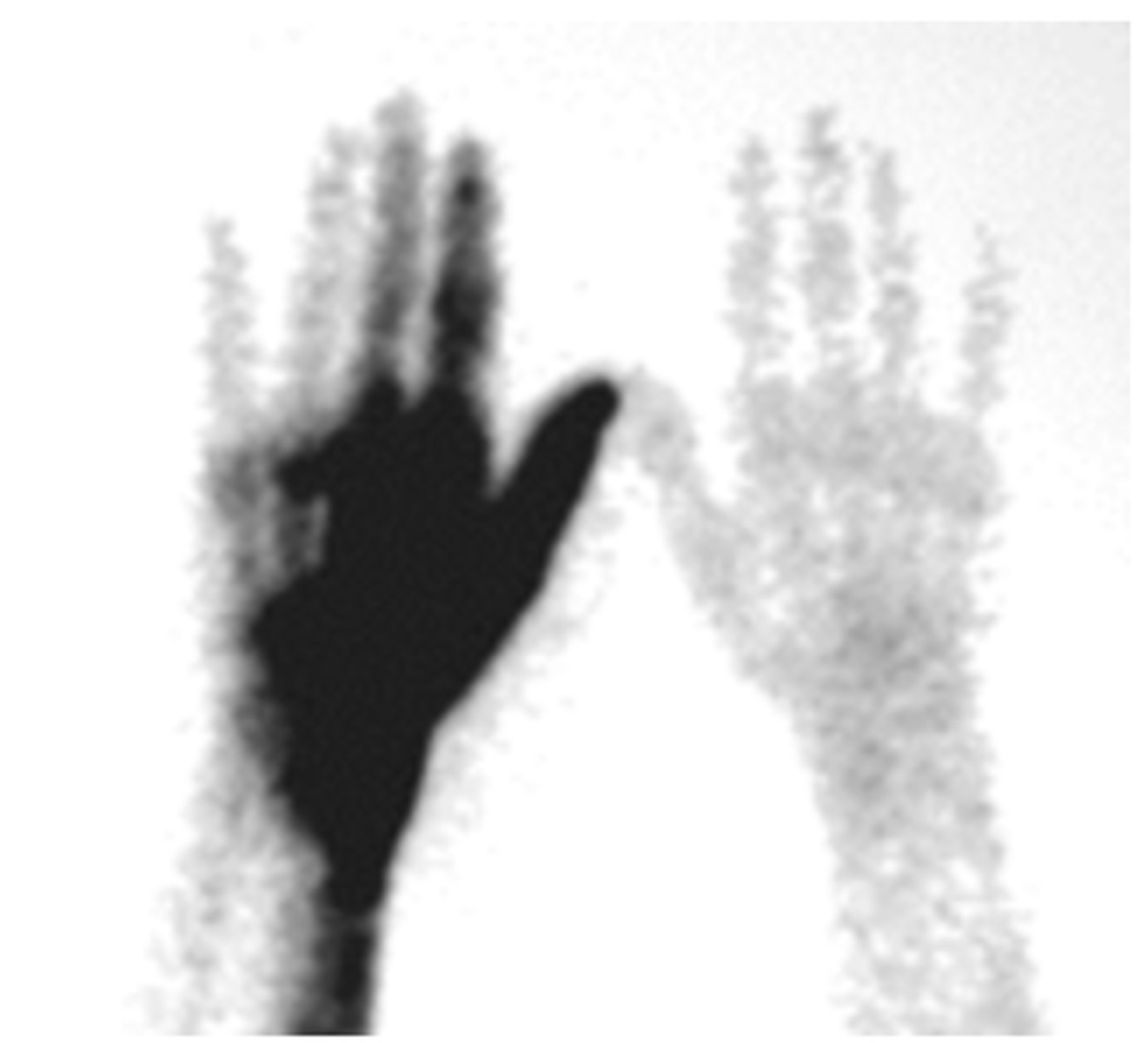
On the following image, what is the most likely cause for the increased activity?
the hand on the left side of the screen is on an arm where the radiopharmaceutical was injected into an artery instead of a vein
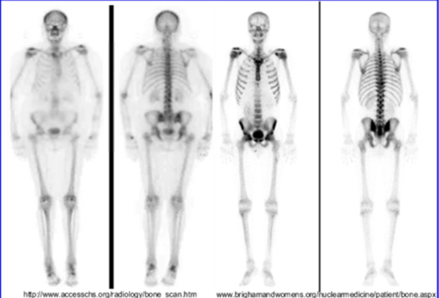
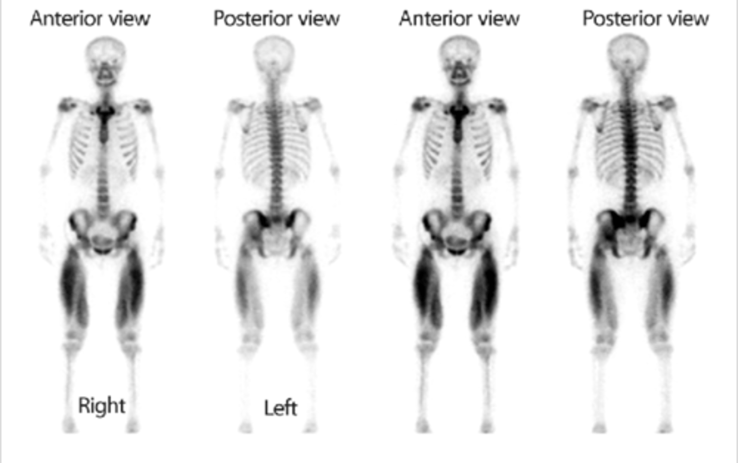
Given the following image of 2 different patients imaged using the same protocol and the same amount of radiopharmaceutical, considering that both scans are considered normal, what is the most likely reason that the patient on the left has a lower quality image than the patient on the right?
the patient is larger which is causing more scatter
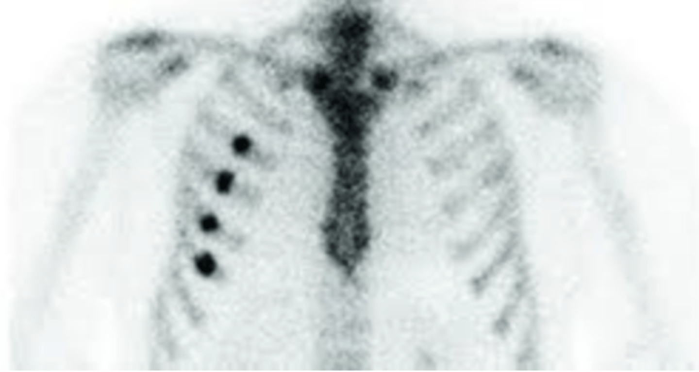
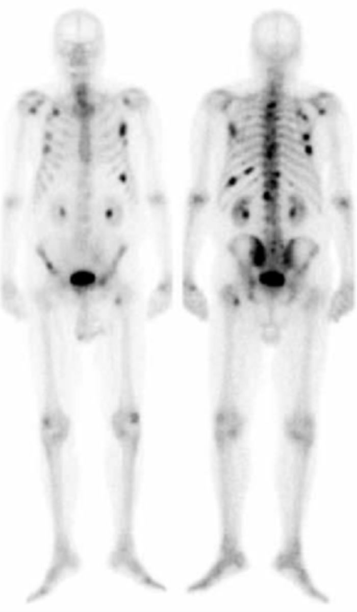
Given the following image, what is the most likely cause of the areas of increased activity?
rib fractures after a trauma
Given the following image, what is the most likely cause of the areas of increased activity?
Paget's disease
A typical dose for a Tc-99m HDP bone scan is
20-25 mCi
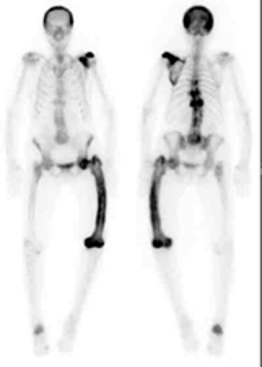
An extremely intense uptake of the radiopharmaceutical in an entire bone or major portion of a few bones while the majority of the rest of the skeleton appears normal is typical of
Paget's disease
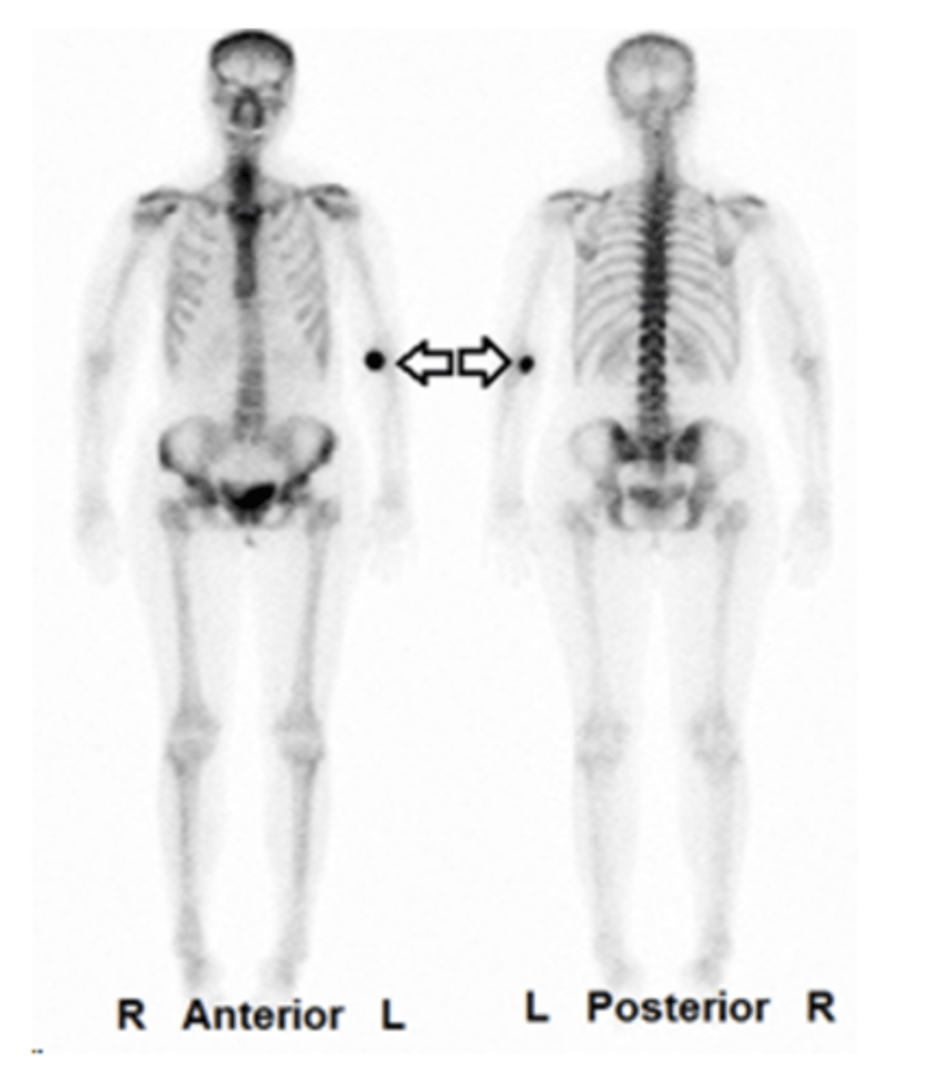
The most likely cause of this abnormal bone scan would be
the patient has widespread metastatic disease
These are normal images of the legs with no significant pathologies being shown (the patient is 8 years old) (true/false)
True
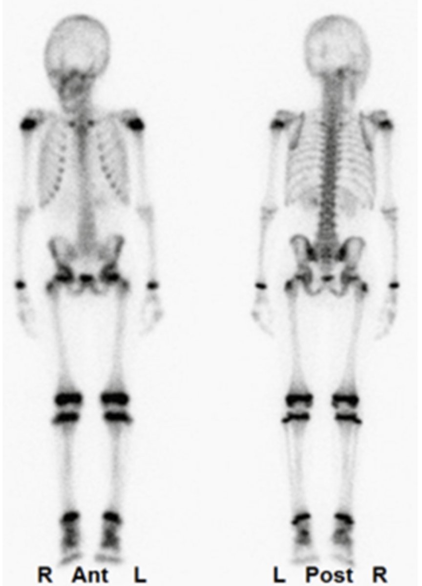
This bone scan was performed on a weightlifter/runner who was complaining of lower back pain. The diagnosis would most likely be a normal bone scan (true/false)
False
Accumulation of radionuclides in the bone is related to both the rate of bone production (osteoblastic activity) and vascularity (true/false)
True
Patients having a bone scan should be hydrated between injection and delayed imaging in order to aid clearance of the radiopharmaceutical from the body (true/false)
True
Information density (equal time imaging) for static bone spot imaging refers to the technique in which one area is imaged for present counts with the same time recorded and all subsequent images are taken for that same time interval (true/false)
True
Typically, skeletal metastatic disease has multiple focal lesions throughout the skeletal, with greatest involvement generally in the axial skeleton (true/false)
True