Human Anatomy & Physiology II: Digestive System
1/44
Earn XP
Description and Tags
This set of vocabulary flashcards covers both the upper and lower digestive systems, including organs of the alimentary canal, accessory organs, specific cellular functions, and the structural features of the intestines.
Name | Mastery | Learn | Test | Matching | Spaced | Call with Kai |
|---|
No analytics yet
Send a link to your students to track their progress
45 Terms
Alimentary Canal
A continuous tube that food and drink travel through, starting at the mouth and ending at the anus; it includes the oral cavity, pharynx, esophagus, stomach, small intestine, and large intestine.
Accessory Digestive Organs
Organs connected with the alimentary canal that assist with the breakdown of food, such as the teeth, tongue, salivary glands, liver, gall bladder, and pancreas.
Starch
A complex carbohydrate that must be converted into a simple carbohydrate, such as glucose (a monosaccharide), to be used by cells.
Cellulose
A major component of plants that is not digestible by humans and passes through the digestive system unchanged to be removed in the feces.
Salivary Glands
Three pairs of accessory organs (Parotid, Sublingual, and Submandibular) that secrete saliva containing enzymes for chemical breakdown in the oral cavity.
Hard Palate
The structure at the top of the mouth against which the tongue presses food to assist with mechanical breakdown.
Pharynx
The throat, which is divided into three regions: the nasopharynx (upper), oropharynx (middle), and laryngopharynx (final).
Epiglottis
A valve present in the laryngopharynx that directs swallowed food away from the respiratory tract and toward the esophagus.
Chyme
A semifluid mass into which ingested food is converted through chemical and mechanical digestion within the stomach.
Stomach Muscle Layers
Three layers—oblique, circular, and longitudinal—whose contractions cause the stomach to churn and mix food with liquids.
Chief Cells
Cells in the stomach that secrete the enzymes pepsin (for protein breakdown) and lipase (for fat breakdown).
Stomach Acidity
The internal environment of the stomach which maintains a pH=1.5−2 to help break down food.
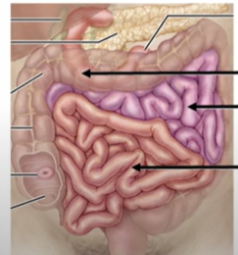
Small Intestine
A long tube connecting the stomach to the large intestine that performs chemical digestion and absorbs almost all nutrients into the blood.
Duodenum
The first segment of the small intestine specialized in chemical digestion using secretions from the liver, gall bladder, and pancreas.
Pyloric Sphincter
A valve that periodically opens to allow chyme from the stomach to enter the duodenum.
Bile
A fluid produced by the liver that serves to emulsify fats, breaking large droplets into smaller, more easily absorbed droplets.
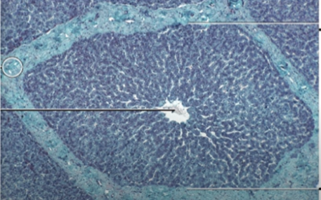
Hepatocytes
Cells in the liver organized into hexagonal stacks called liver lobules that produce bile.
Portal Triads
A set of three tubes located at the corner of each liver lobule, composed of a vein, an artery, and a bile duct.
Gall Bladder
An accessory organ that stores bile produced in the liver.
Cystic Duct
The tube that connects the gall bladder to the shared pathway for bile transport.
Common Bile Duct
The tube through which bile released from the gall bladder enters the duodenum.
Exocrine Pancreas
The portion of the pancreas consisting of acinar cells that secrete digestive enzymes (amylases, lipases, proteases, and nucleases) into the duodenum.
Endocrine Pancreas
The portion of the pancreas consisting of islets that release hormones like insulin into the blood.
Jejunum
The middle segment of the small intestine specialized in nutrient absorption by maximizing surface area.
Villi
Finger-like projections covering the circular folds of the jejunum that increase surface area and contain blood vessels for nutrient transport.
Microvilli
Also known as a brush border, these are tiny finger-like projections on the surface of villi that further increase surface area for absorption.
Ileocecal Valve
The valve at the end of the ileum that connects with the large intestine and controls the entry of materials.
Large Intestine Functions
Absorption of some water, electrolytes (Na+, Cl−), and vitamins; holding the gut microbiome; and moving indigestible materials to be removed as feces.
Cecum
A dead-end tube in the large intestine near the connection with the ileum that contains the appendix.
Colon Sections
The four parts of the colon: Ascending (upward), Transverse (across), Descending (downward), and Sigmoid (S-shaped).
Haustra
Segments of the colon formed by rings of smooth muscle that contract like drawstrings to push materials through the tract.
Anal Sphincter
Muscular structure at the end of the rectum that controls the removal of feces through the anus.
The jejunum has circular folds (plicae circulares), villi, and microvilli that all increase its surface area for nutrient absorption.
Presence of villi (finger-like projections)
Between the ileum (small intestine) and cecum (large intestine). Controls movement of material into the large intestine
Cecum, colon, rectum
Small tube-like projection off the cecum, attached to the cecum
The four sections of the colon are the ascending colon, transverse colon, descending colon, and sigmoid colon.
Segmented pouches of the large intestine Contracts to move material through the colon
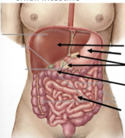
Liver – upper right abdomen
Gallbladder – under liver
Pancreas – behind stomach
Stomach – upper left abdomen
Small intestine – center of abdomen
Large intestine – surrounds small intestine
Food passes through the duodenum, jejunum, ileum in the small intestine, followed by the cecum, ascending colon, transverse colon, descending colon, sigmoid colon, and finally the rectum in the large intestine.
Full path from small → large intestine?
Duodenum → Jejunum → Ileum → Ileocecal valve → Cecum → Ascending → Transverse → Descending → Sigmoid → Rectum → Anus
What is this?
LIVER: HEPATOCYTES ARE ORGANIZED INTO HEZAGONAL STACK CALLED LIVER LOBULES
LABEL
LABEL