3.2.4 More than Meets the Eye
1/25
There's no tags or description
Looks like no tags are added yet.
Name | Mastery | Learn | Test | Matching | Spaced | Call with Kai |
|---|
No analytics yet
Send a link to your students to track their progress
26 Terms
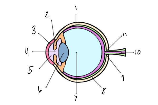
Label the eye
Retina
Iris
Cornea
Pupil
Aqueous Humor
Lens
Vitreous Humor
Sclera
Tapetum
Optic Nerve
Blind Spot
Retina
The light-sensitive layers of nerve tissue lining the most posterior chamber of the vertebrate eye
What is the function of the retina?
It receives images and send them as electric signals through the optic nerve to the brain
What is the function of the iris?
Determines how much light to let in the eye
Cornea
The transparent part of the coat of the eyeball that covers the iris and pupil
What is the function of the cornea?
It admits light to the interior eye
What is the function of the pupil?
Admits light into the interior of the vertebrate
Aqueous Humor
The fluid that fills the space between the lens and the cornea
What is the function of the aqueous humor?
It maintains intraocular pressure, keeping the eye’s shape
What is the function of the lens?
Refracts light to be focused on the retina
Vitreous Humor
The clear, viscous substance that fills the eyeball behind the lens
What is the function of the vitreous humor?
It maintains the eyeball’s spherical shape, supports the retina, and acts as a shock absorber
Sclera
The white external layer of the eyeball
What is the function of the sclera?
Protects the inner eye from trauma, maintains the shape of the eye against external pressure, and provides an attachment point for the extraocular muscles
What is the function of the tapetum?
Enhances night vision by acting as a mirror
What is the function of the optic nerve?
The vital cable connecting the eye to the brain, transmitting visual information via electrical impulses
Blind Spot
The small circular area in the retina where the optic nerve enters the eye that is devoid of rods and cones and is insensitive to light
What is the function of the blind spot?
It provides necessary pathways for nerve fibers to carry visual information to the brain
What does the brain have to do due to the curves of the eyes?
The curves of the eye changes the direction of light entering it, so the image projected on the retina is upside down, so the brain has to invert it. The brain also has to layer the two images from your retinas to create the 3D image in your mind and give you depth perception
Why is the retina so important?
It is the sensory layer of the eyeball, which contains receptors for sight: rods and cones
Rods
Any of the long rod-shaped photosensitive receptors in the retina responsive to faint light
Cones
Any of the conical photosensitive receptor cells of the vertebrate retina that function in color vision
Choroid
A spongy membrane full of vessels located underneath the sclera
Vitreous Body
A transparent gel-like substance that maintains eye shape, filling the space inside the globe of the eye
Fovea Centralis
A small area with a high density of cone photoreceptor cells, allowing for sharpness of vision
Conjunctiva
Helps protect the eye and plays a role in tear production, with very small blood vessels within it