Gen Path Exam 1 - Picture based questions (IP5)
1/90
There's no tags or description
Looks like no tags are added yet.
Name | Mastery | Learn | Test | Matching | Spaced | Call with Kai |
|---|
No analytics yet
Send a link to your students to track their progress
91 Terms
Edema
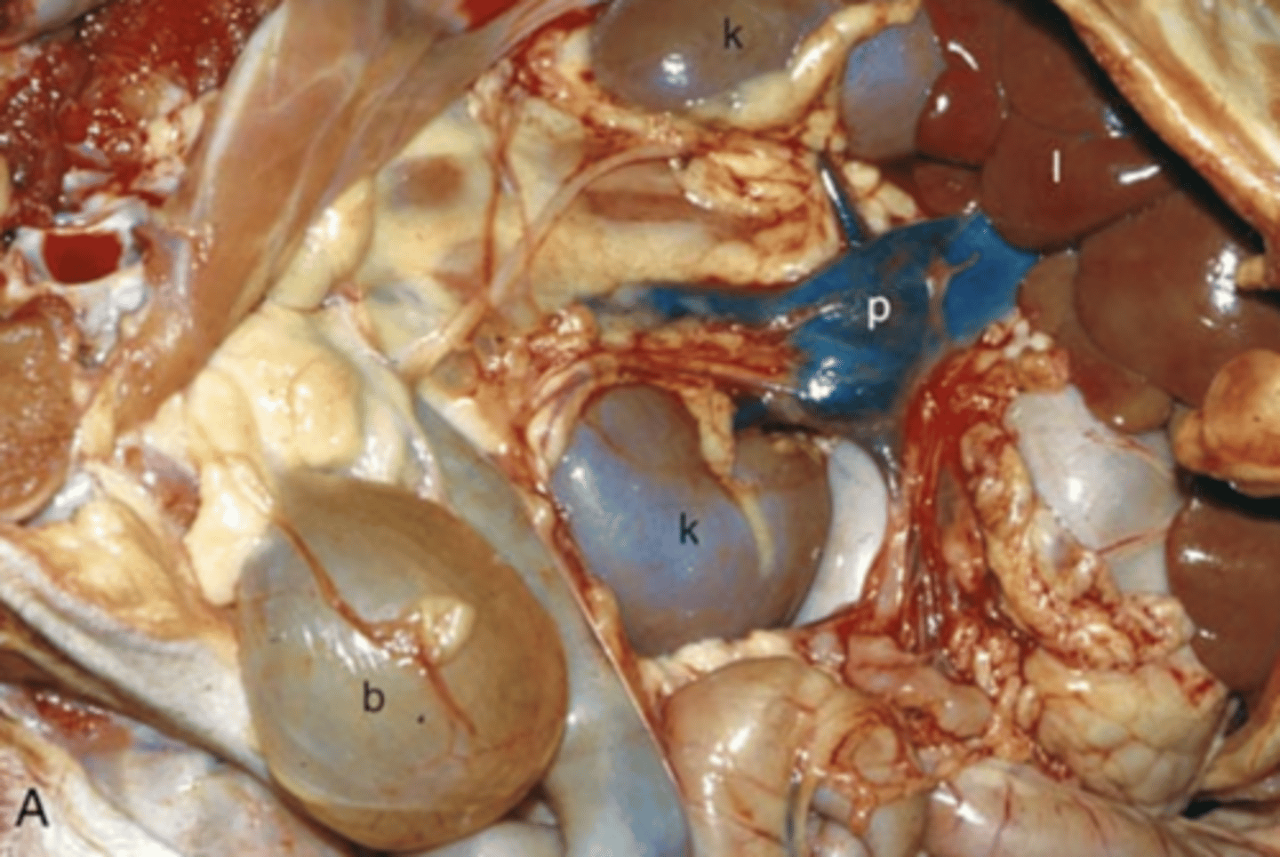
What is being shown in this photo?
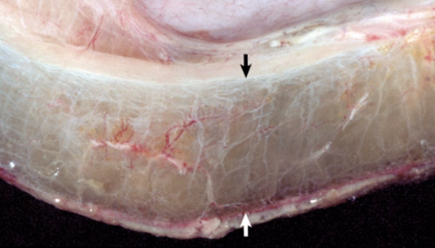
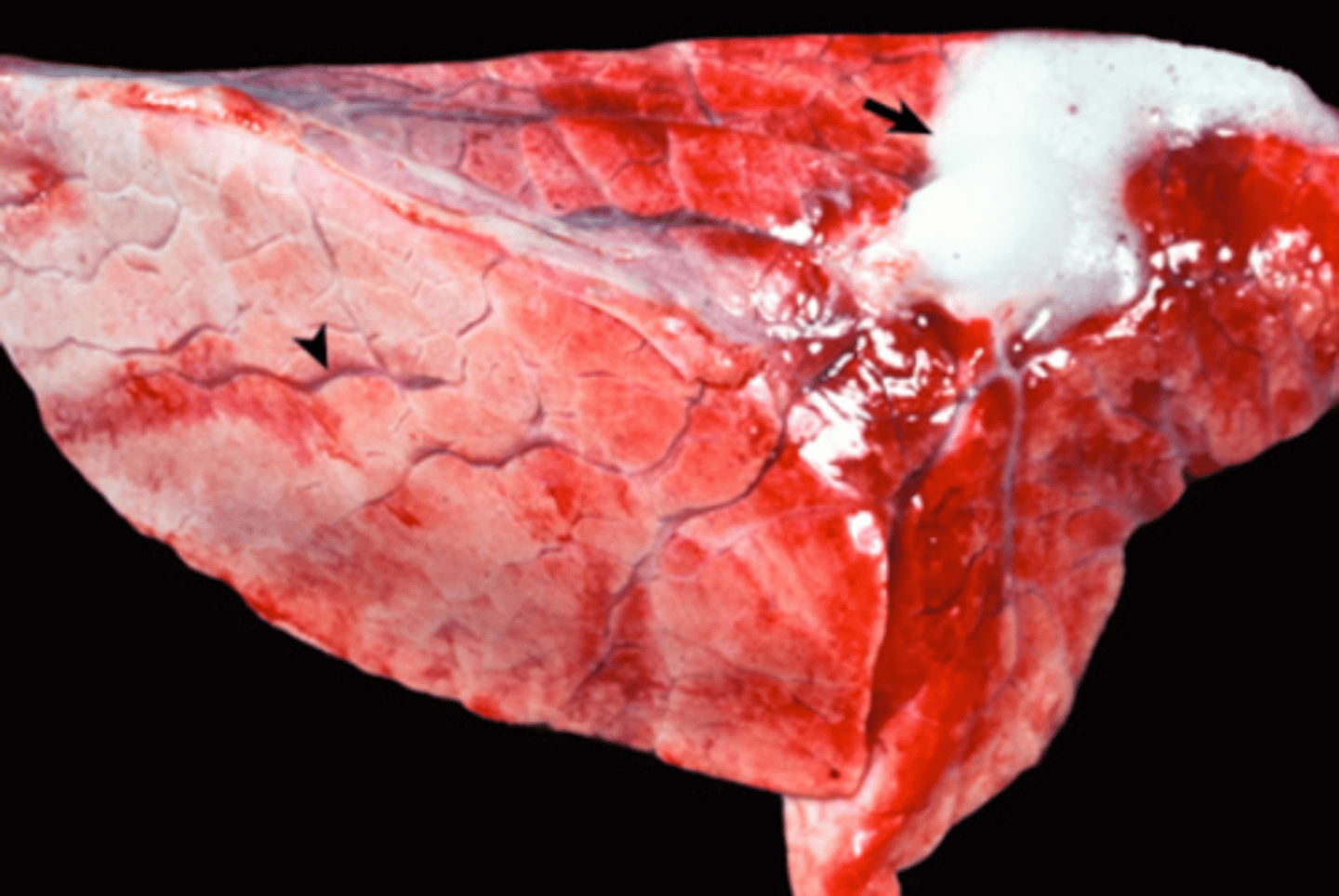
Pulmonary edema
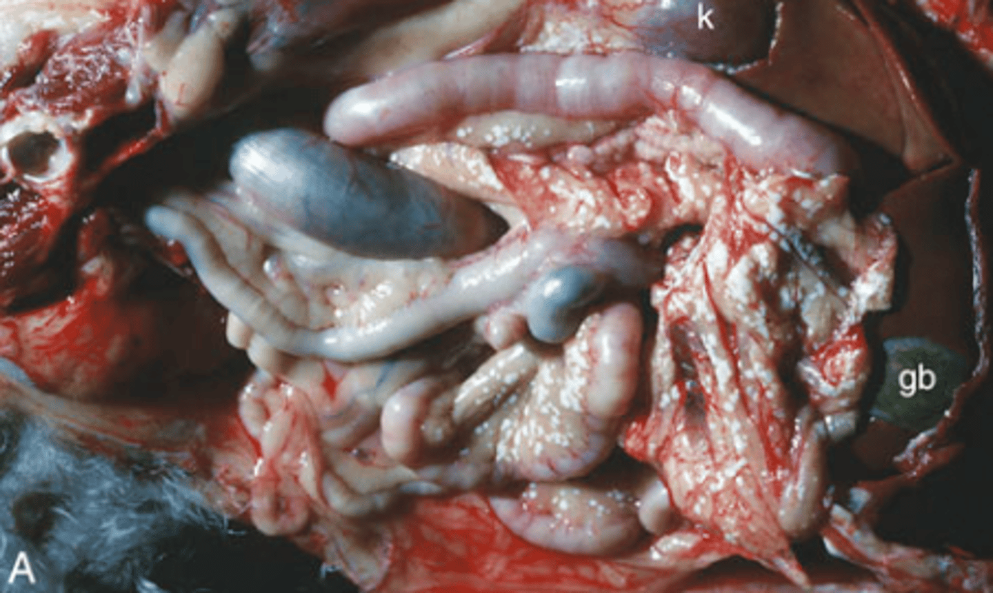
What is being shown in this photo?
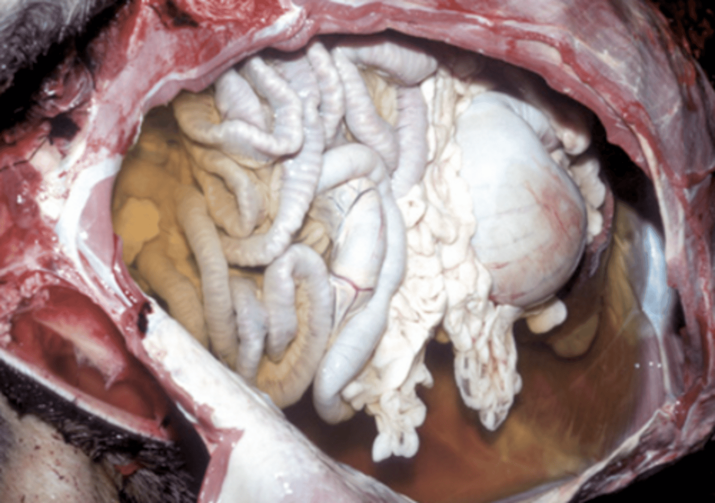
Ascites (Hydroperitoneum)
What is being shown in this photo?
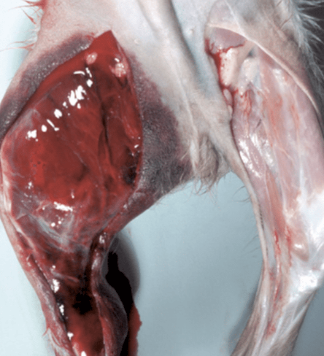
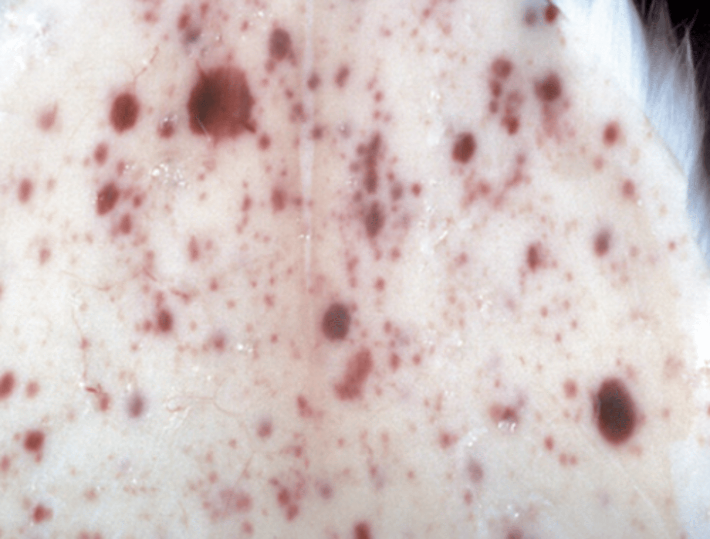
Petechiae & Eccymoses
This is a photo of hemorrhage. What type(s) of hemorrhages are seen?
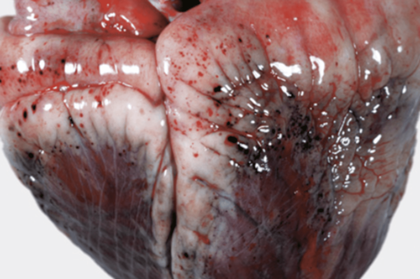
Hemorrhage
What is being shown in this photo?
Ecchymoses
What type of hemorrhages are being shown?
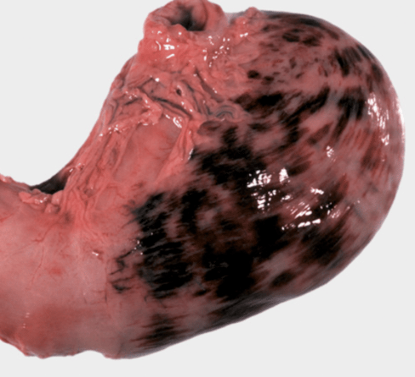
Suffusive
What type of hemorrhages are being shown?
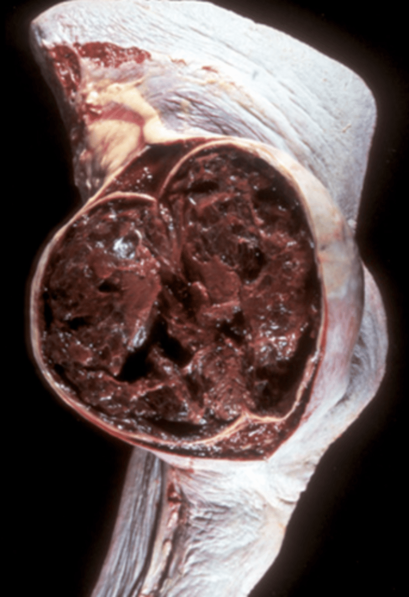
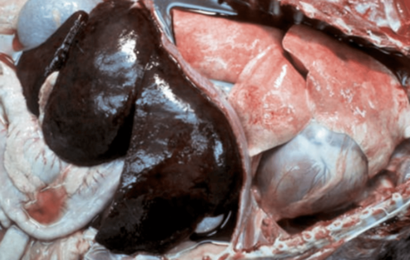
Hematoma in the spleen
This is a result to damage to the splenic red pulp and its vessels. What is being shown in this photo?
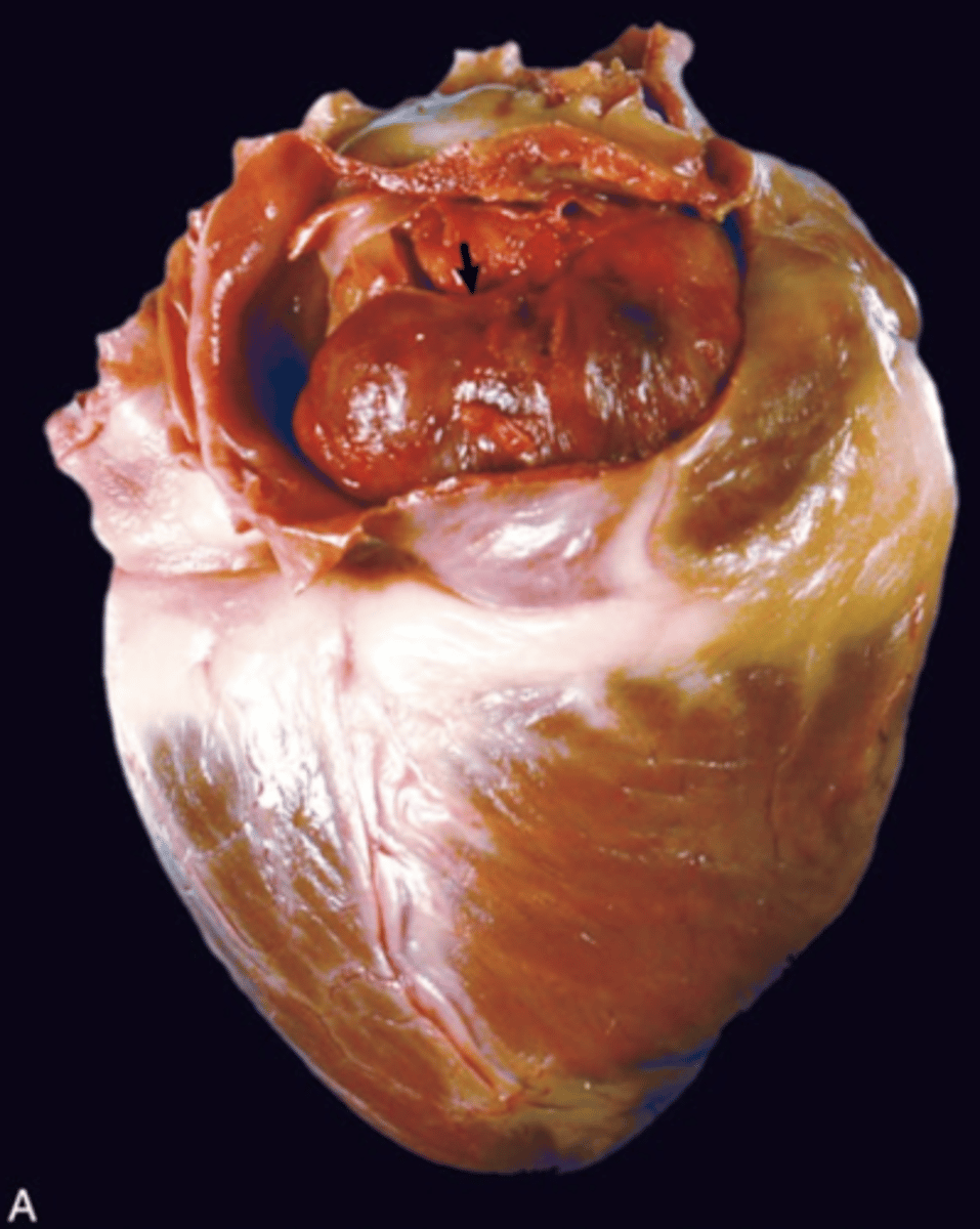
Hemopericardium
What is being shown in this photo?

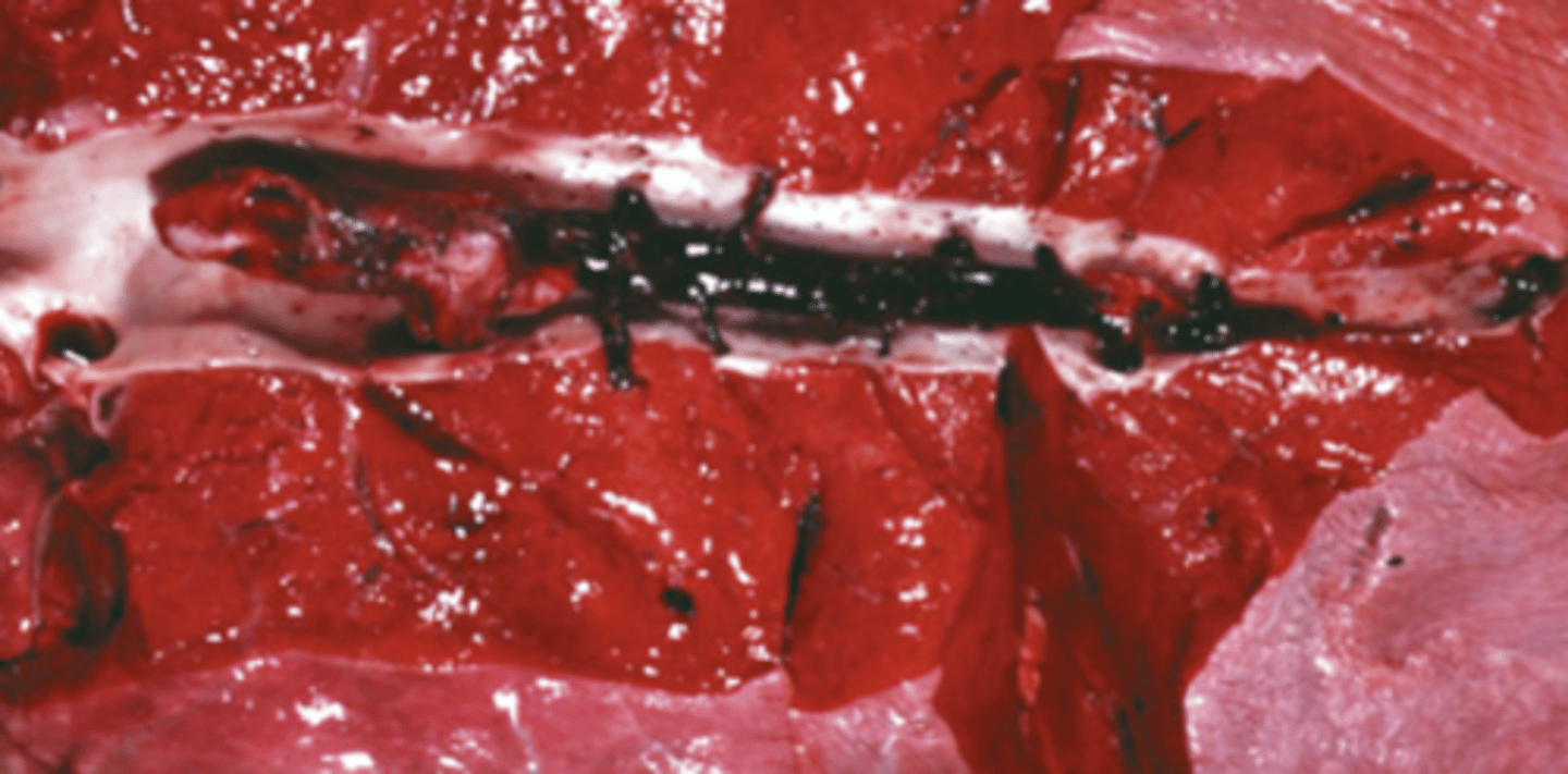
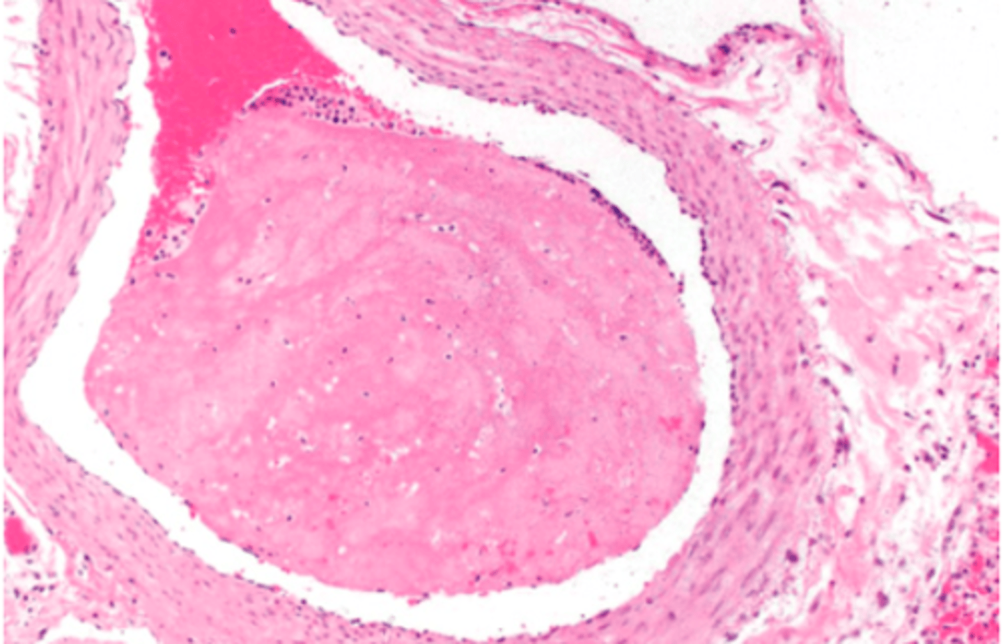
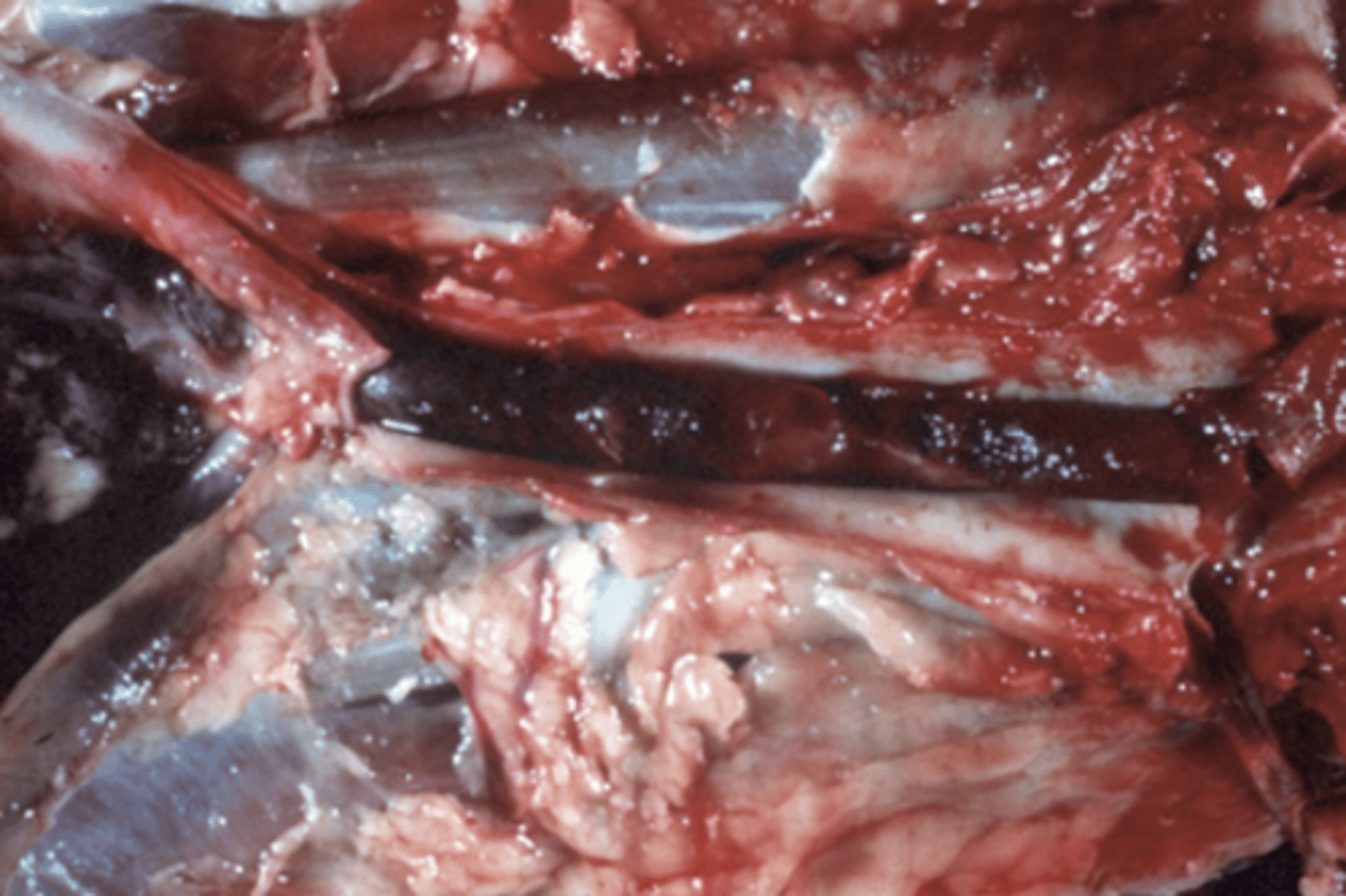
Thrombus
What is being shown in this photo?
arterial
What type of thrombus is being shown in this photo?
arterial or venous
venous
What type of thrombus is being shown?
arterial or venous
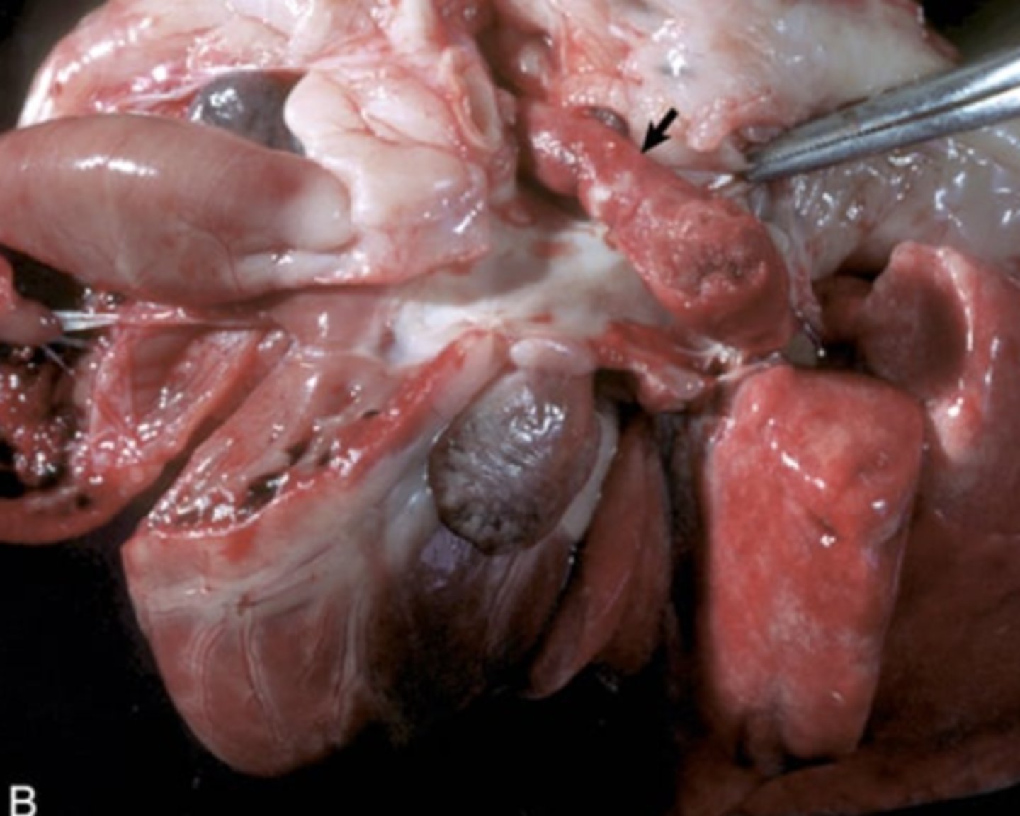
Thrombus
What is being shown in this photo?
Saddle Thrombus
What is being shown in this photo?
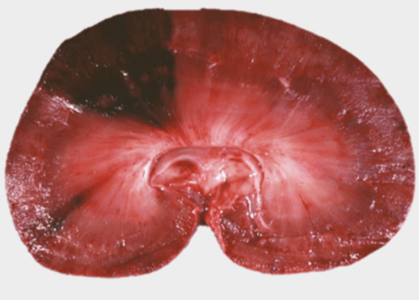
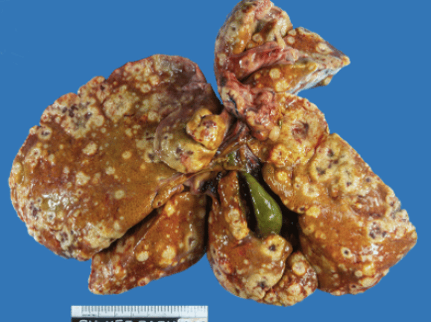
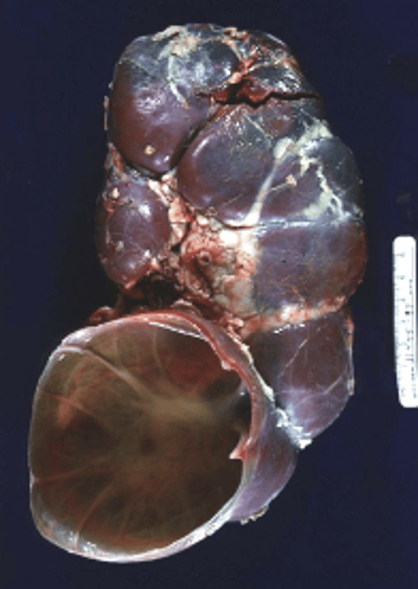
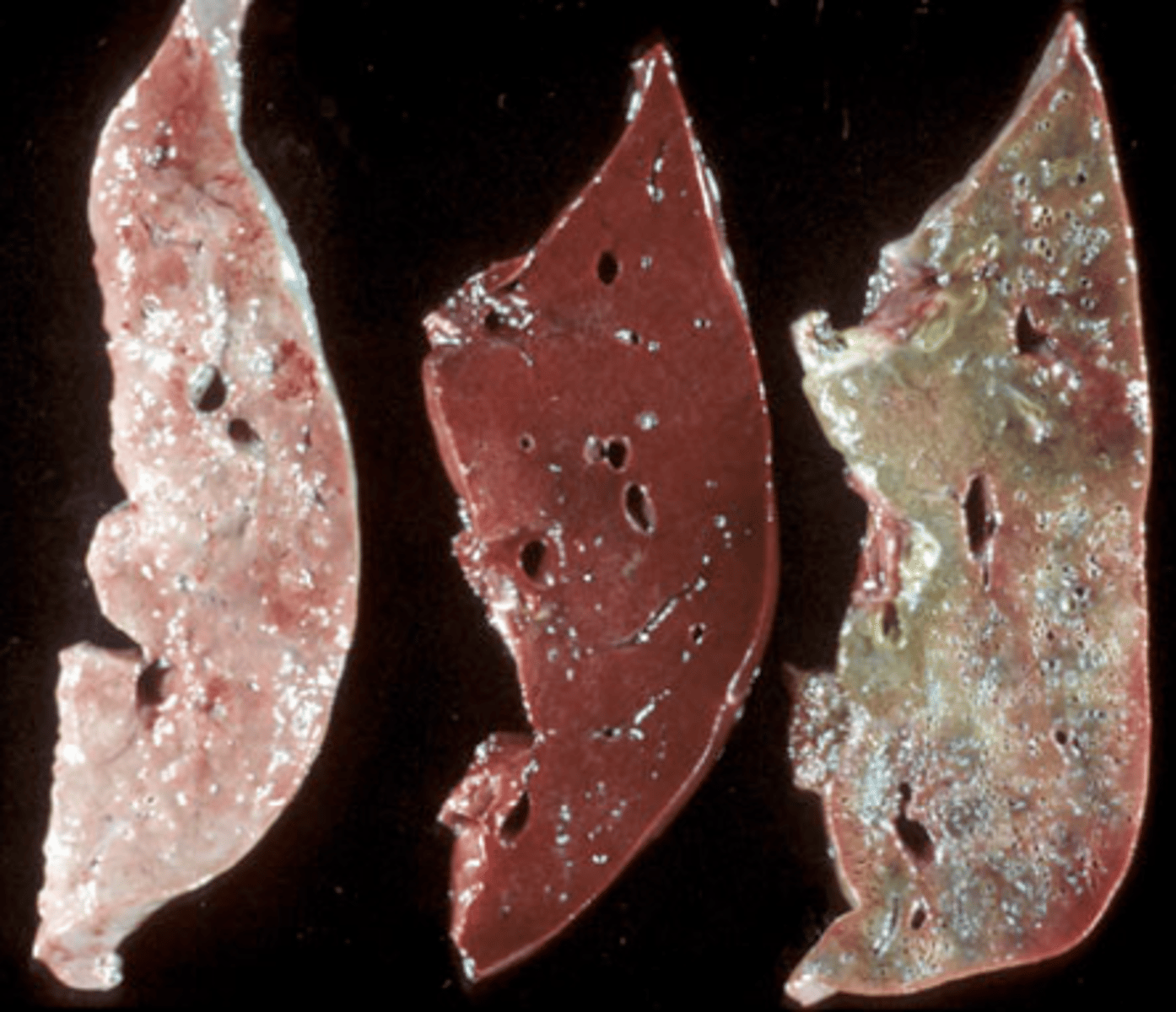
Passive Congestion of the liver
What is being shown in this photo?
Passive Congestion of the liver (chronic - this is nutmeg liver)
What is being shown in this photo?
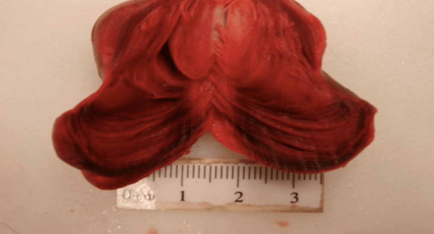
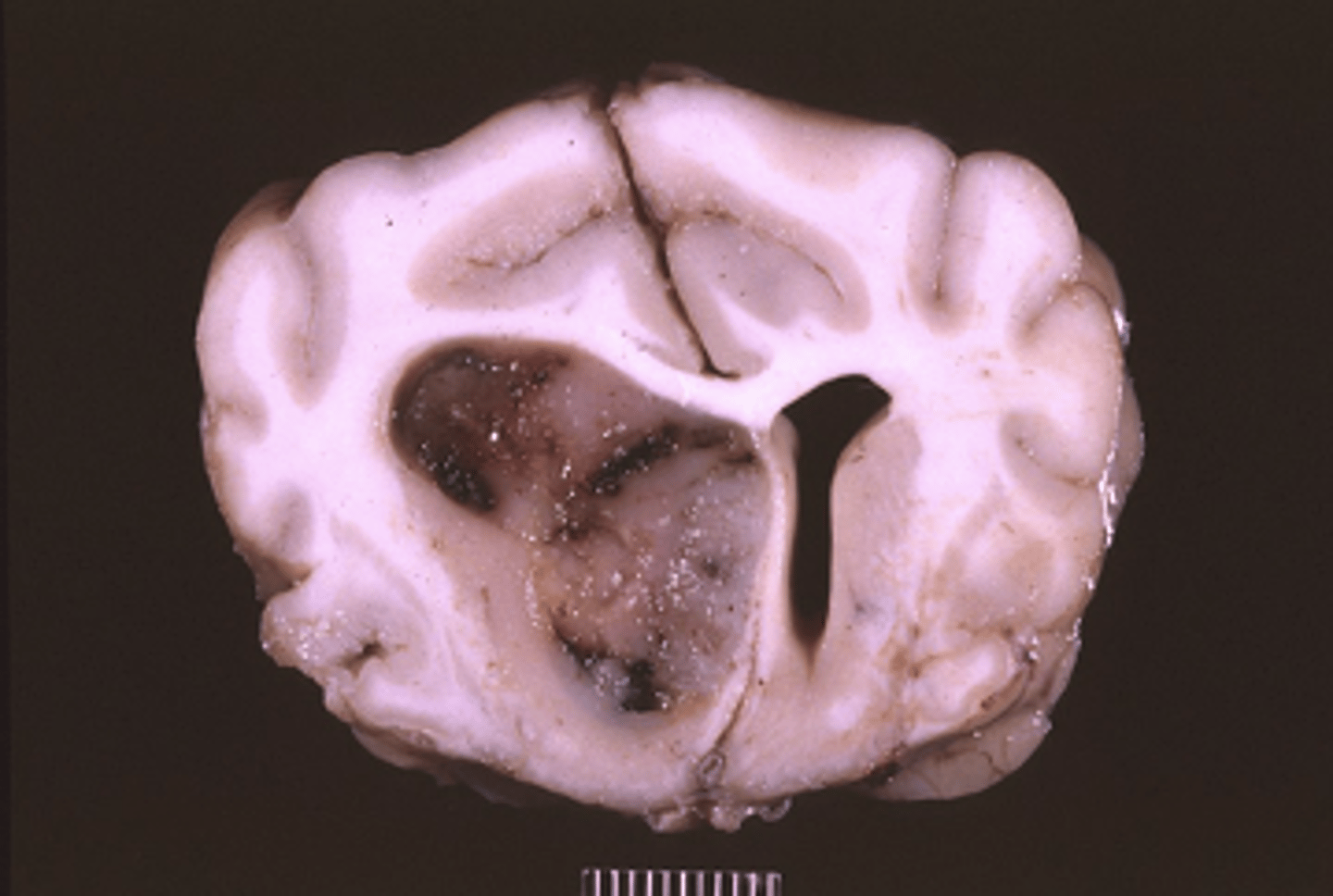
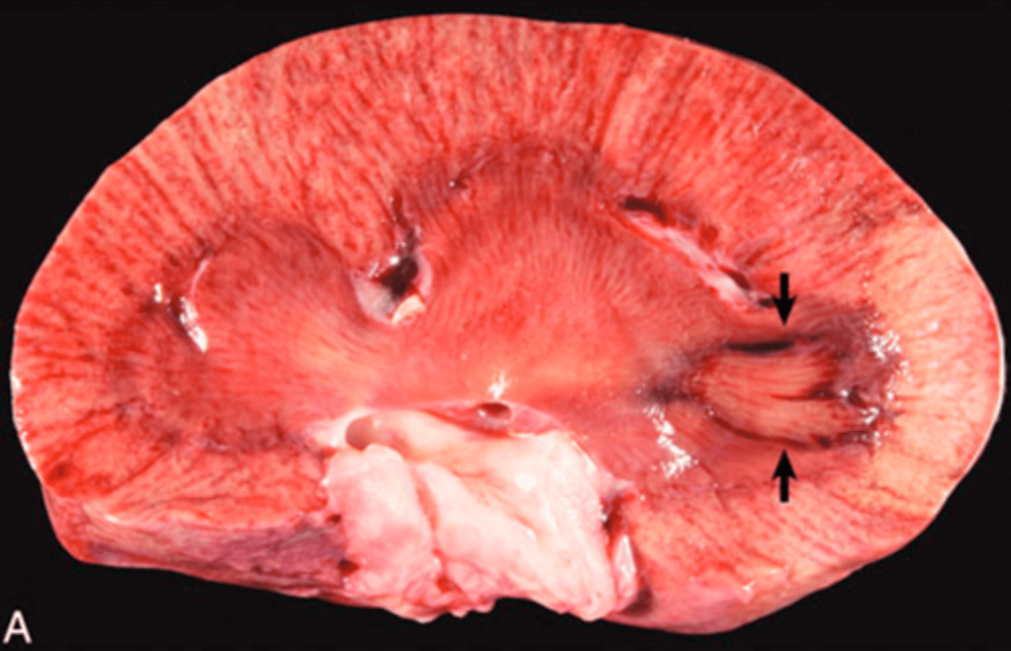
Hemorrhagic Infarct
What is being shown in this photo?
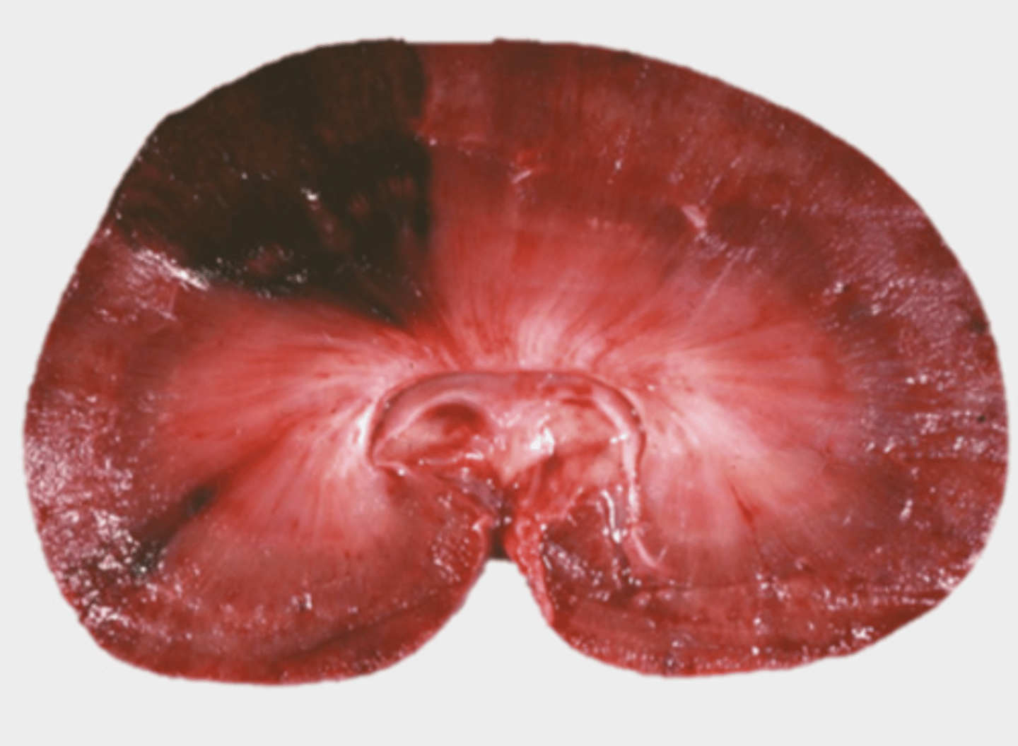
focal
What is the distrubution this hemorrhagic infarct?
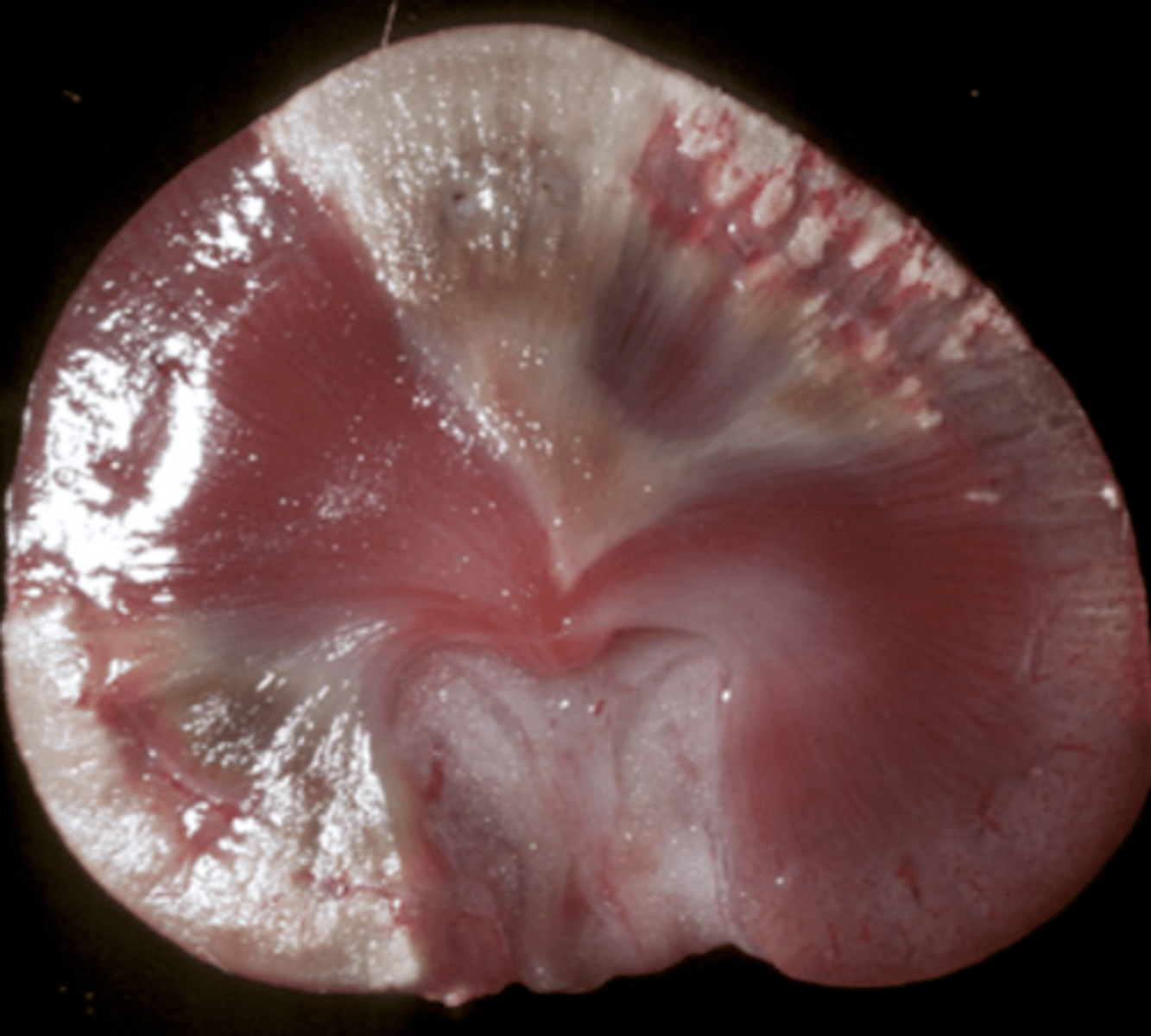
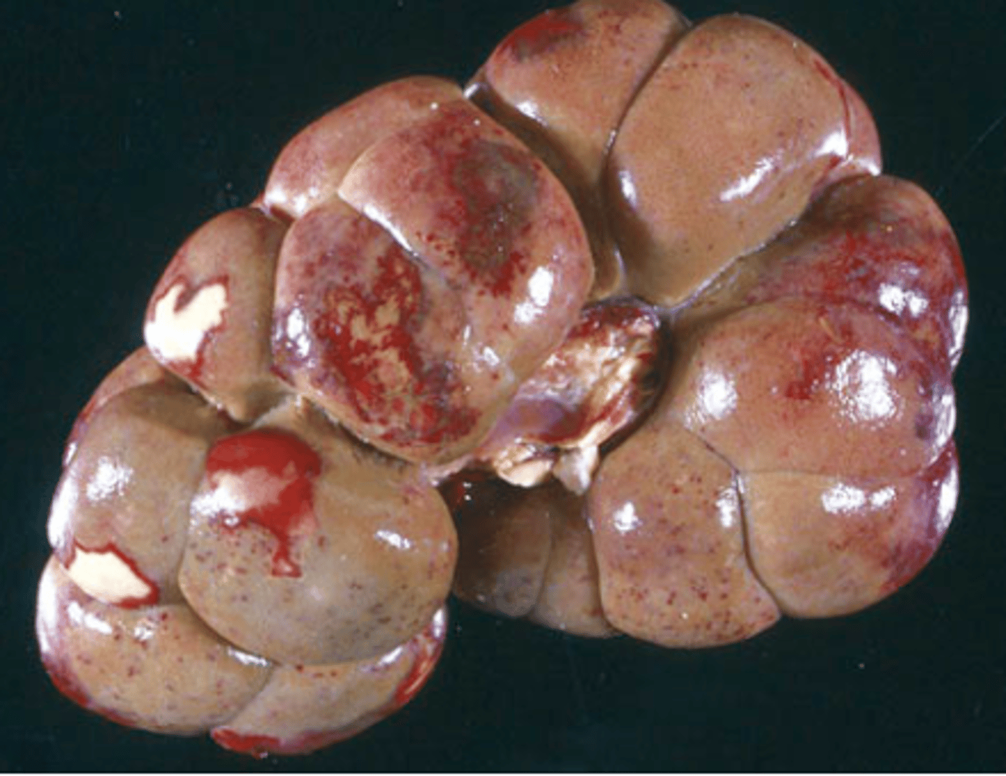
Pale Infarcts
What is being shown in this photo?
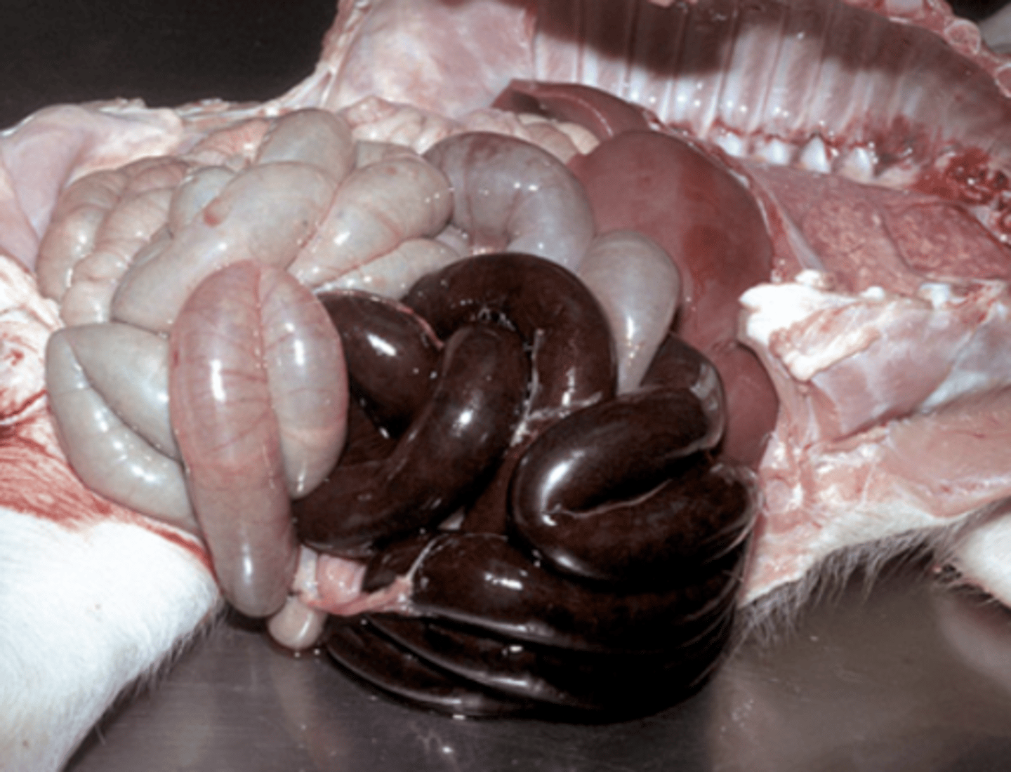
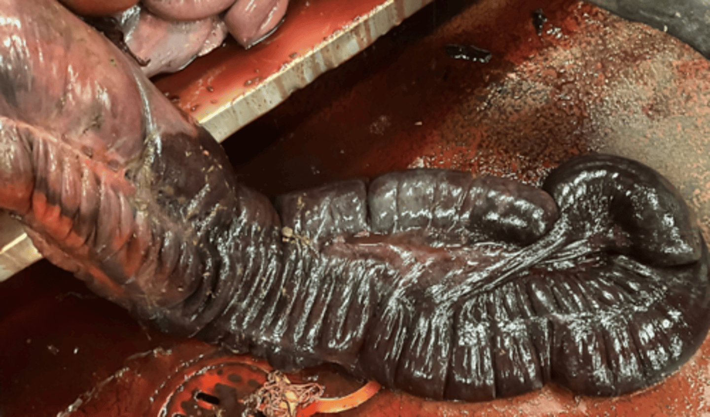
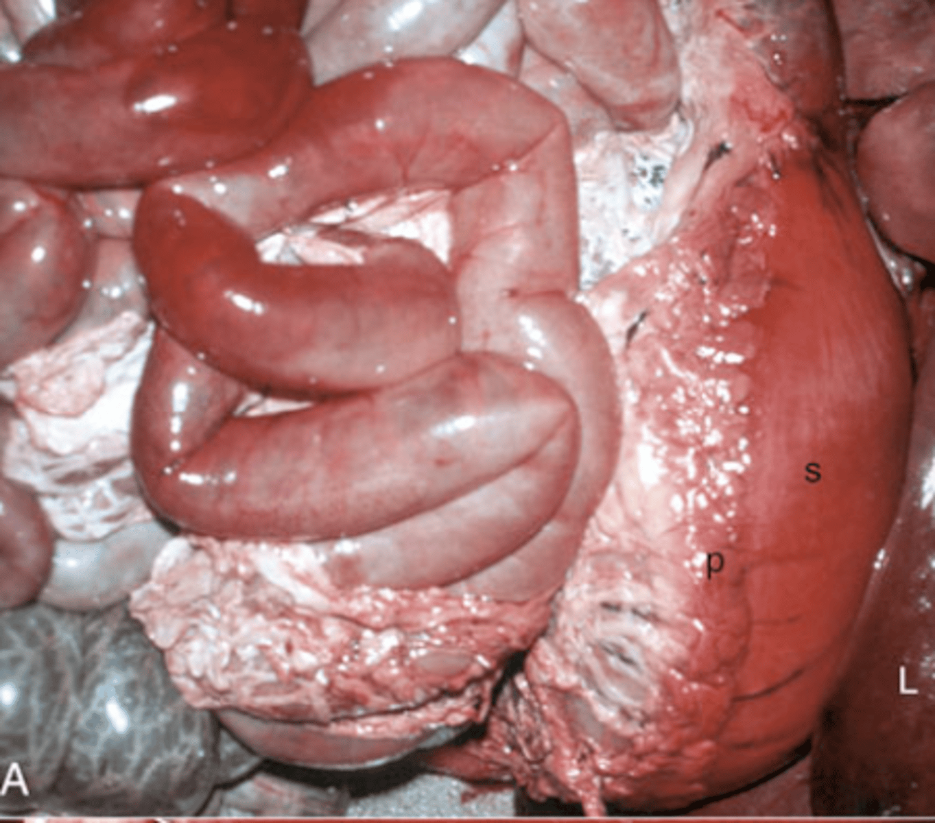
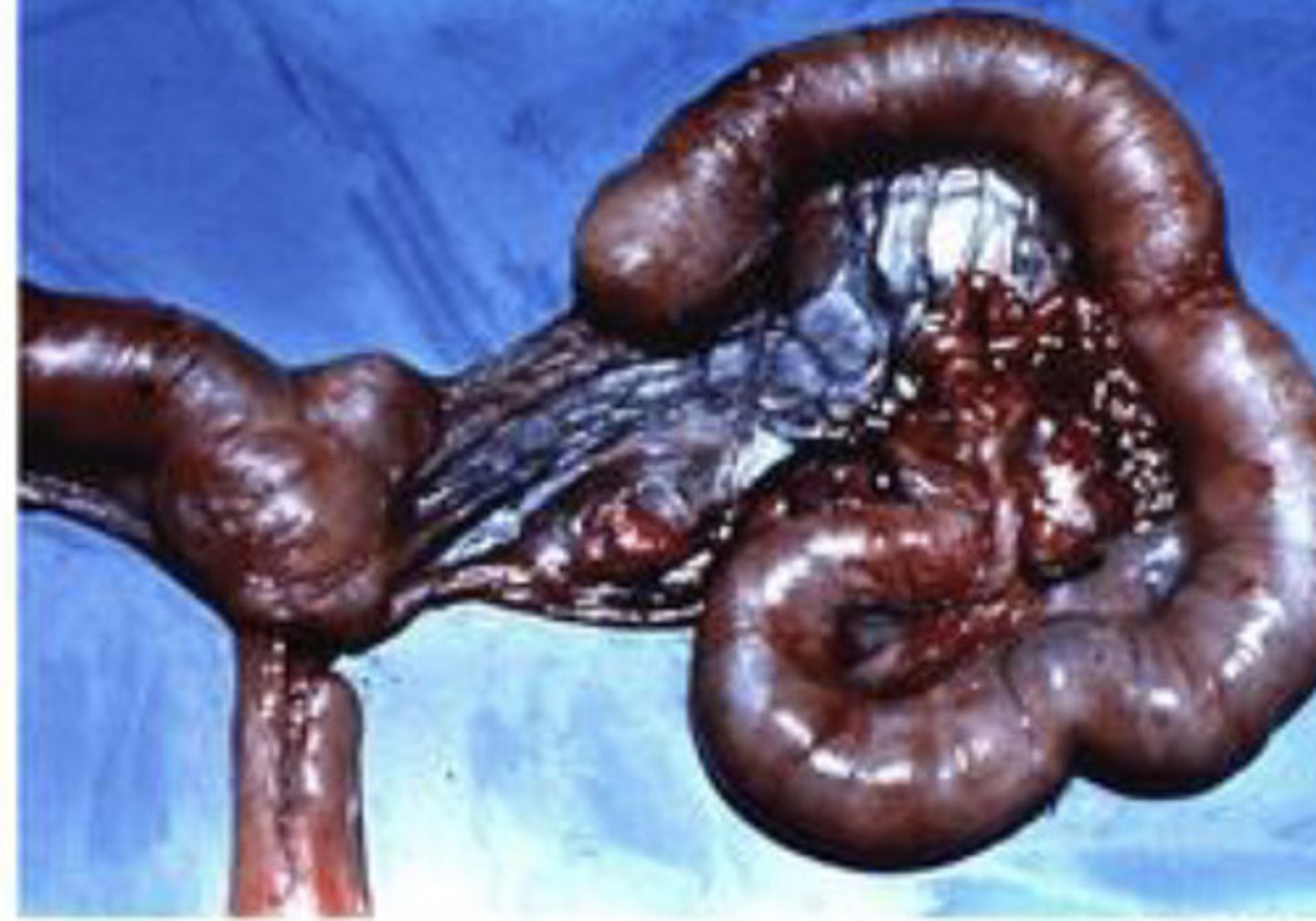
venous infarction
This is an image of intestinal volvulus, what is being shown?
arterial thrombus
What type of thrombus is being shown
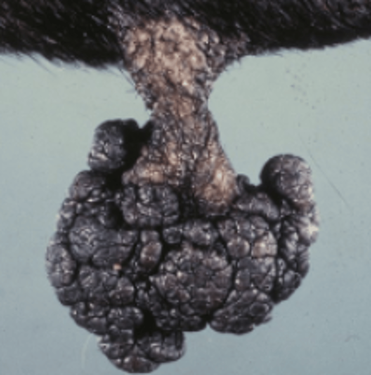
Exophytic
How would you describe this lesion based on the shape?
pedunculated
How would you describe this lesion based on the shape?
umbilicated
How would you describe this lesion based on the shape?
good margins
Describe the margination of this lesion
Cyst
Classify this lesion:
Cyst
Abscess
Polyp
Ulcer
Absecess
Classify this lesion:
Cyst
Abscess
Polyp
Ulcer
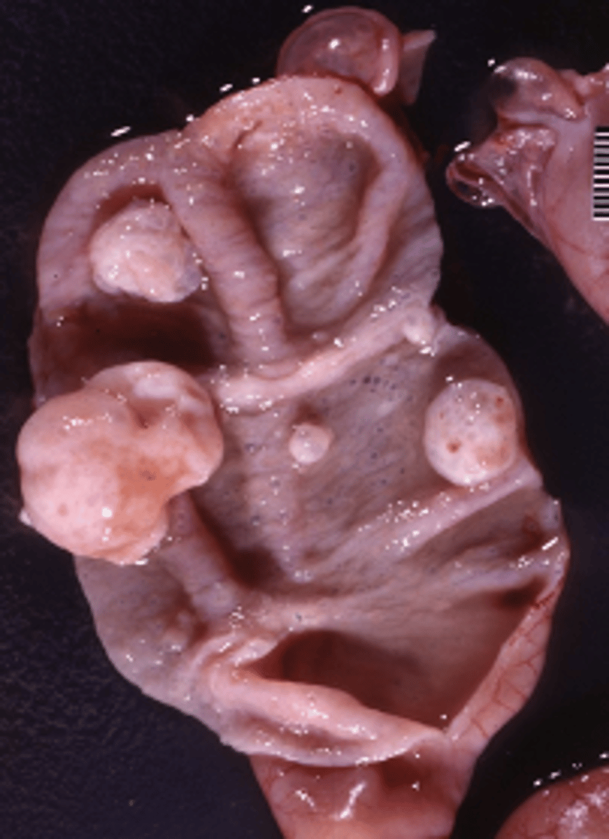
polyp
Classify this lesion:
Cyst
Abscess
Polyp
Ulcer
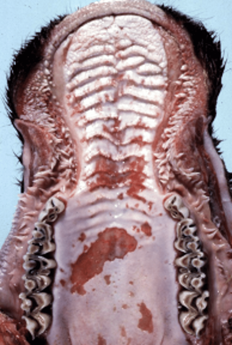
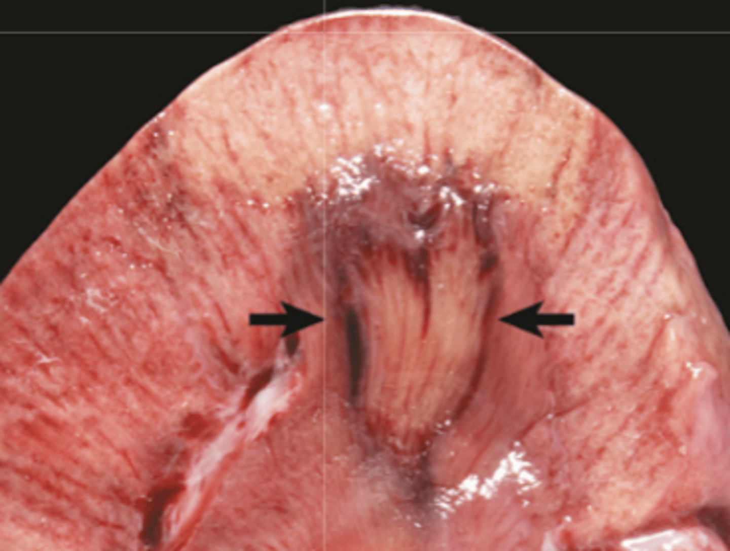
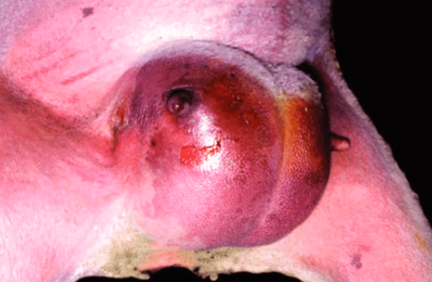
ulcer
Classify this lesion:
Cyst
Abscess
Polyp
Ulcer
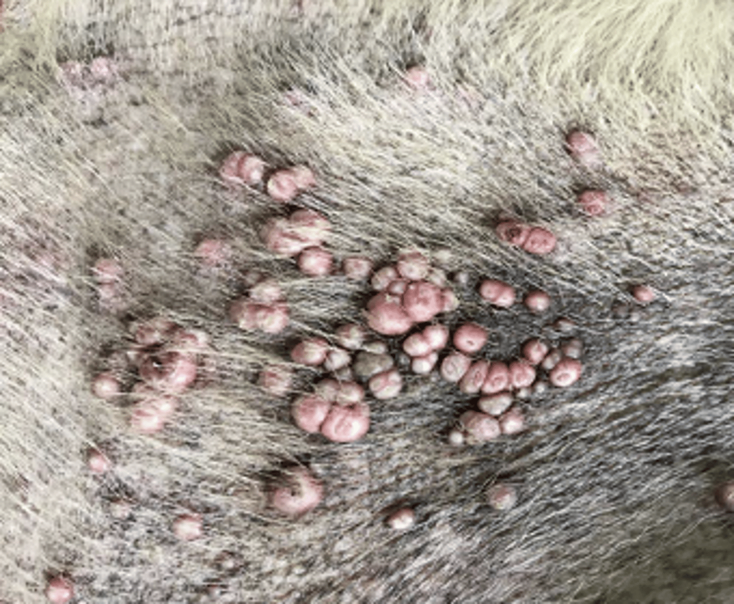
Multifocal
What is the distribution of this lesion
oxygen depletion
What was the most likely cause of this cell injury
Acute cell swelling
What type of reversible cell injury is shown in these images?
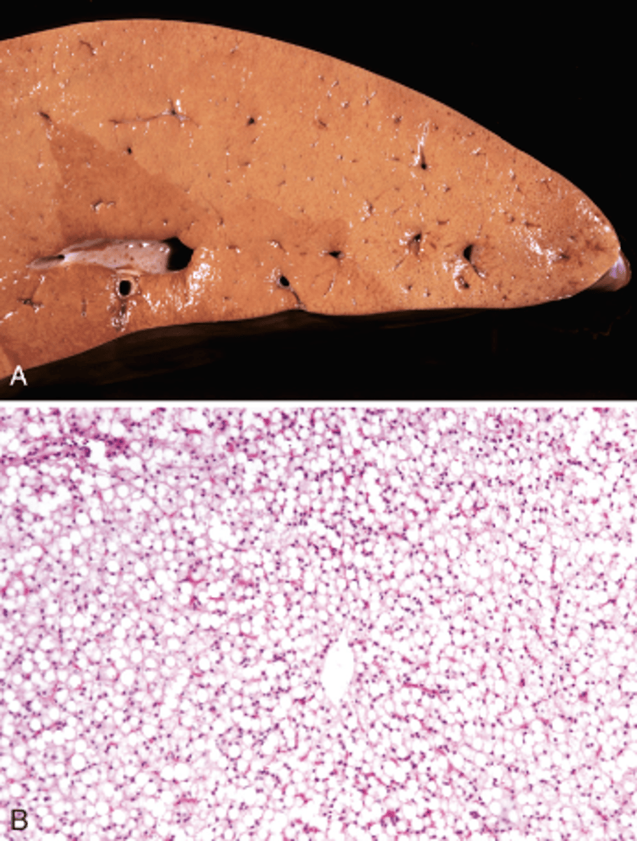
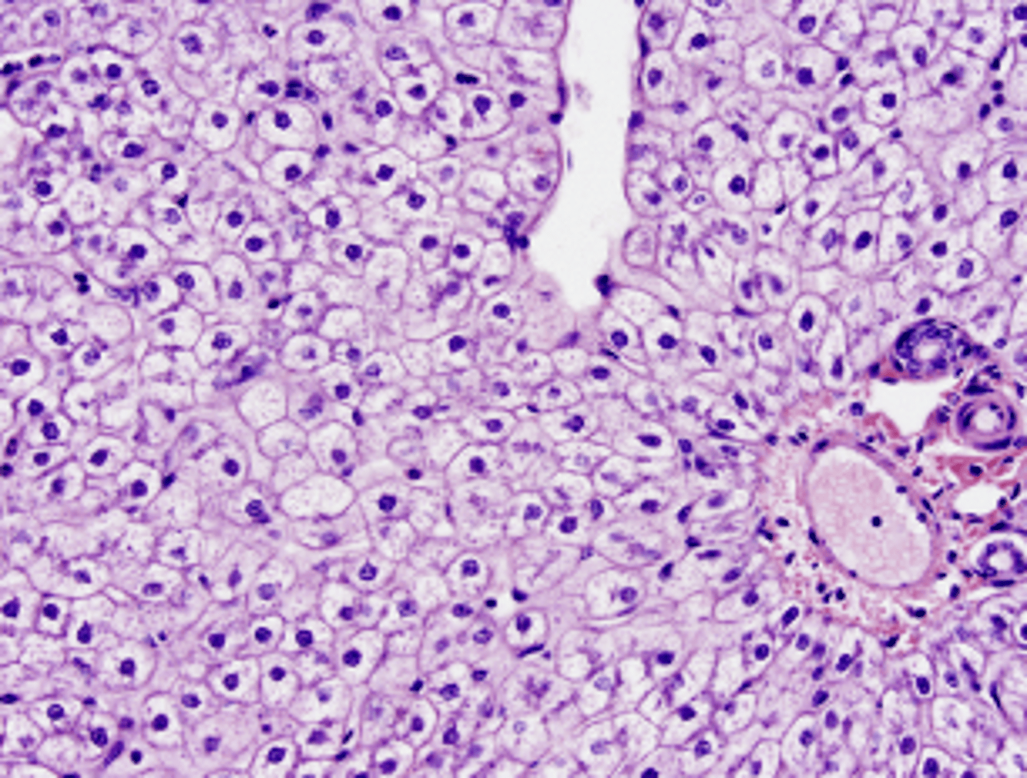
Lipidosis
What type of reversible cell injury is shown in these images?
Glycogen accumulation
What type of reversible cell injury is shown in this image?
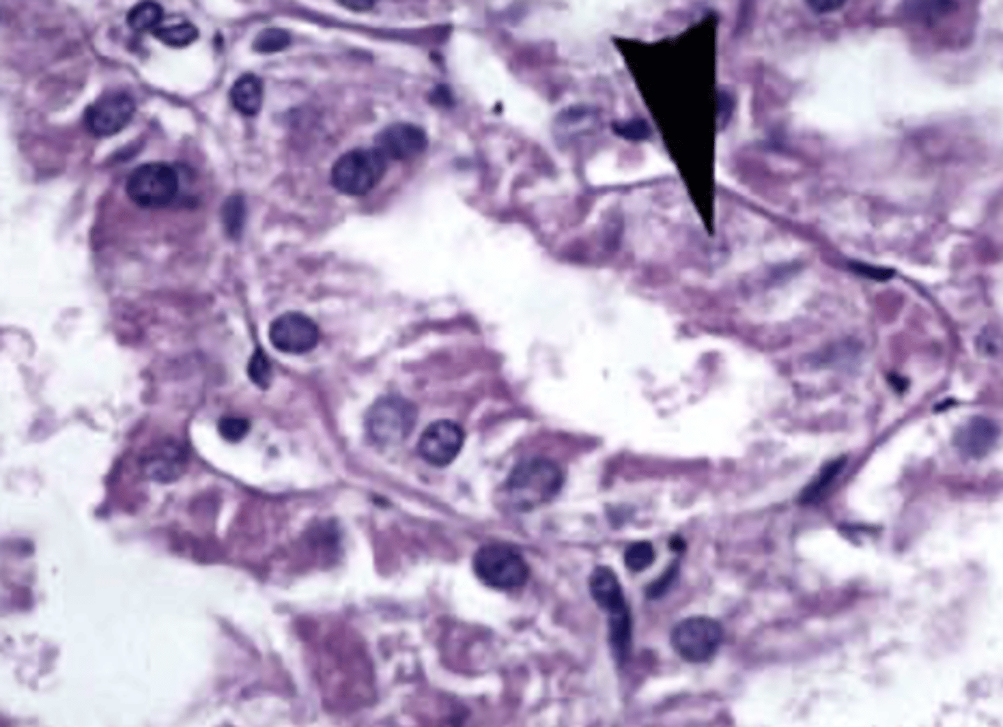
necrosis
This image is showing
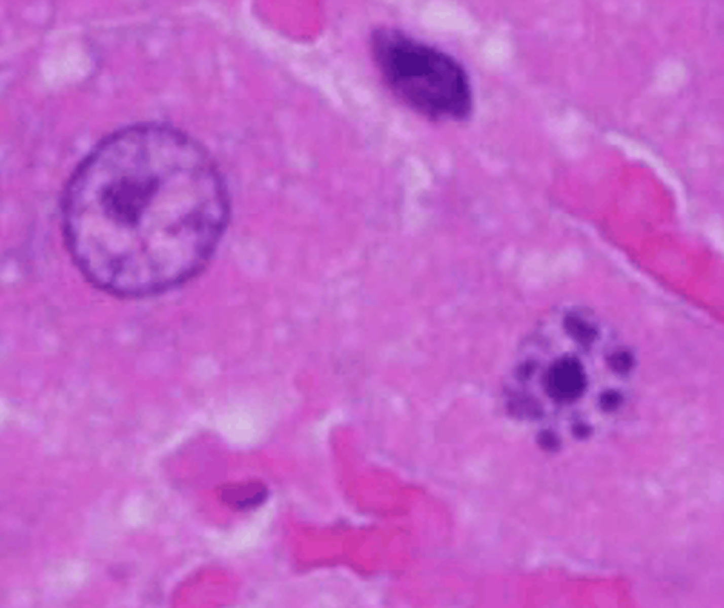
pyknosis
This is an image showing a cell undergoing necrosis. What type of nuclear changes are occuring?
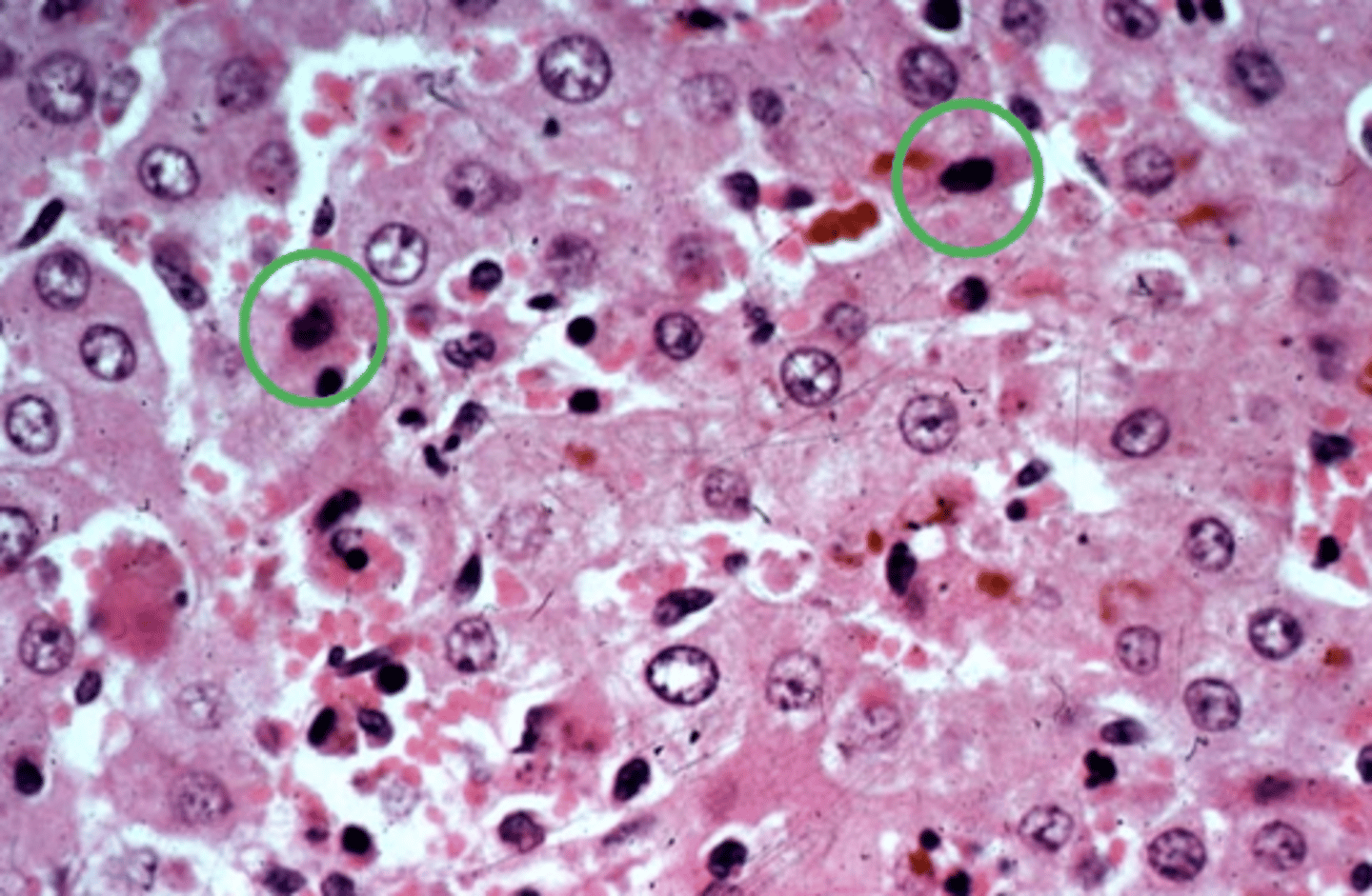
Karyorrhexis
This is an image showing a cell undergoing necrosis. What type of nuclear changes are occuring?
Karyolysis
This is an image showing a cell undergoing necrosis. What type of nuclear changes are occuring?
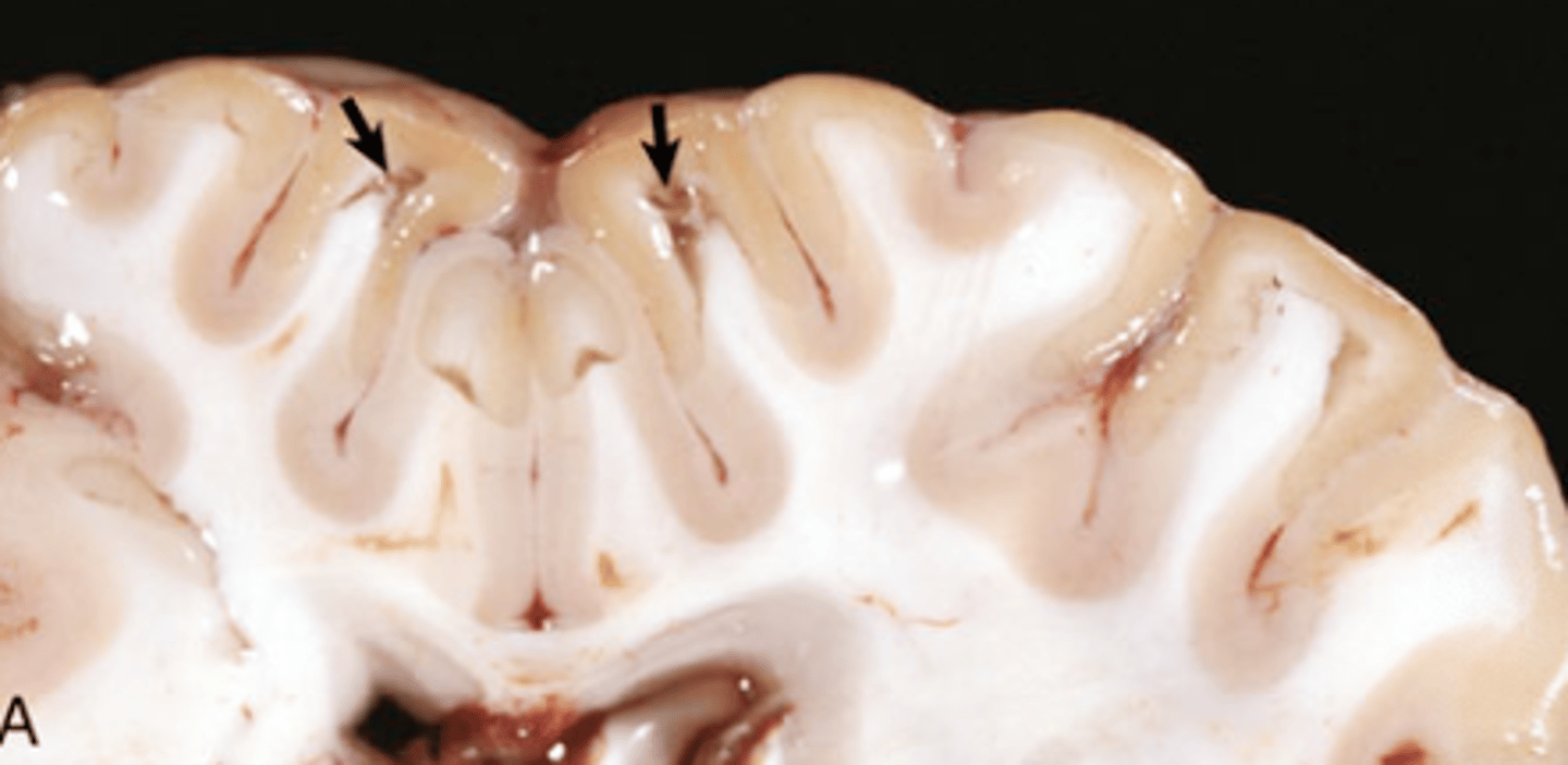
coagulative
What type of necrosis is being shown in this photo?
coagulative
What type of necrosis is being shown in this photo?
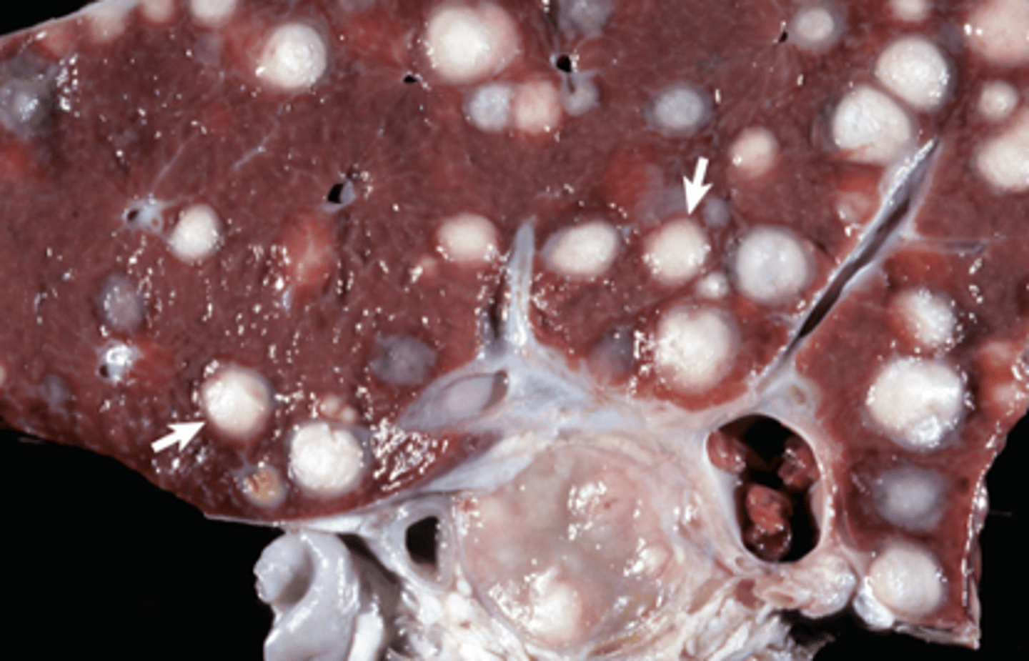
caseous
What type of necrosis is being shown in this photo?
caseous
What type of necrosis is being shown in this photo?
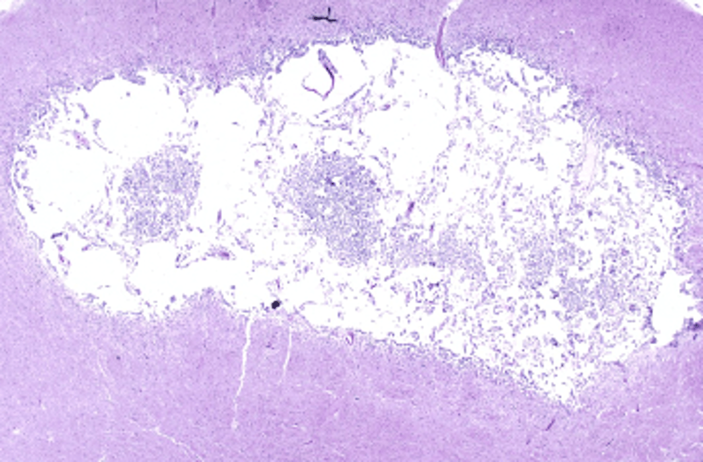
liquefactive
What type of necrosis is being shown in this photo?
liquefactive
What type of necrosis is being shown in this photo?
lytic
What type of necrosis is being shown in this photo?
lytic
What type of necrosis is being shown in this photo?
fat
What type of necrosis is being shown in this photo?
apoptosis
What type of cell death is shown here?
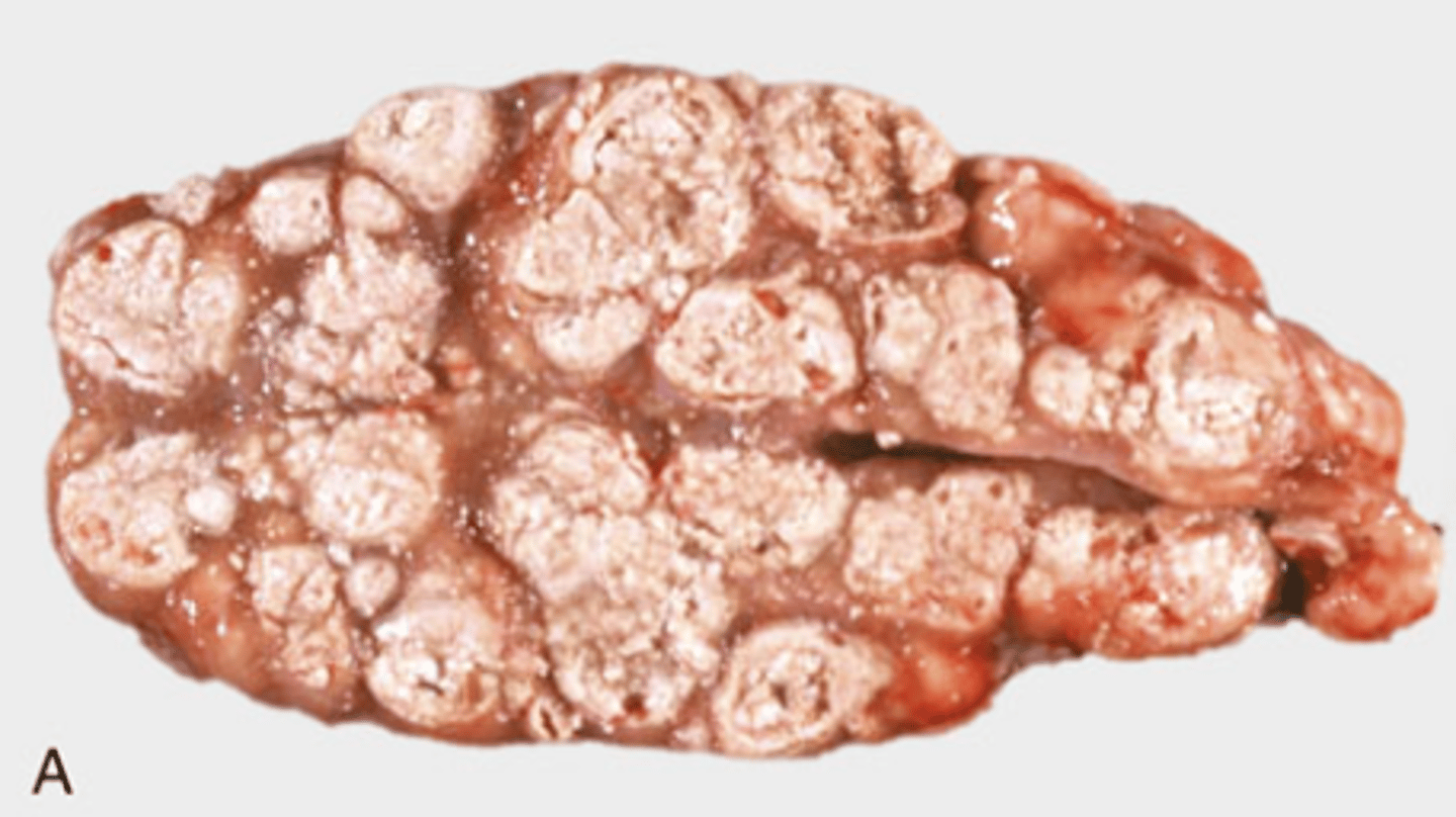
Nodular hyperplasia
What type of cellular adaptation is shown in this photo?
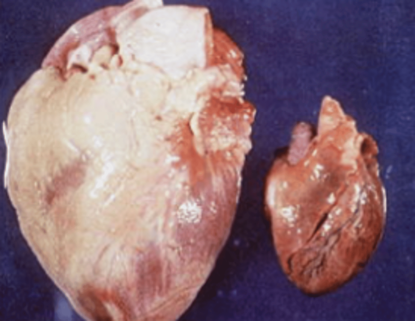
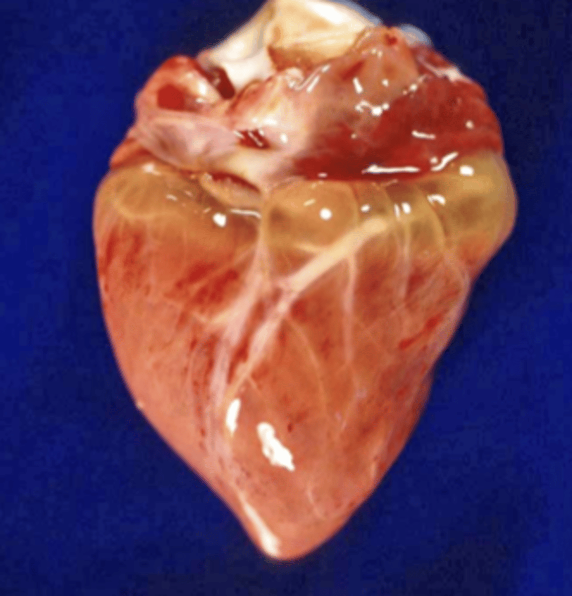
Hypertrophy
What type of cellular adaptation is shown in this photo?
Atrophy
What type of cellular adaptation is shown in this photo?
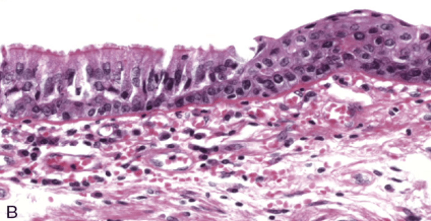
Normal is on the left
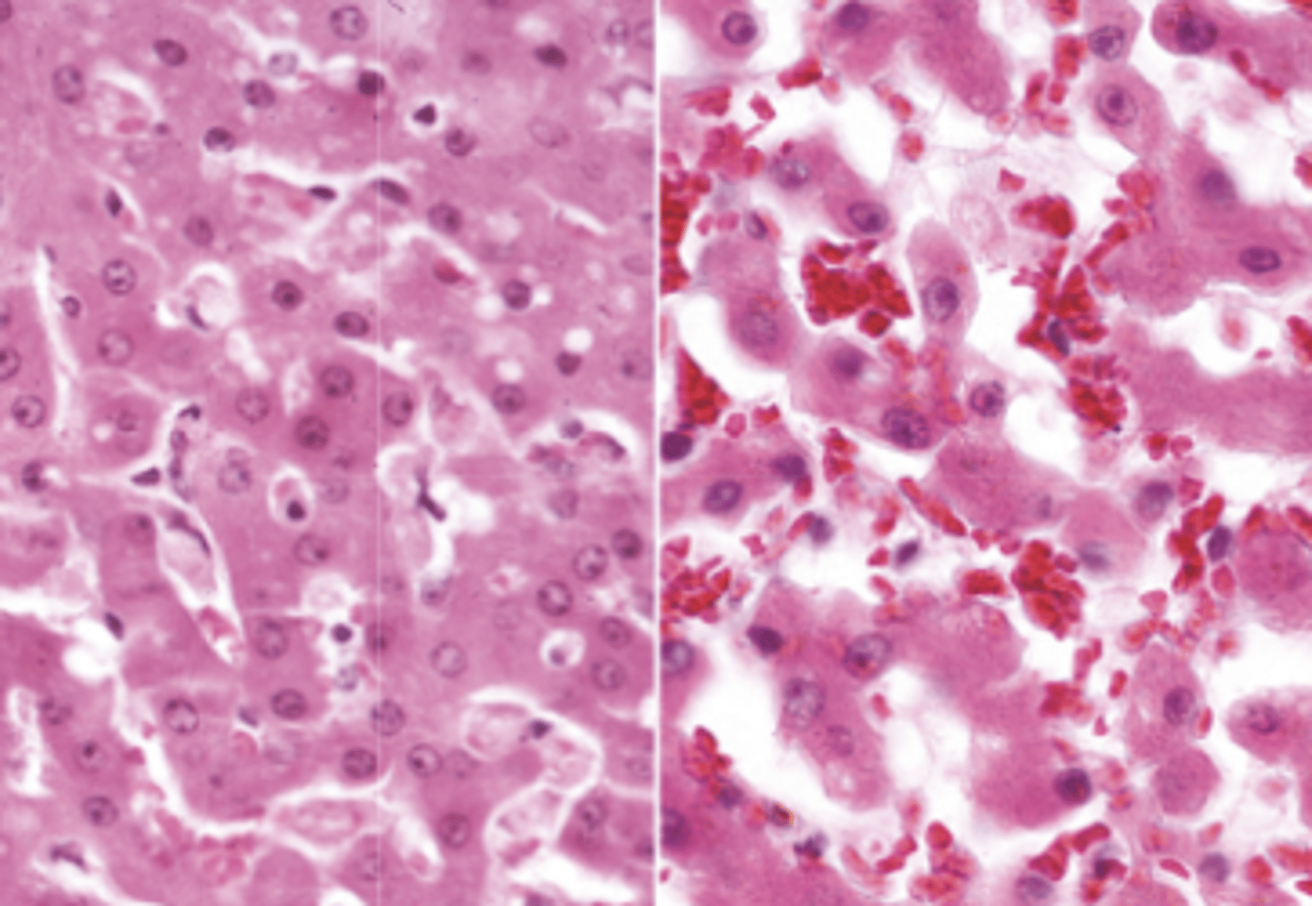
metaplasia
What type of cellular adaptation is shown in this photo?
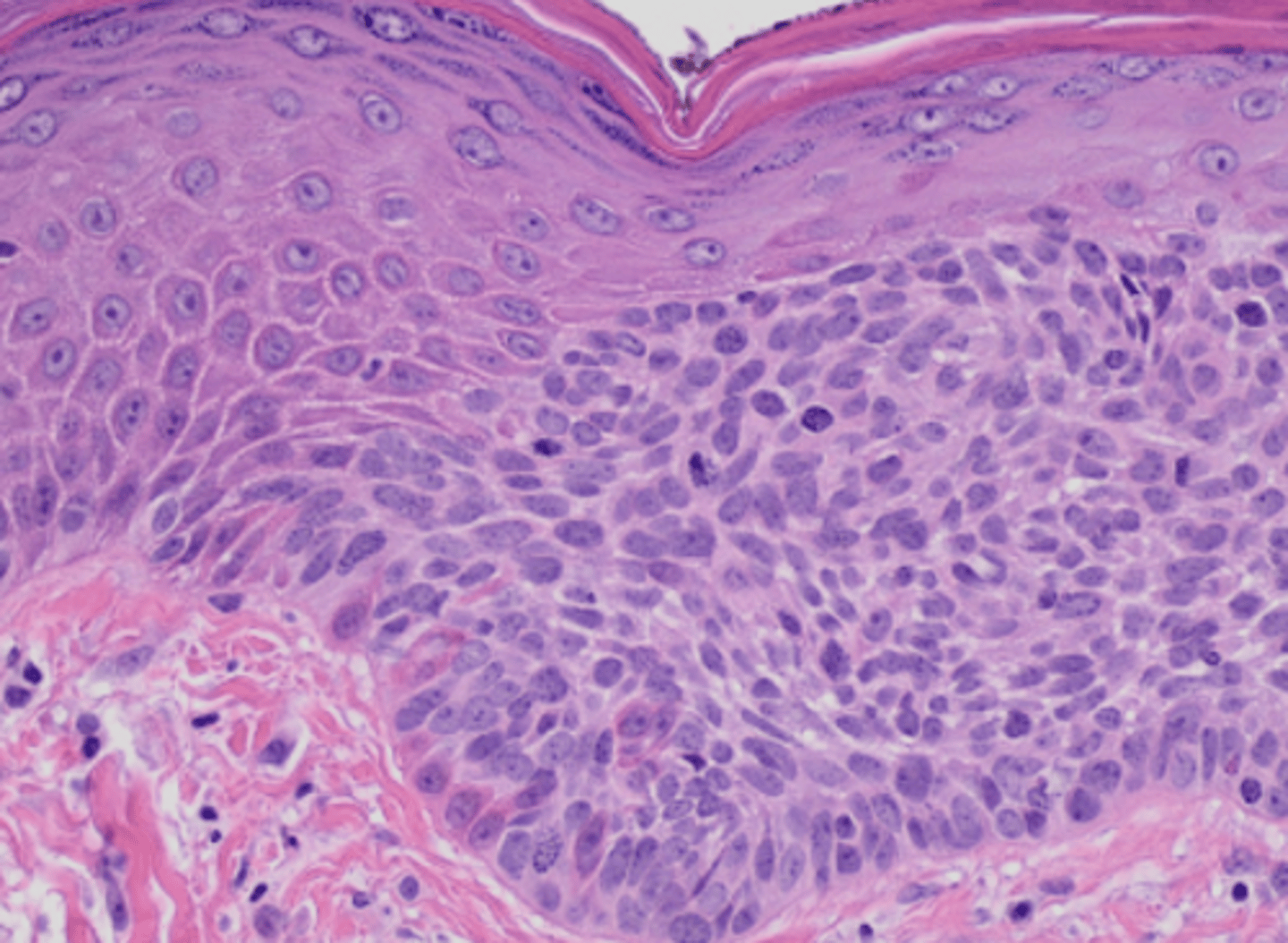
Dysplasia
What type of cellular adaptation is shown in this photo?
Diffuse hyperplasia
What type of cellular adaptation is shown in this photo?
Diffuse hyperplasia
What type of cellular adaptation is shown in this photo?
serous atrophy of fat
What type of cellular adaptation is shown in this photo?
Hydropic degeneration
What type of acute cell swelling is being shown in this image?
1. Cytoxic edema
2. Balloning degernation
3. Hydropic degeneration
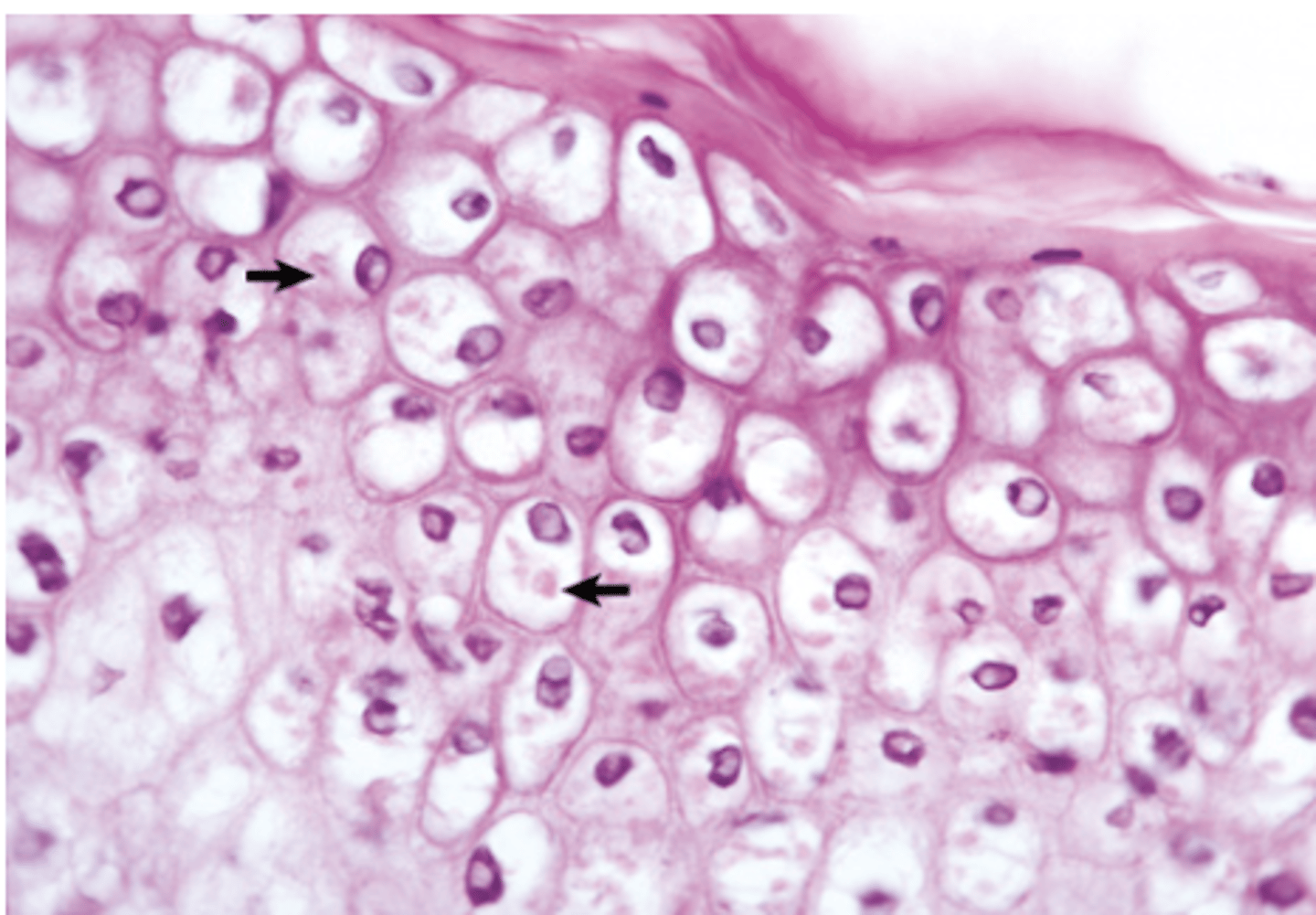
Ballooning degernation
What type of acute cell swelling is being shown in this image?
1. Cytoxic edema
2. Balloning degernation
3. Hydropic degeneration
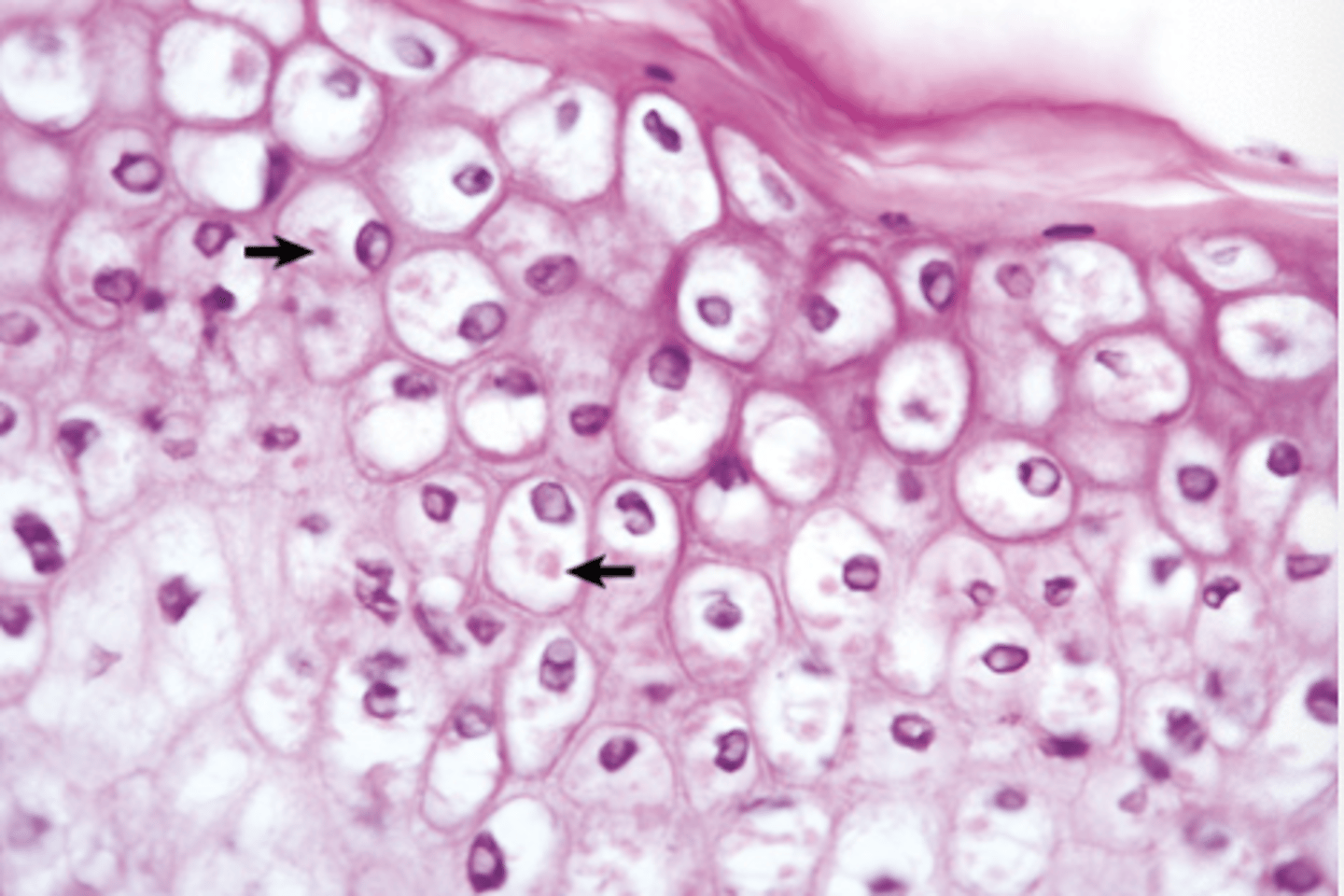
poxviral
This type of acute cell swelling is reserved for talking about what type of infection?
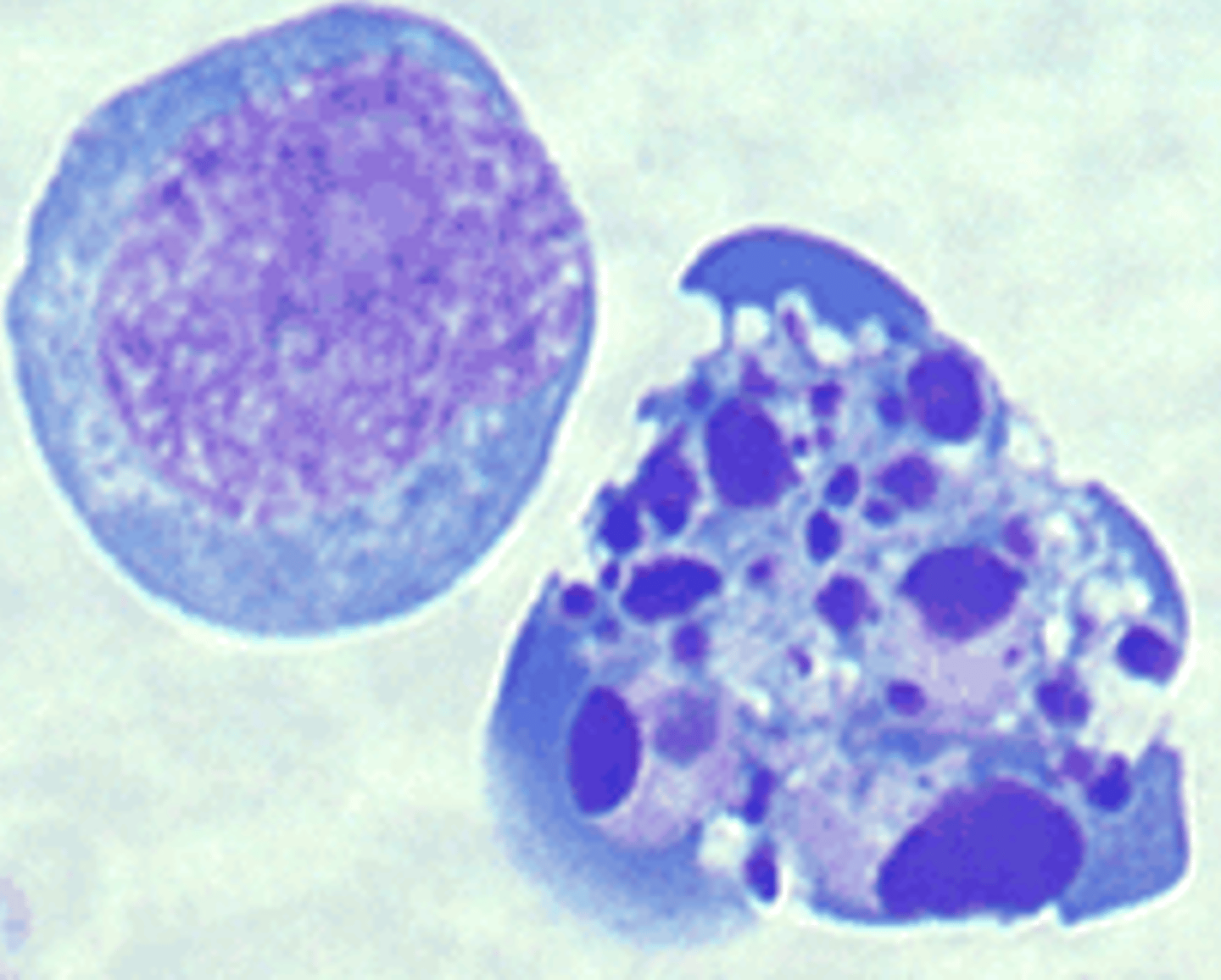
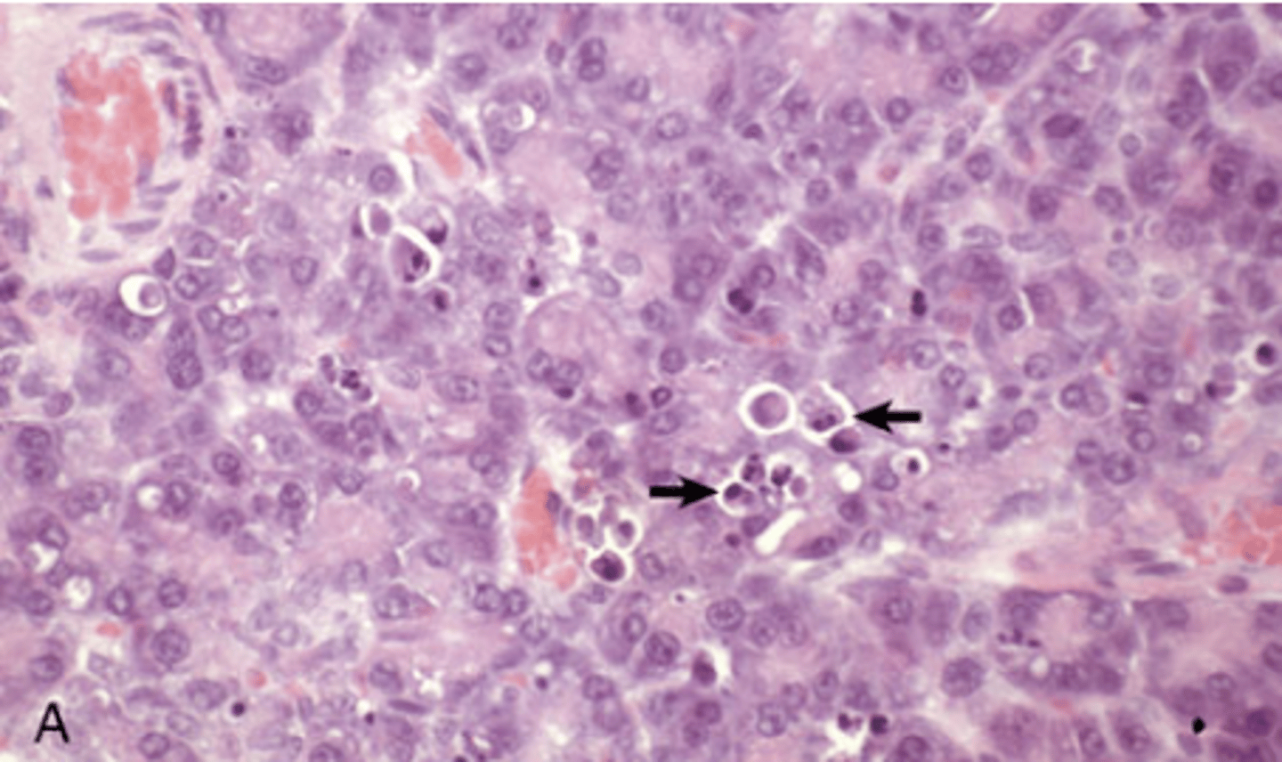
Apoptosis
What type of cell death is being shown in this image? Necrosis or apoptosis?
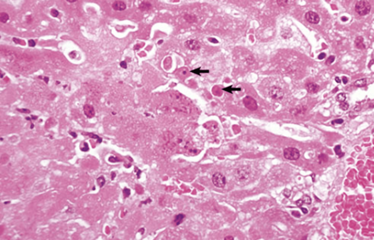
Necrosis
What type of cell death is being shown in this image? Necrosis or apoptosis?
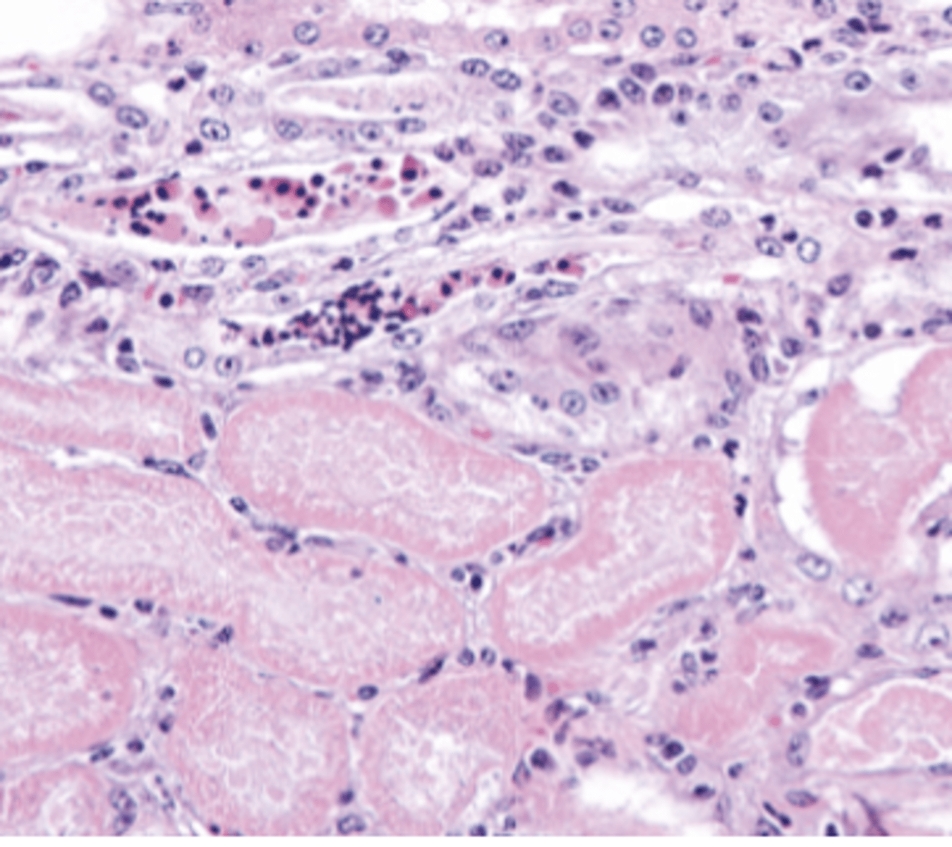
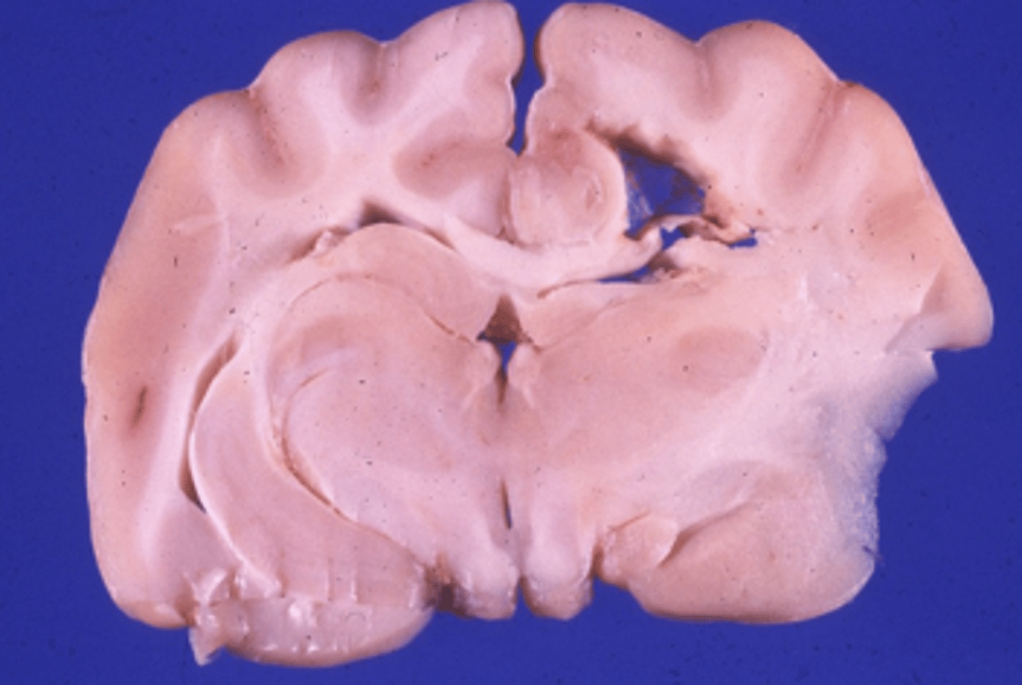
Coagulative
What type of necrosis is being shown in this image?
Coagulative
What type of necrosis is being shown in this image?
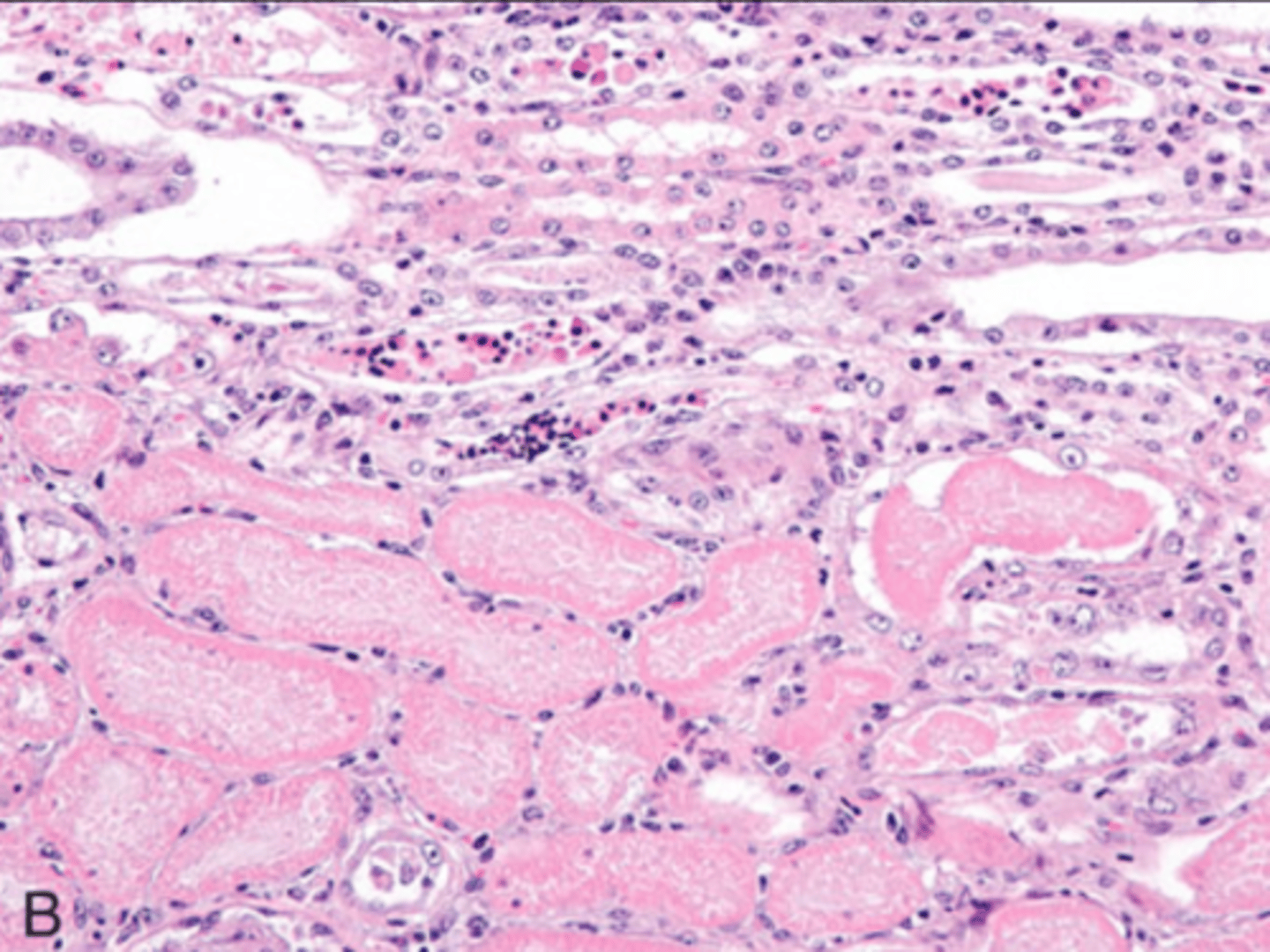
Coagulation
*Notice the acute coagulation necrosis surrounded by the red rim of hyperemia and inflammation
What type of necrosis is being shown in this image?
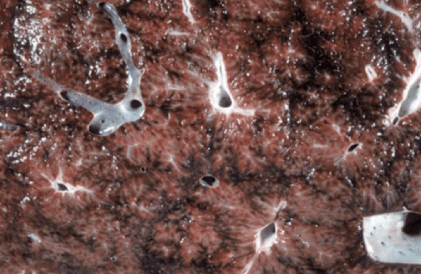
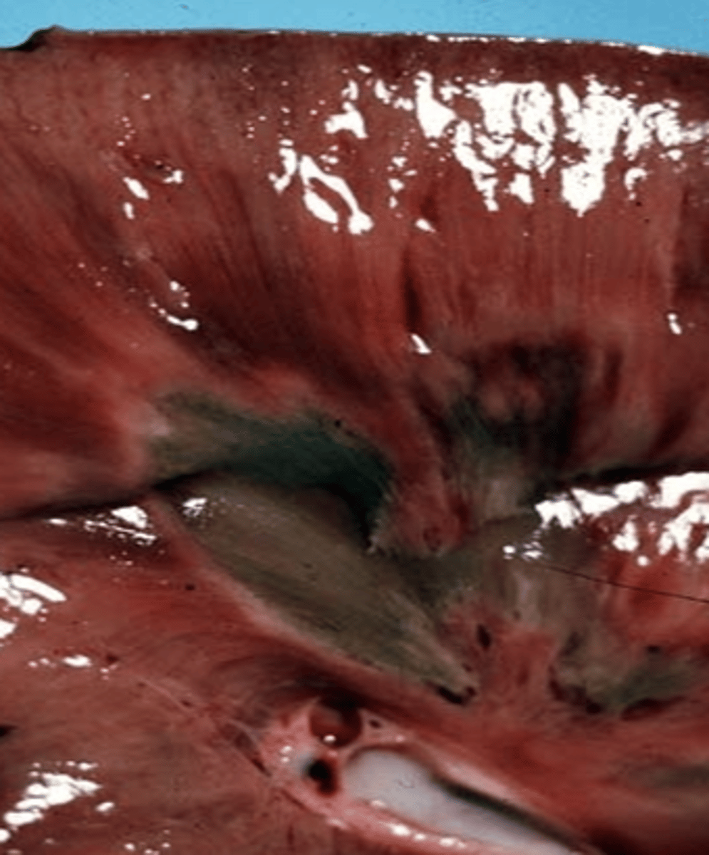
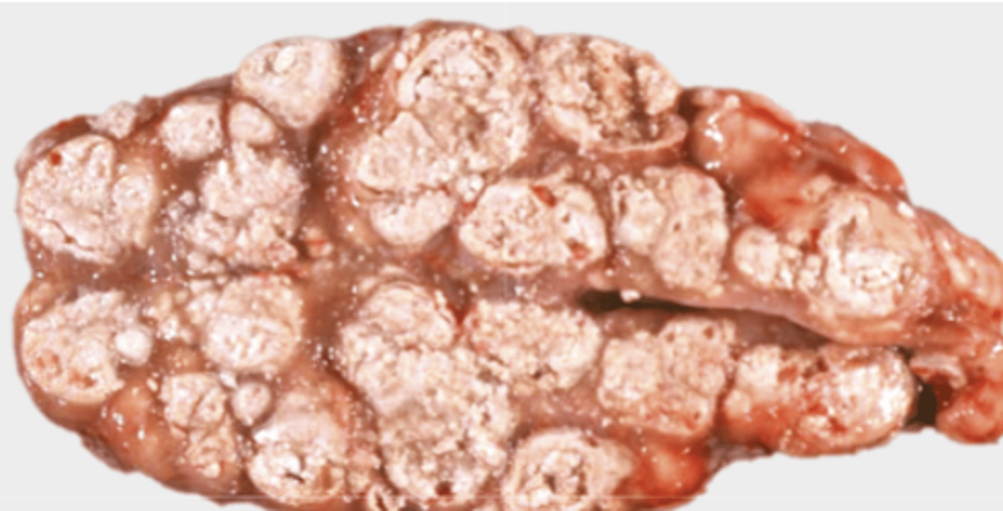
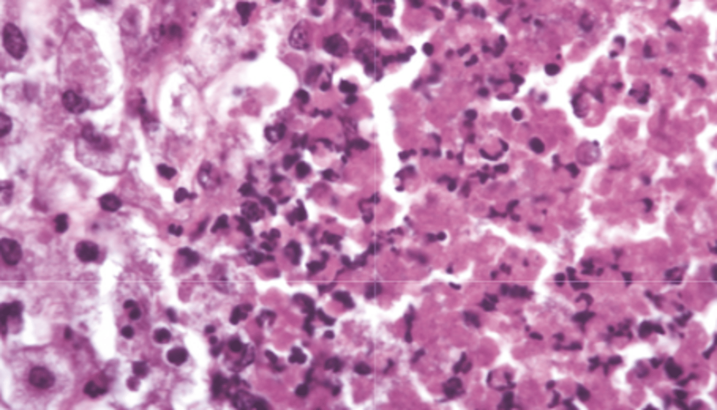
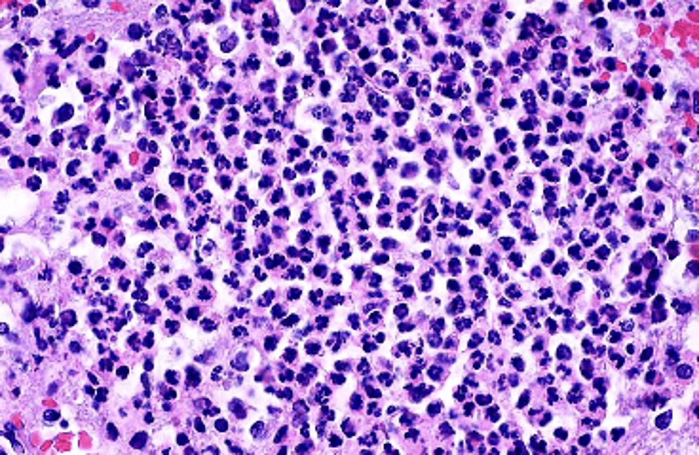
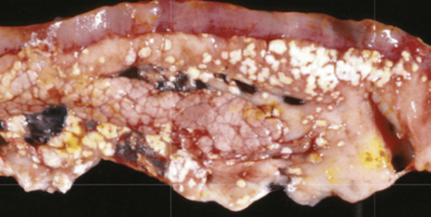
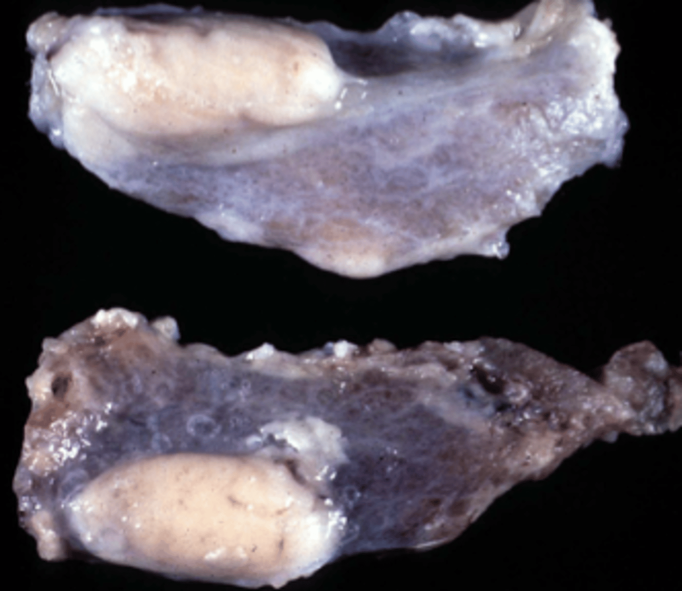
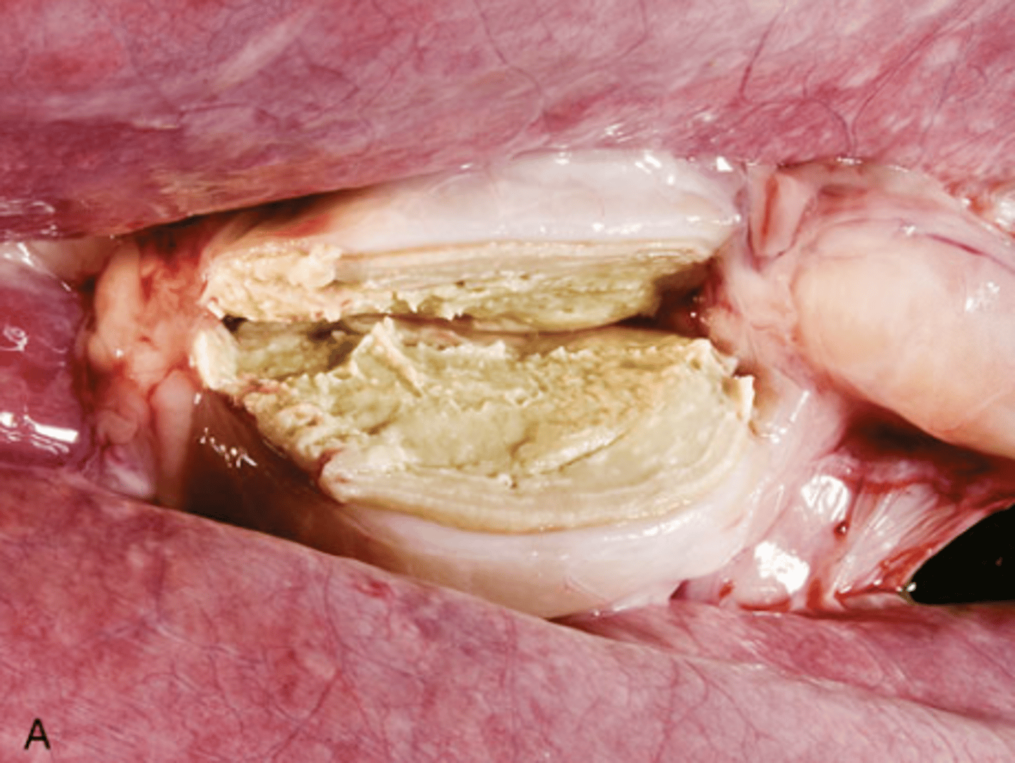
Caseous necrosis
What type of necrosis is being shown in this image?
caseous necrosis
*in the lymph node, caseous necrosis appears as inspissated yellow white and concentrically laminated exudate
What type of necrosis is being shown in this image?
liquidative necrosis
What type of necrosis is being shown in this image?
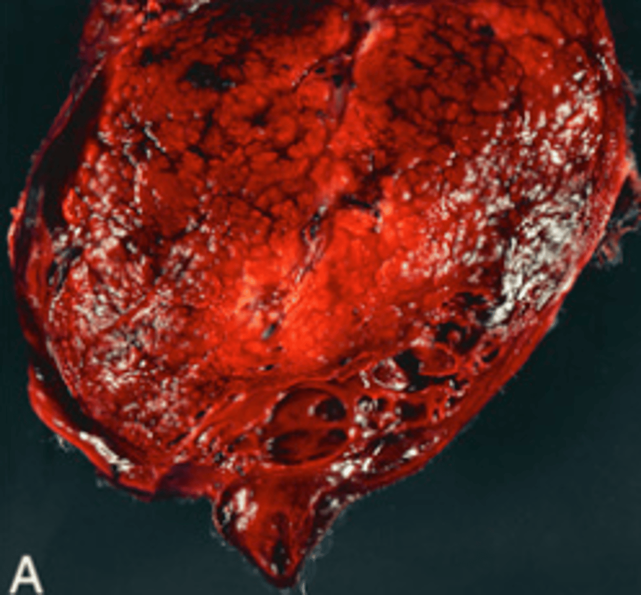
Wet Gangrene necrosis
What type of necrosis is being shown in this image?
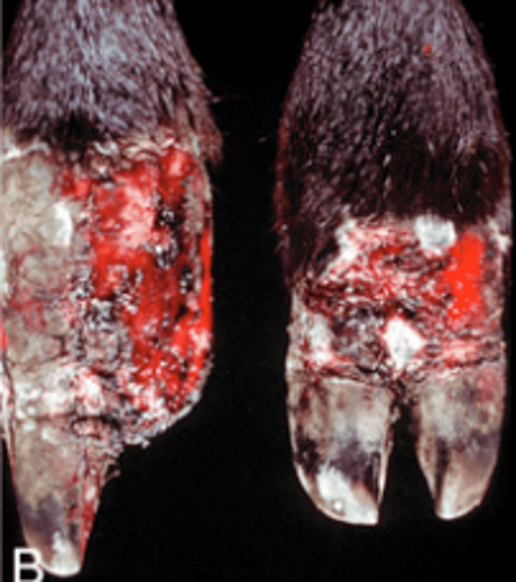
Dry Gangrene necrosis
What type of necrosis is being shown in this image?
Wet Gangrene
What type of necrosis is being shown in this image?
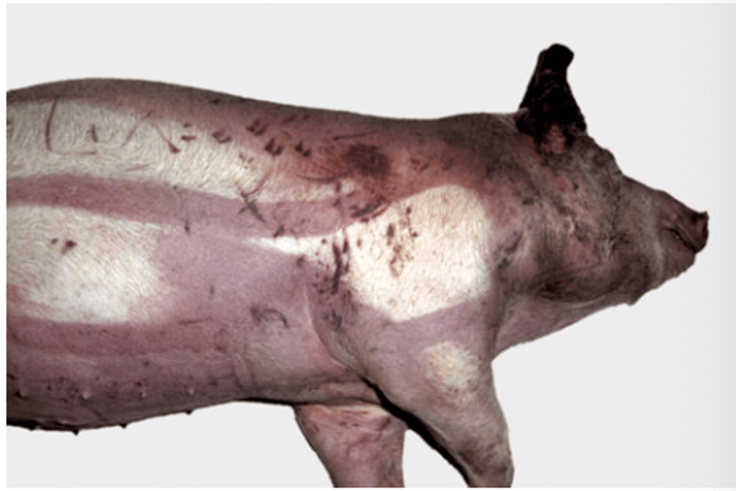
Livor Mortis
What type of postmortem change is shown?
A. Rigor mortis
B. Algor mortis
C. Livor mortis
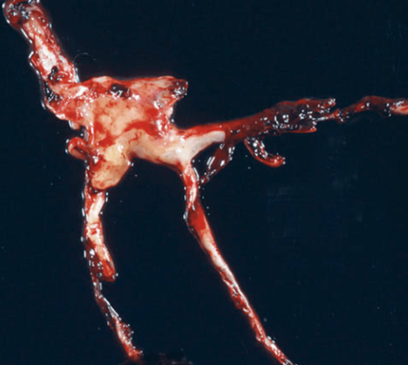
1. Chicken fat clot (the off white to yellow)
2. Currant Jelly clot (the shiny dark red)
This is an image of a postmortem clot. What are the two types of postmortem clots?
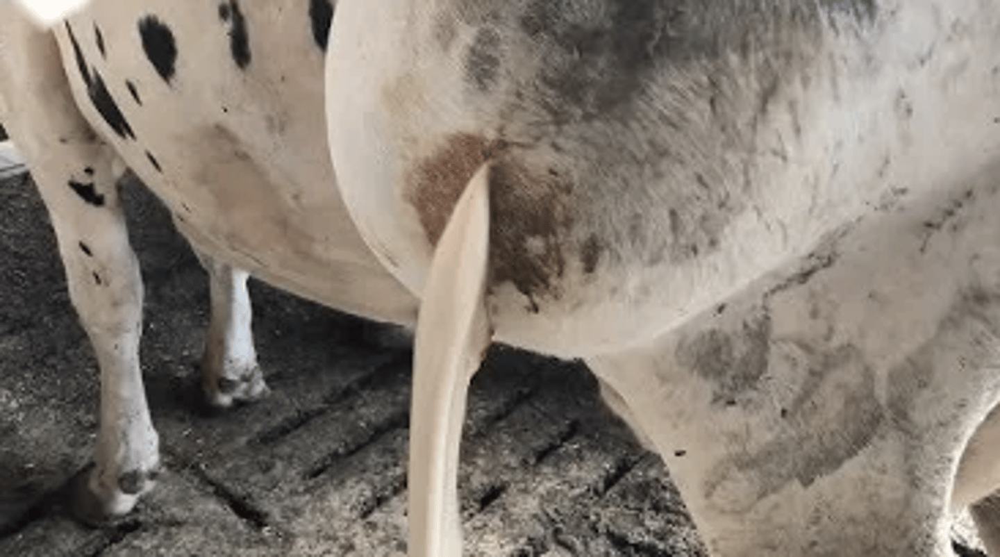
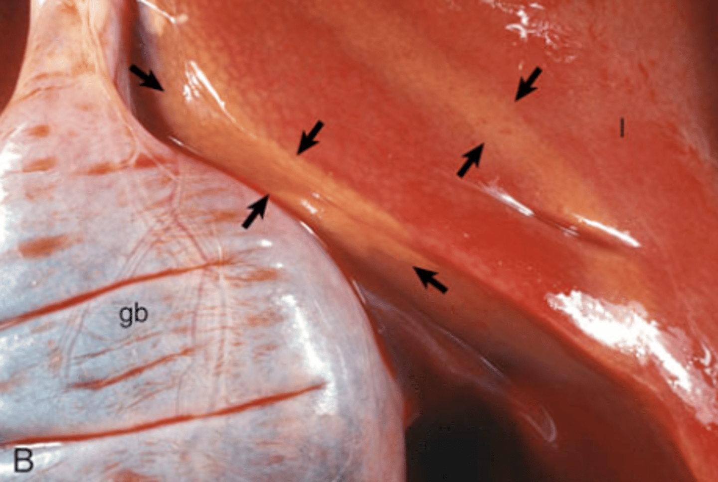
Hemoglobin imbibition
*Dark pink discoloration of serosal surfaces
What type of imbibition is shown in this image?
Hemoglobin or Bile?
Bile imbibition
Orange discoloration due to staining by bile
What type of imbibition is shown in this image?
Hemoglobin or Bile?
Bile imbibition
What type of imbibition is shown in this image?
Hemoglobin or Bile?
Enzymatic
Due to lipases being injured from pancreatic acinar cells
This is an image of Fat Necrosis. What type of Fat Necrosis is seen? What causes this kind of fat necrosis?
Enzymatic Necrosis of Fat
This is an image of Fat Necrosis. What type of Fat Necrosis is seen? What causes this kind of fat necrosis?

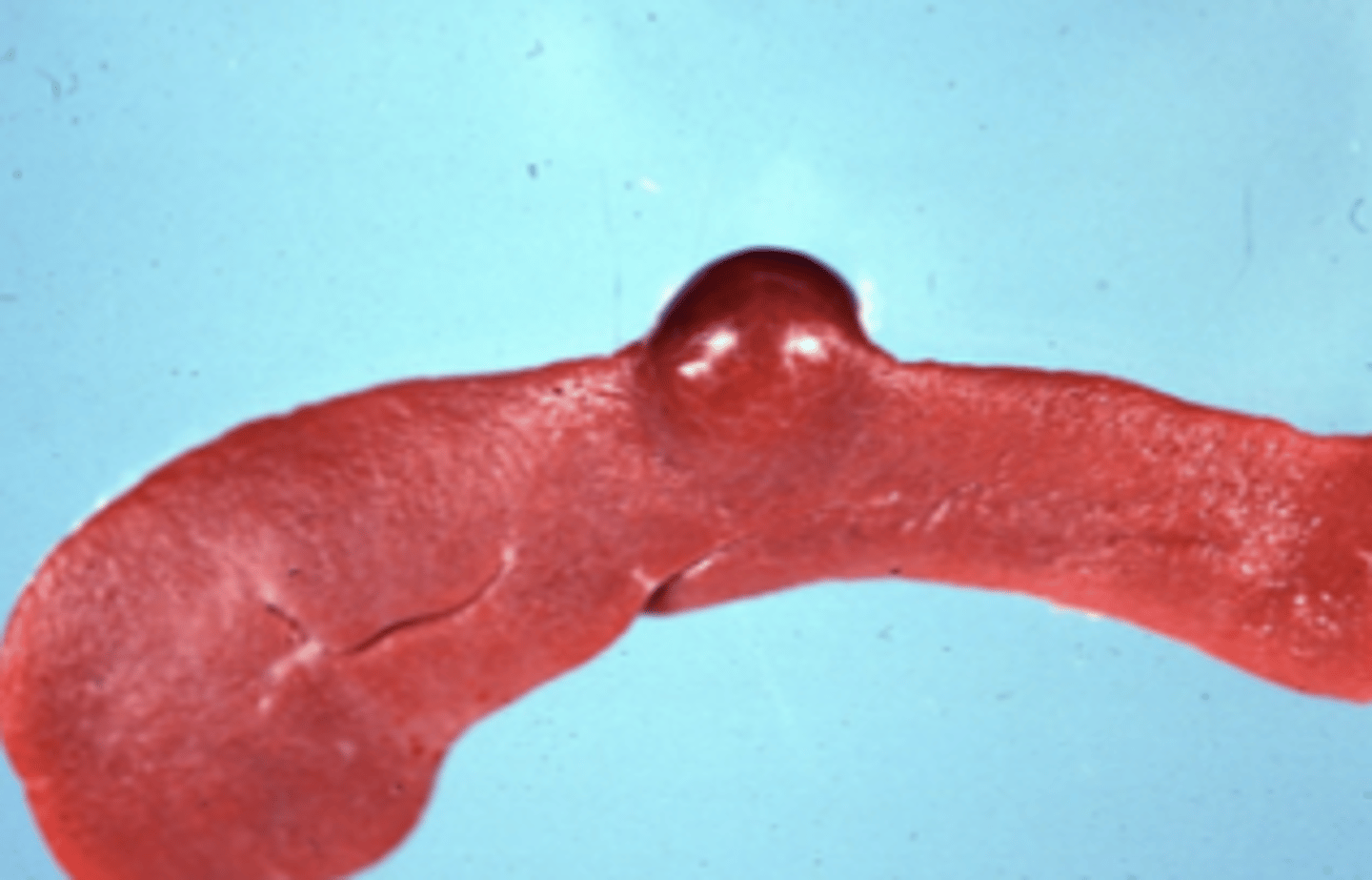
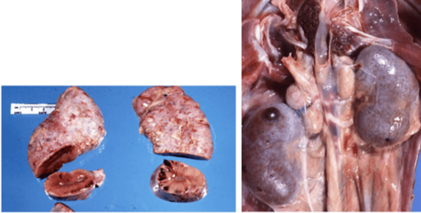
Atrophy
Note the small size (under the liver) but it is the normal color
What type of cellular adaptation has the liver undergone in this photo and how do you know?
Segmental
What is the best term for the distribution of this lesion?
A. Focal
B. Segmental
C. Multifocal to coalescing
D. Disseminated
E. Diffuse
E. infectious
Lymph obstruction
How would you classify the eitology of this lesion?
A. toxic
B. iatrogenic
C. neoplastic
D. degenerative
E. infectious
A. acute
A 3 day old lesion would be best described as
A. acute
B. chronic
C. peracute
D. subacute
D. infarction
Which term refers to death of a tissue caused by interruption of its blood supply?
A. anoxia
B. hypoxia
C. ischemia
D. infarction
E. ulceration
D. Diabetes mellitus
Which of these can cause both hepatic lipidosis and glycogen hepatopathy?
A. ketosis
B. dietary excess
C. poxviral infection
D. Diabetes mellitus
E. hyperadrenocorticism
hypoxia
Coagulative necrosis is associated with which of the following?
A. hypoxia
B. ulceration
C. bacterial infection
D. vit E deficiency
E. granulomatous inflammation
D. pseudomelanosis
Which of these color changes is caused by the production of hydrogen sulfide by bacteria?
A. bloat line
B. livor mortis
C. bile imbibtion
D. pseudomelanosis
E. hemoglobin imbibition
autoimmune disease
Which is one possible consequence of defective apoptosis?
A. lipidosis
B. dry gangrene
C. inflammation
D. pathological atrophy
E. autoimmune disease
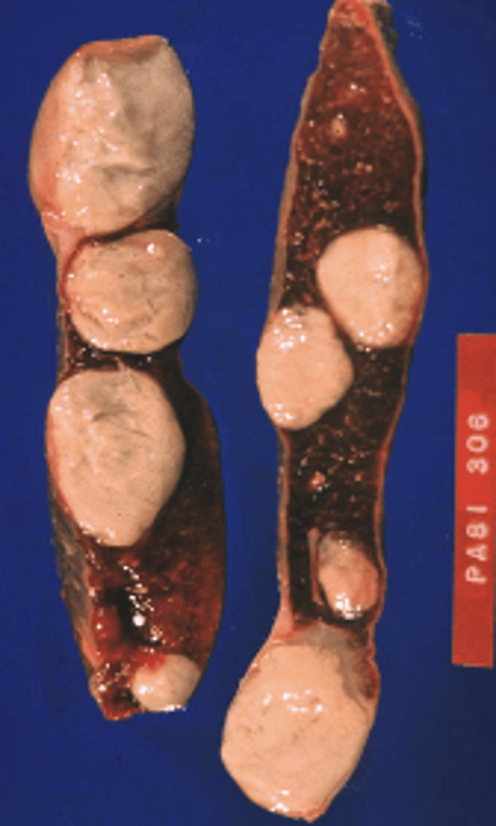
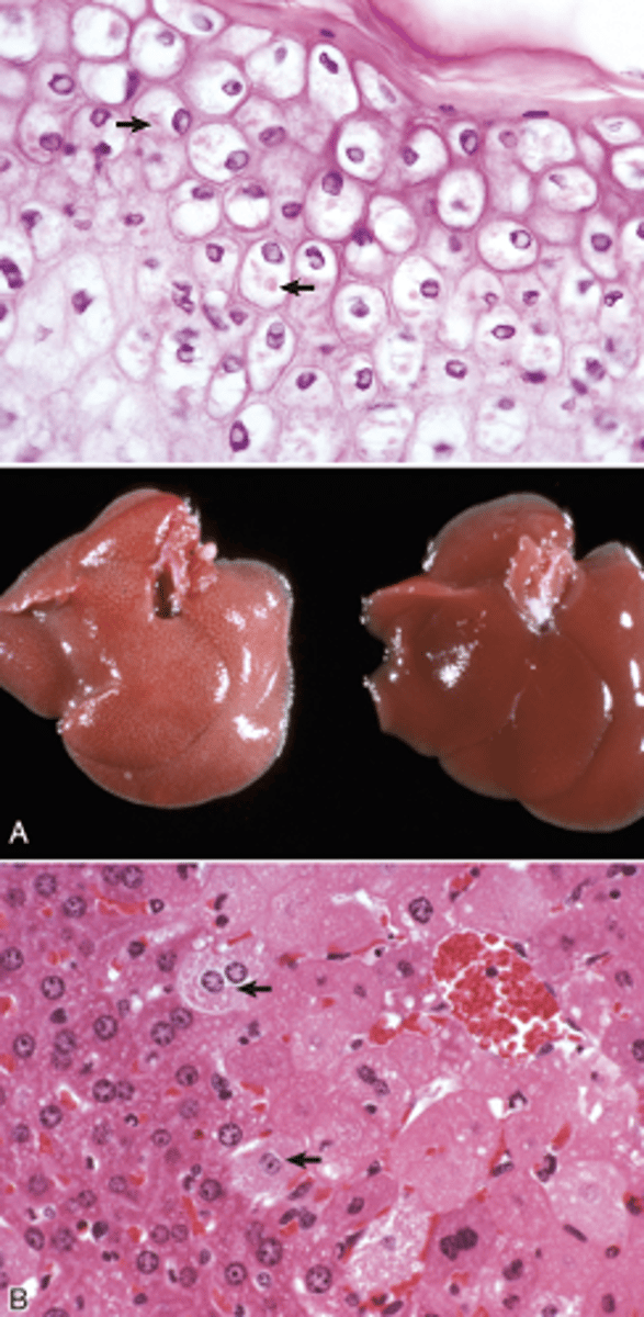
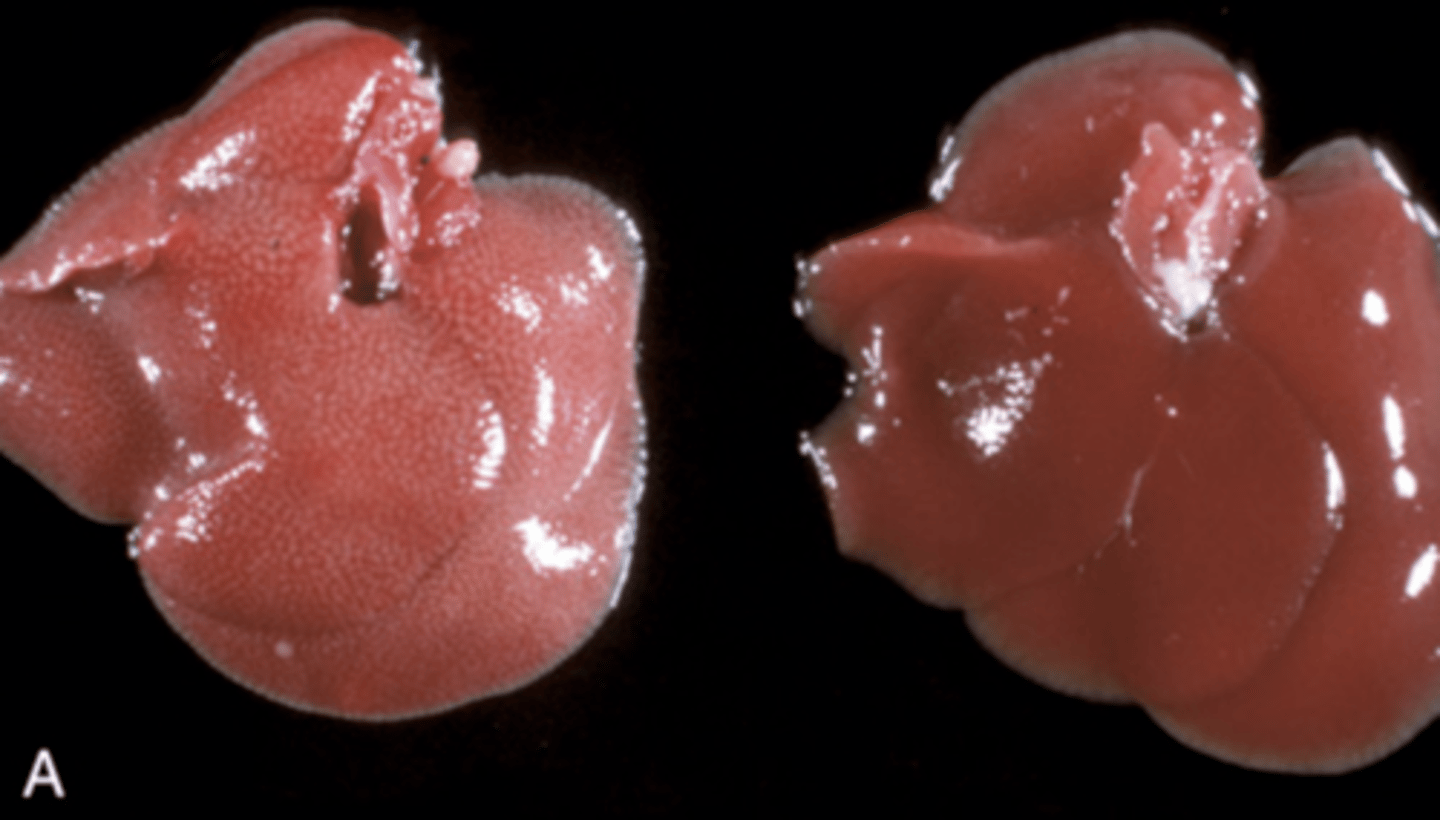
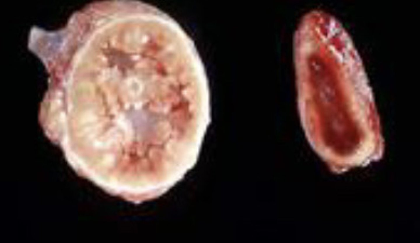
C. left is hyperplastic, right is normal
These two adrenal glands are from the same dog? Which is abnormal?
A. Right is atrophic, left is normal
B. left is dysplastic, right is normal
C. left is hyperplastic, right is normal
D. left is hypertrophic, right is normal
Repetitive trauma
Which of these results in dystrophic calcification?
A. Vitamin D toxicity
B. Repetitive trauma
C. Hyperparathyroidism
D. Chronic inflammation
E. Elevated Ca-P product
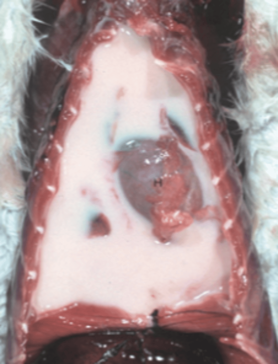
d. pulmonary edema
You have a canine patient in left sided heart failure. Which of the following do you expect to see at necropsy?
A. ascites
B. hemothorax
C. pleural effusion
d. pulmonary edema
E. subcutaneous edema
pleural effusion
You have a feline patient in left sided heart failure. What additional lesion might you see that would not be seen in a dog?
A. ascites
B. hemothorax
C. pleural effusion
d. pulmonary edema
E. subcutaneous edema
C. inflammation
Which of the following is a possible cause of hyperemia?
A. GI torsion
B. Livor mortis
C. inflammation
D. hemoglobin imbibition
E. right sided heart failure