Spinal Cord
1/12
There's no tags or description
Looks like no tags are added yet.
Name | Mastery | Learn | Test | Matching | Spaced | Call with Kai |
|---|
No analytics yet
Send a link to your students to track their progress
13 Terms
Spinal Chord
neural tissue originating from medulla oblongata
protected by meninges
33 vertebrae forming the vertebral column
functions of the spinal chord
relays information between body and brain
communicates with rest of body through spinal nerves
bypass brain → spinal reflex actions
structure of spinal chord (What does it contain)
surrounded by white matter - myelinated, high lipid content
H shaped inside part - grey matter, high concentration of cell bodies and dendrites
dorsal root (one at the top),
ventral root (one at the bottom)
dorsal root ganglion → Swelling in dorsal root due to aggregation of cell bodies
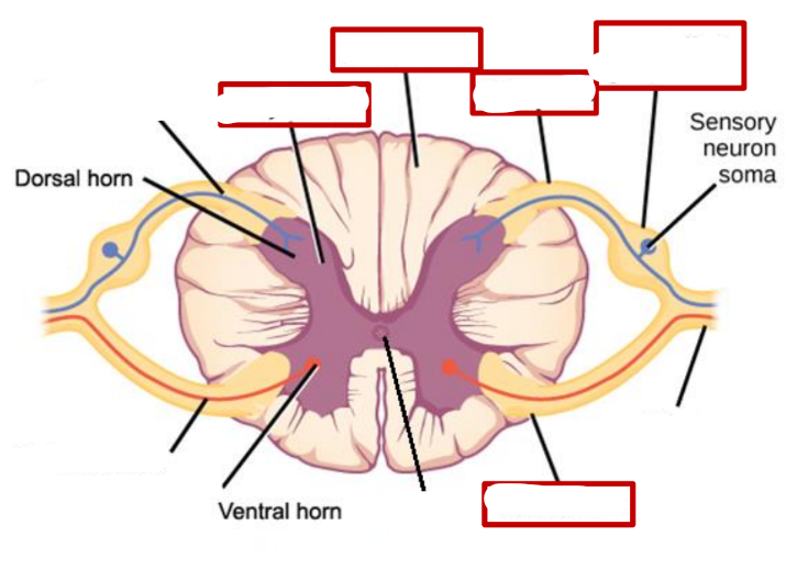
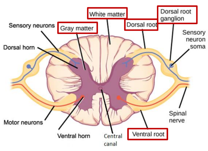
Passage of Neurons through the Spinal Cord
Sensory neurons enter through the dorsal root and have their cell bodies in the dorsal root ganglion.
enters the dorsal horn in the grey matter where it synapses with interneurons which are unmyelinated allowing a decision to be taken
Motor neurons leave the cord through the ventral root, and are unmyelinated
reflex action
rapid automatic response to a stimulus which is not under voluntary control of the brain
these are not learnt → function is protection
same stimulus same response
Cranial Reflexes
sneezing, coughing, pupil dilation and blinking
Spinal Nerve Reflexes
vasodilation
vasoconstriction
peristalsis of the gut
reflex arc
the pathway taken by a nerve impulses during a reflex action
→ monosynaptic
→ polysnaptic
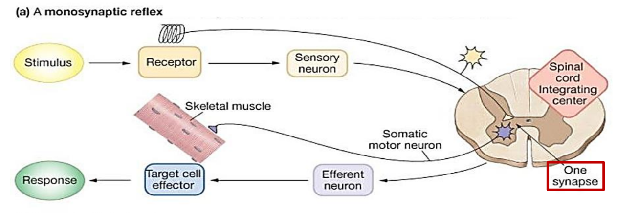
monosynaptic
→ only one synapse
→ knee-jerk reflex - needed to walk
→ sensory neuron synapses directly to motor (quicker)
diagram of monosynaptic reflex
polysynaptic
→ more than 1 synapse
→ removing hand from hot object or pin
→ relay neurons are required between sensory and motor
ascending vs descending nerves
ascending - carry sensory information to the brain
descending - carry motor information away from the brain and to the spinal cord.