Amide proton exchange and in vivo folding
1/6
There's no tags or description
Looks like no tags are added yet.
Name | Mastery | Learn | Test | Matching | Spaced | Call with Kai |
|---|
No analytics yet
Send a link to your students to track their progress
7 Terms
What is the amide proton exhcnage?
NH (protein) + D2O ⇌ ND (protein) + HDO
Proteins are not static as they change conformations. We can measure these structural fluctuations by observing the exchange of hydrogen atoms on the peptide backbone with deuterium (D2O) from the solvent
What are monitoring methods of amide protin exchnage?
NMR (detecting the disappearance of 1H signals)
Mass Spec (detecting the increase in mass as H is replaced by D)
Experimental setup for amide proton exchange
Isotopic labelling: bacteria (E. coli) are grown on media containing 15N ammonium salts to incorporate it into every peptide bond during protein synthesis
The exchange: the labelled protein is extracted and purified, and then dissolved in a buffer made with D2O
Recordings of HSQC (Heteronuclear single quantum correlation) NMR spectra continue over minutes, hours, or days
In HSQC spectrum, 15N-1H pair produces a specific cross-peak
Deuterium is NMR silent, so when an amide H is replaced by D in this method to become 15N-D, the signal for that specific residue disappears
Intensity analysis: the decrease in intensity (volume) of each cross-peak over time is analysed and interpreted:
Fast disappearance: residues are on the surface or in flexible loops, and are not protected by hydrogen bonds
Slow disappearance: residues are buried in the hydrophobic core or locked in an alpha-helix/ beta-sheet, so they only exchange when the protein changes conformation/unfolds locally
Fit the curve to a single exponential decay to calculate kex using 𝑦 = 𝐴𝑒−𝑘ext
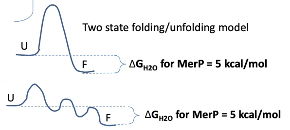
What happens to the structure of the protein during the 3 main ways for the amide proton to be exposed?
Global unfolding (top pic): entire protein cooperative tertiary network completely collapses, opening up the deeply buried core hydrophobic regions to the solvent, exposing almost all backbone amide positions simultaneously.
Partial unfolding: specific domains or standalone sub-structures fold/unfold independently while the remaining body of the protein stays intact
Local fluctuation (bottom pic): protein maintains its overall native fold, but experiences rapid, low-amplitude thermal vibrations/breathing actions. This breaks hydrogen bonds locally, giving solvent molecules momentary access to local amides without altering the macroscopic structure
What are chaperones? Give examples
Specialised proteins that prevent aggregation and assist in the folding process of other proteins without being part of the final structure.
HSP70 system
Chaperoning (e.g., GroEL/GroES)
HSP70 system
Binds to exposed hydrophobic segments of unfolded polypeptides
Prevents aggregation
Uses ATP hydrolysis cycle to release substrate and allow folding attempts
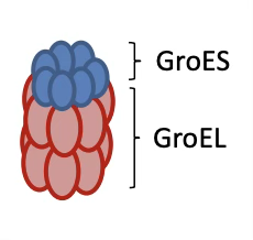
Chaperoning (e.g., GroEL/GroES)
Large barrel-shaped complex
GroEL: a double-ring structure that captures the unfolded protein
GroES: the lid that closes the cage
Encapsulates unfolded protein in a protected chamber, allowing it to fold in isolation, this process is ATP-dependent
Especially important for β-sheet proteins