Head, Skull, Brain, and Arterial Supply
1/74
There's no tags or description
Looks like no tags are added yet.
Name | Mastery | Learn | Test | Matching | Spaced | Call with Kai |
|---|
No analytics yet
Send a link to your students to track their progress
75 Terms
How many bones are in the skull?
8 Cranial Bones, 14 facial bones (Total 22 bones!)
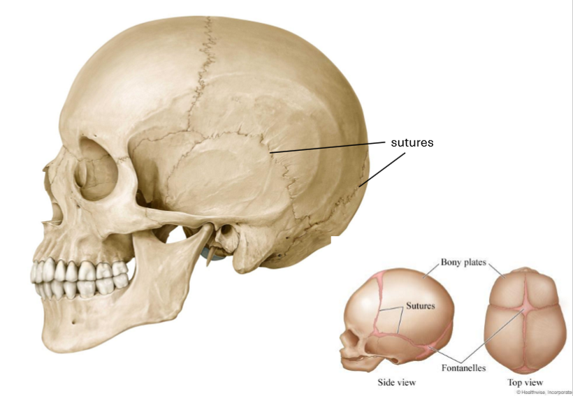
What are sutures?
Joints between the bones of the skull
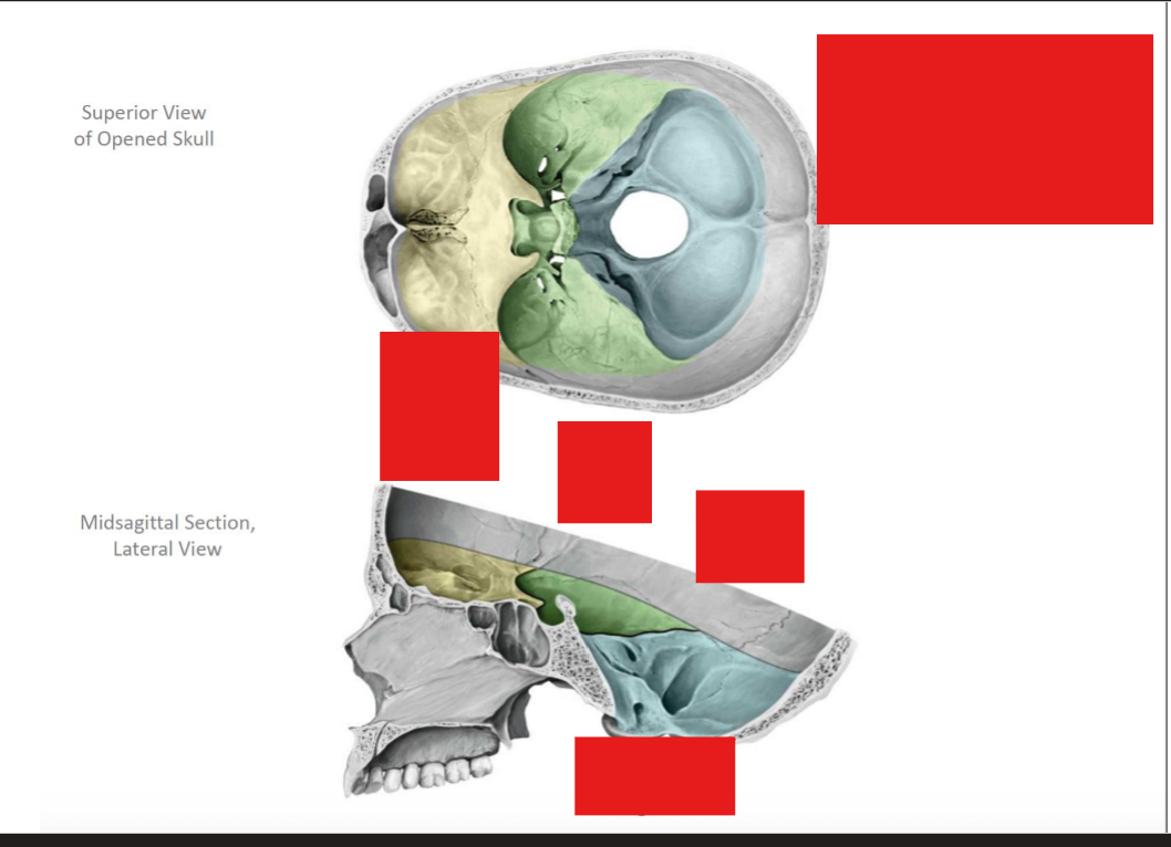
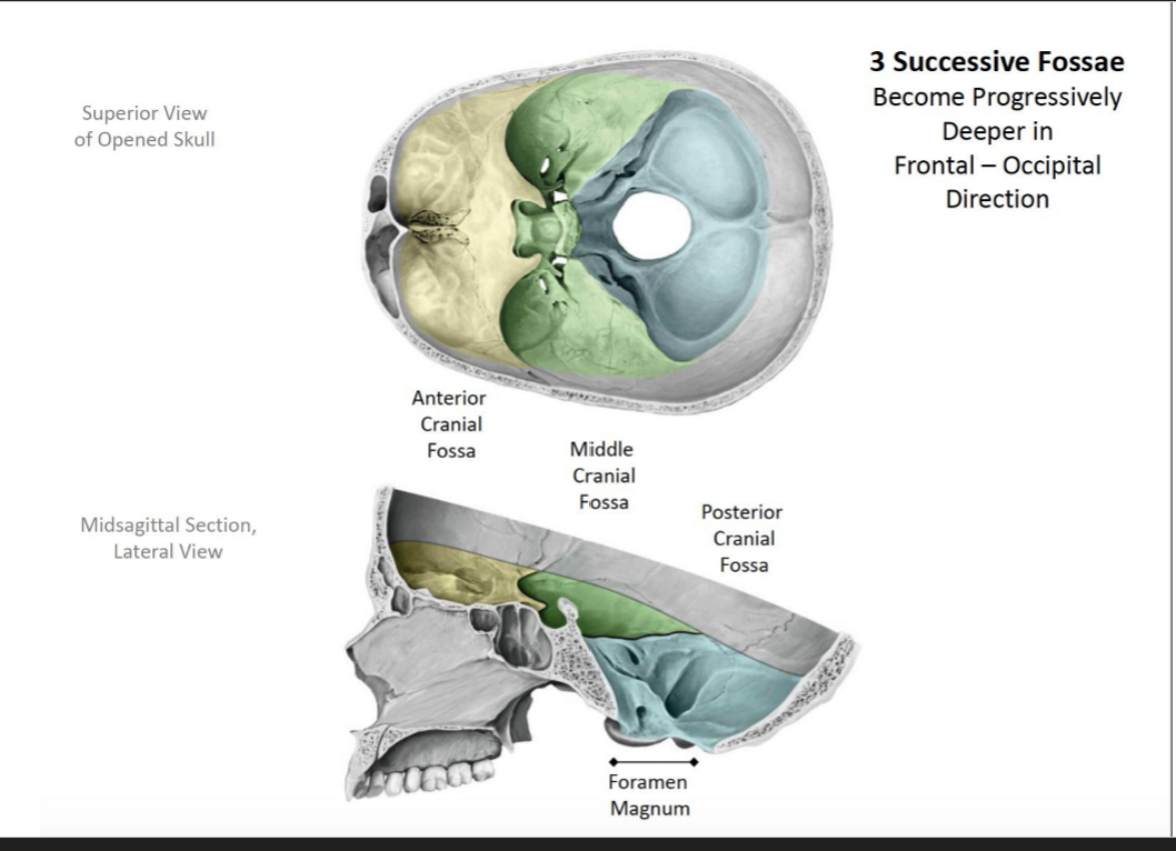
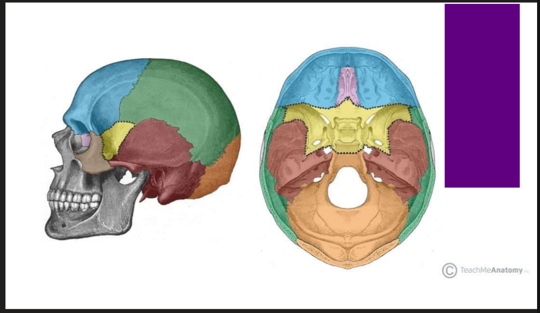
How many fossae are in the skull
Three! Each become progressively deeper in the frontal- > occipital direction
Anterior Cranial Fossa → Middle Cranial Fossa → Posterior Cranial Fossa
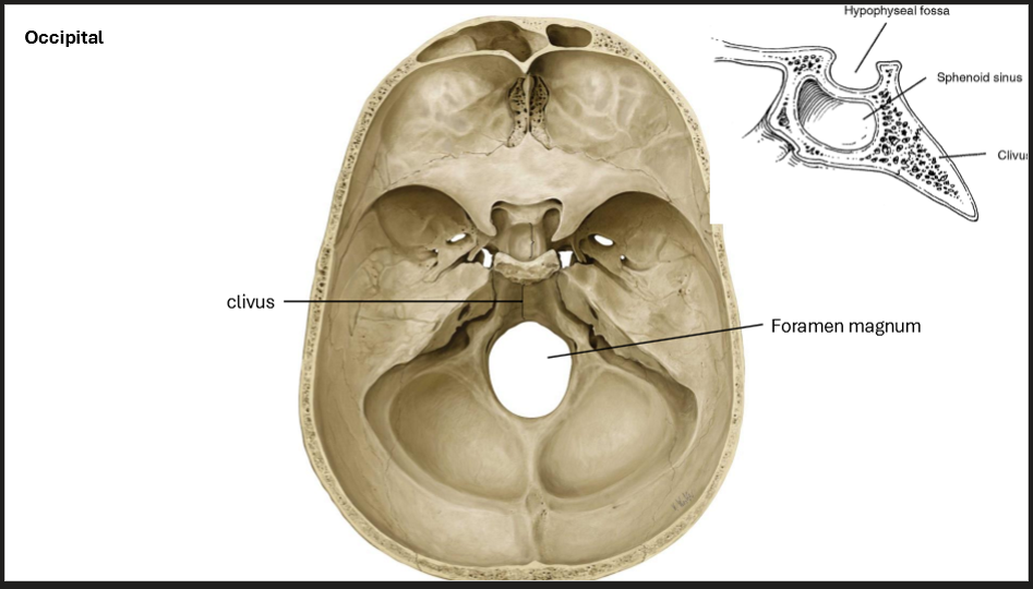
*In the posterior cranial fossa, you’ll find the foramen magnum
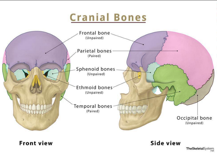
What are the singular bones centered in the midline of the cranial bone
Singular Bones!
Frontal
Ethmoidal
Sphenoidal
Occipital
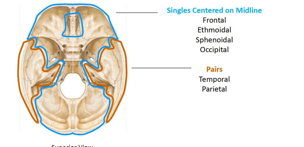
What are the pairs of cranial bones?
The Bilateral Pairs!
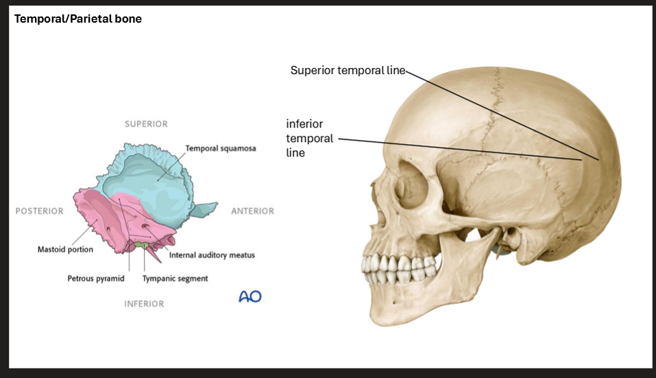
Tempral
Parietal

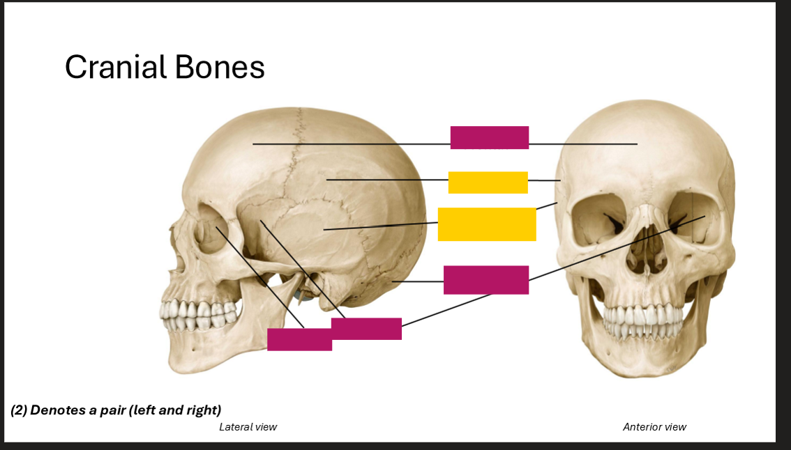
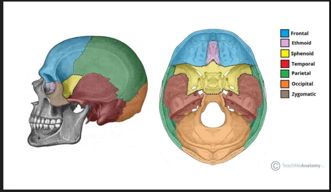
Label the Cranial bones
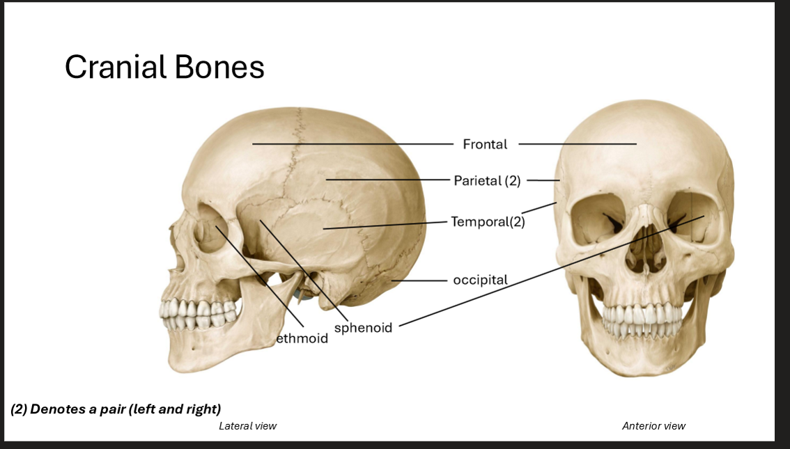
Label the Cranial Bones
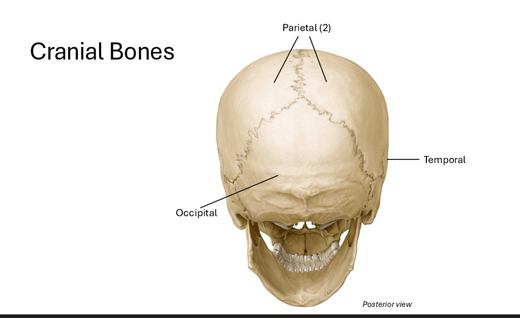
Whats a good mnemonic (from front to back) cranial bones?
Ethan’s Fried Spanish Pasta Tempted Octavia
(Ethmoid, Frontal, Sphenoid, Parietal (2), Temporal (2), Occipital)
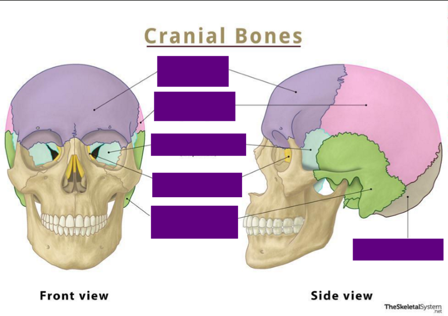
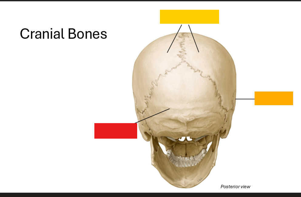
Label the cranial bones
Label the bones!
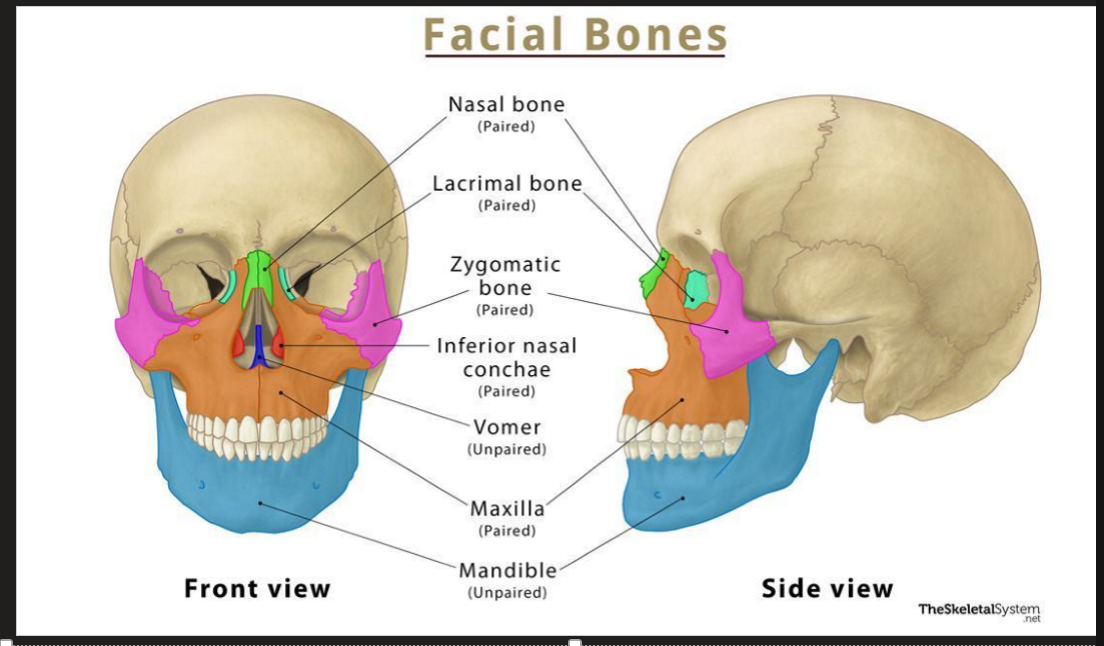
What are the singular facial skeletal bones
Mandible and Vomer
What are the paired facial skeletal bones?
Maxila, Inferior Nasal Concha, Zygomatic, Palatine, Nasal, Lacrimal
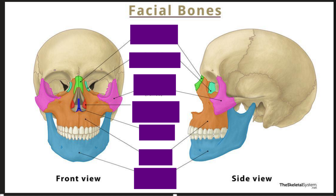
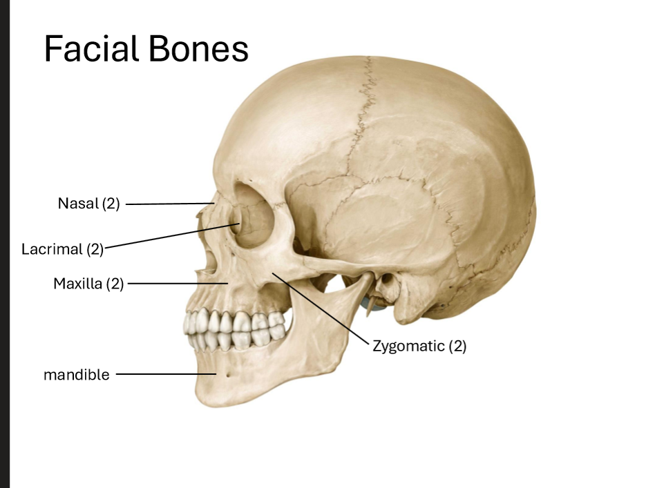
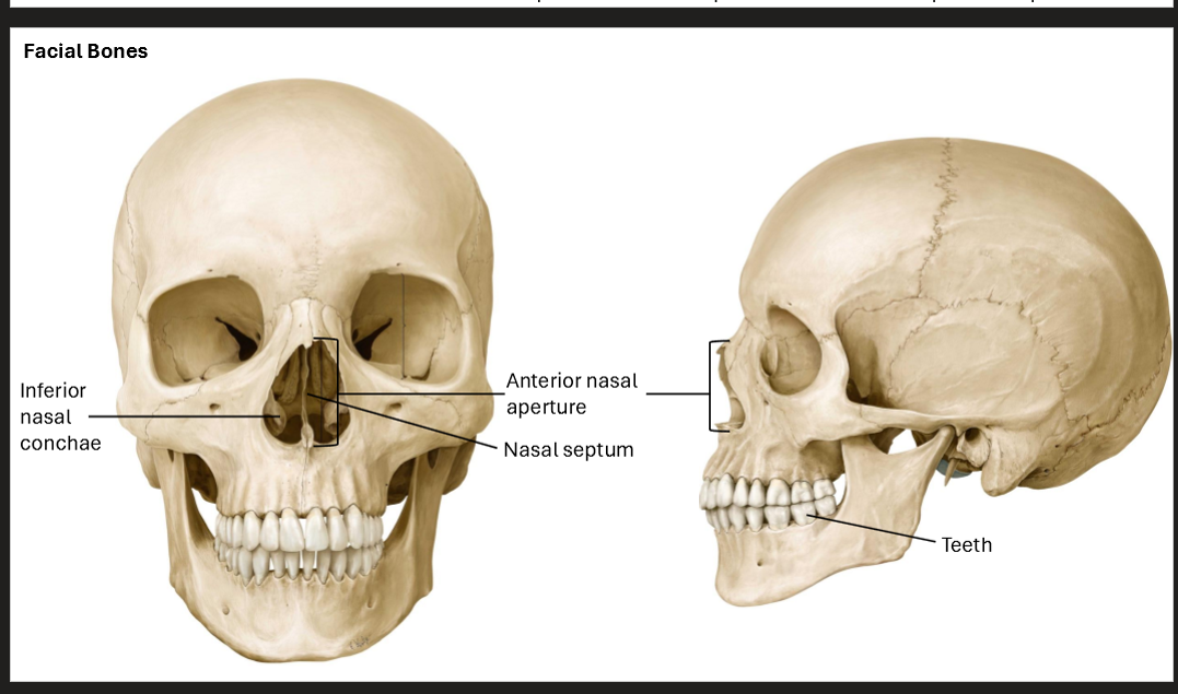
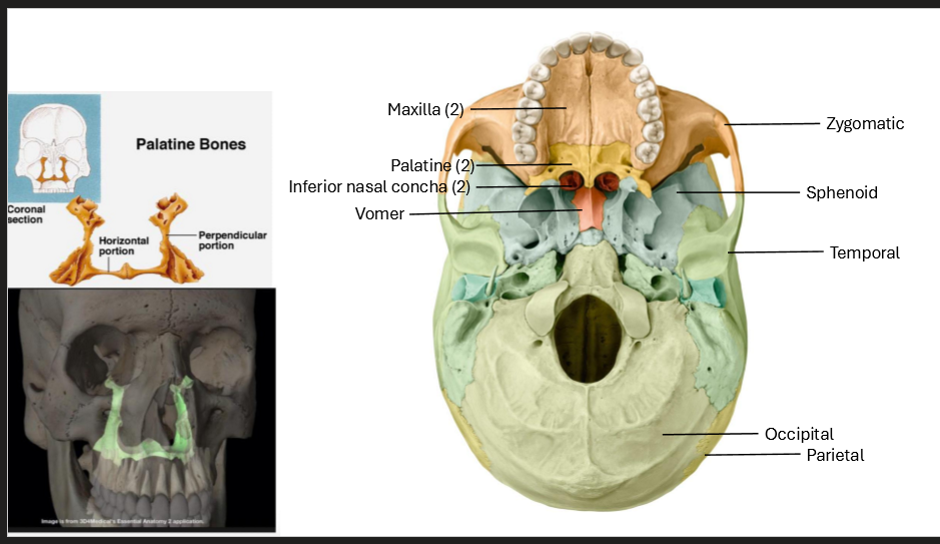
Label the Facial Bones
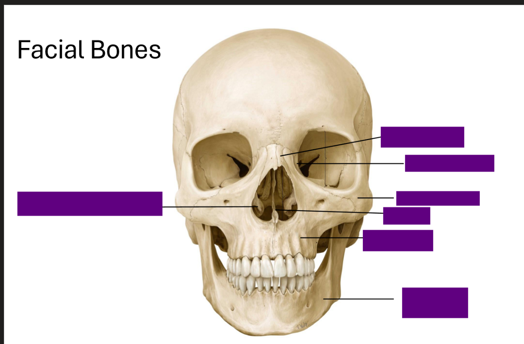
Label the Facial Bones
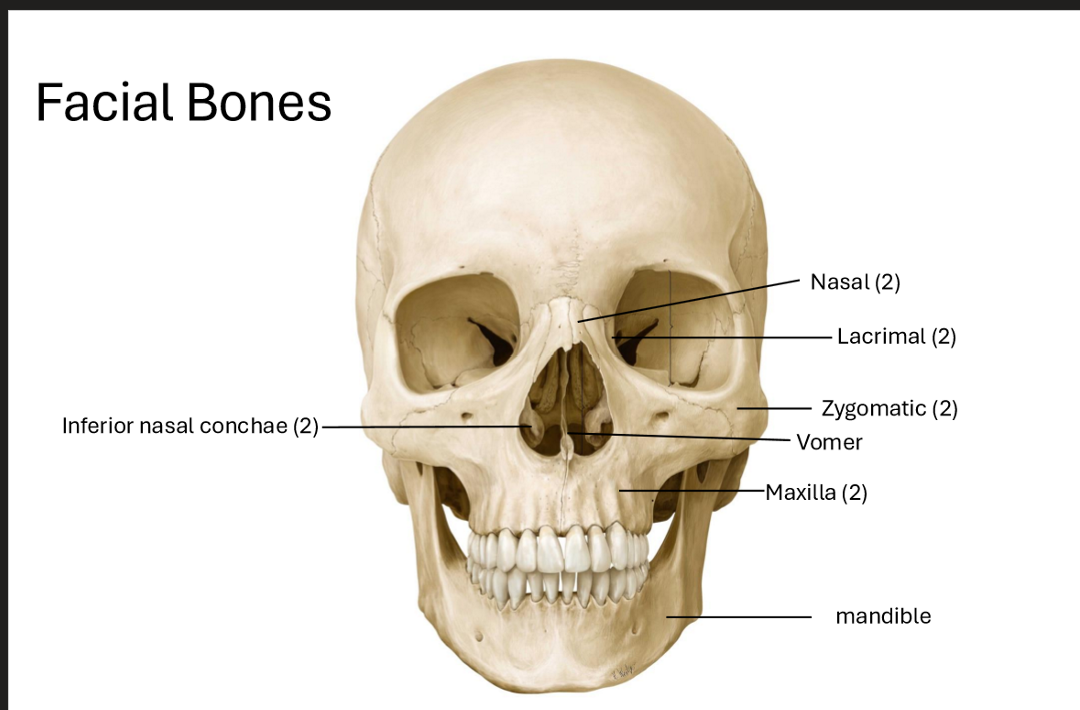
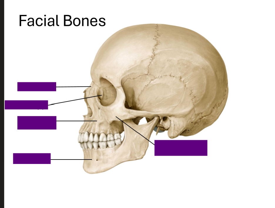
Label the Facial Bones
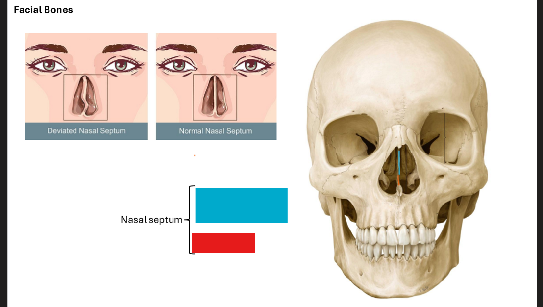
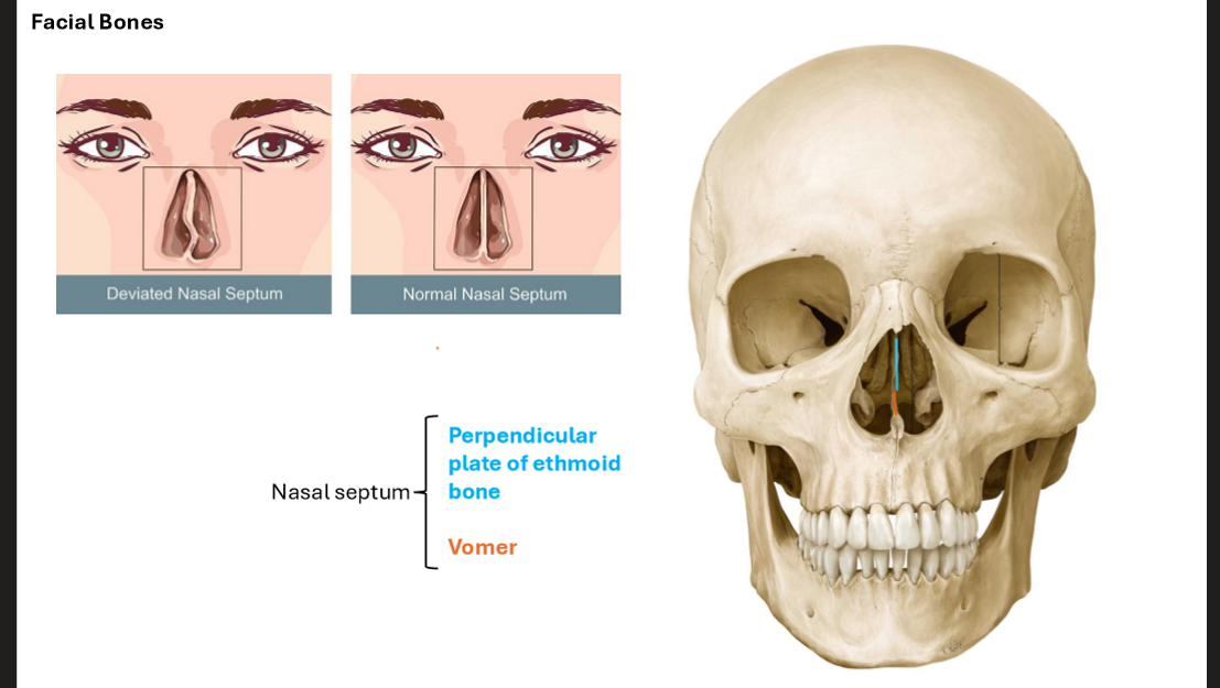
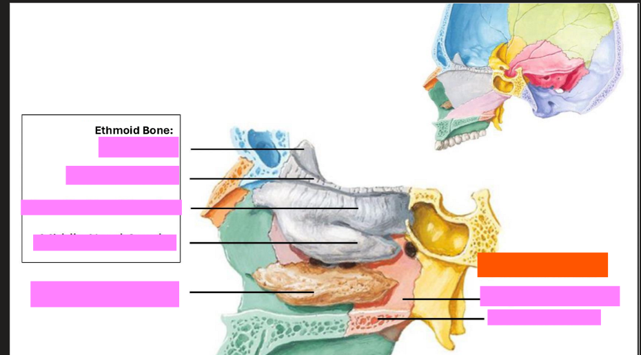
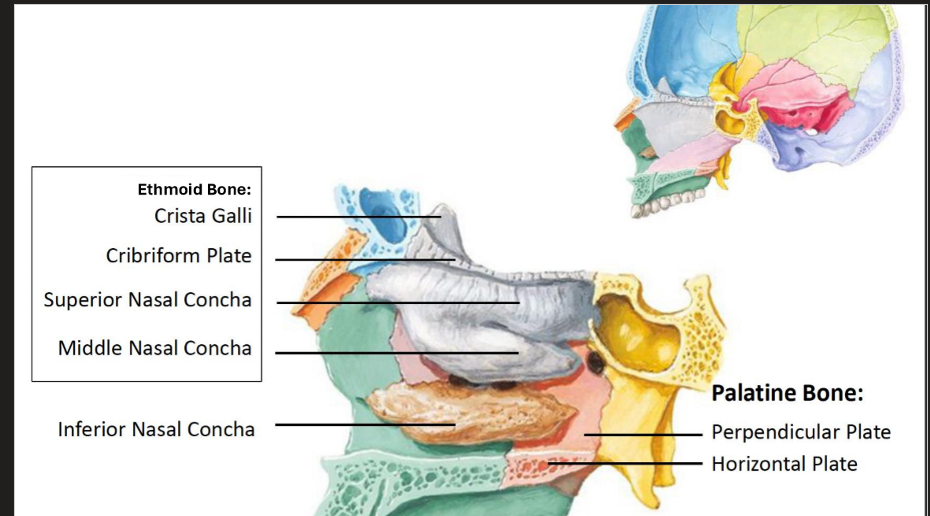
What are the bones of the nasal septum?
Superior → Perpendicular plate of ethmoid bone
Inferior → Vomer
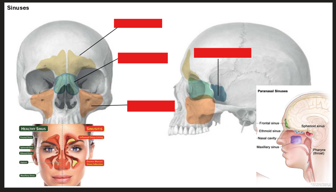
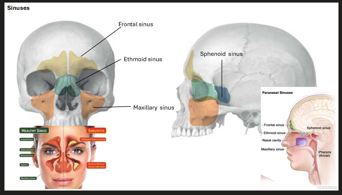
Label the Sinuses
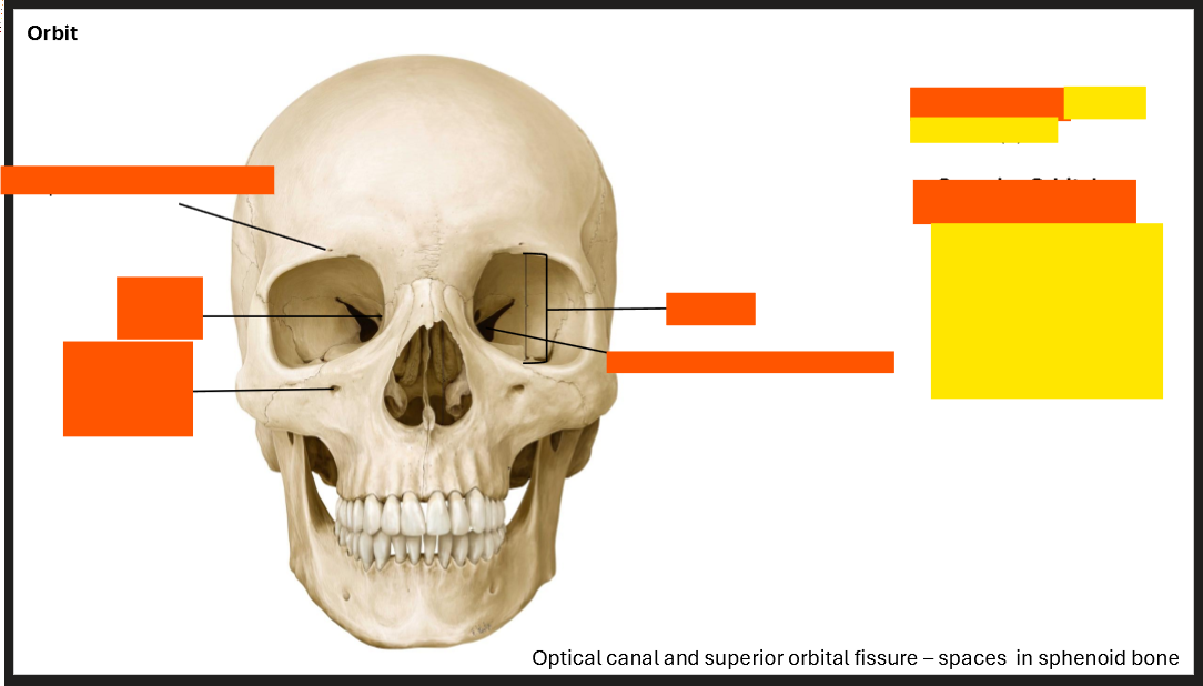
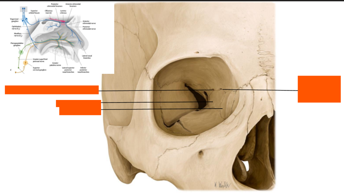
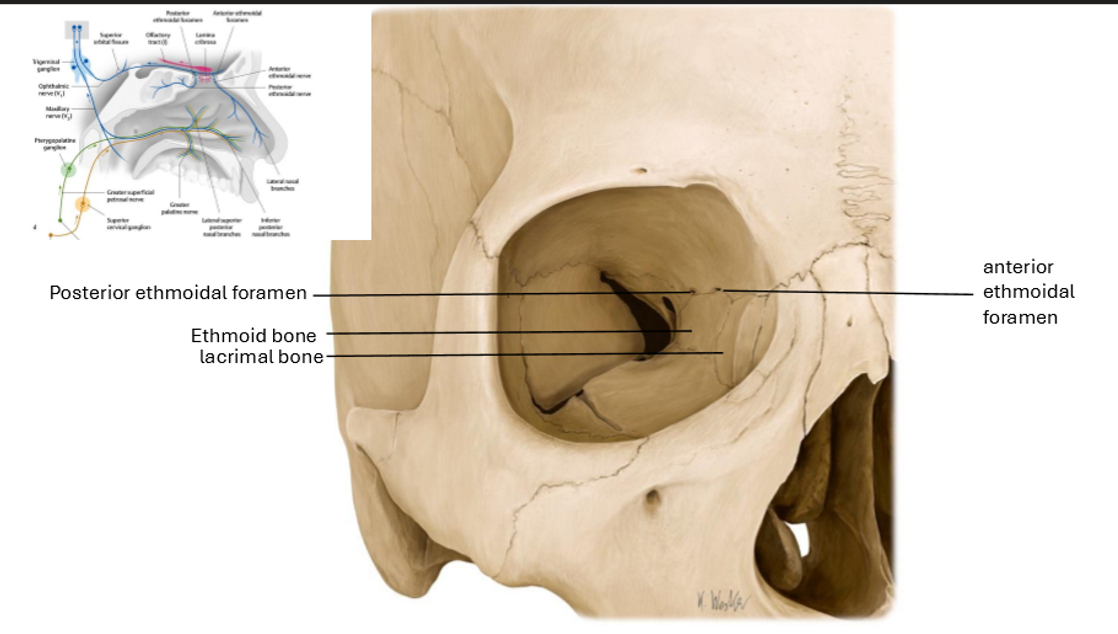
Label the Orbit
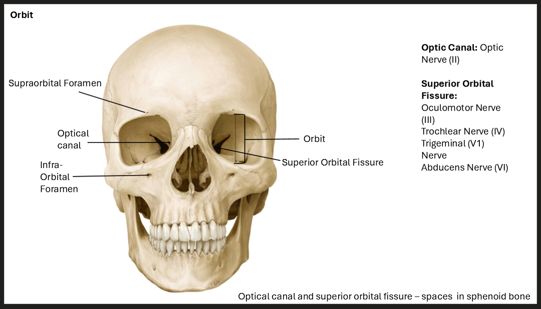
What are the nerve/s that go through the Optic Canal?
The Optic Nerve (II)
What are the nerve/s that go through the Superior Orbital Fissure?
Occulomotor Nerve (III), Trochlear Nerve (IV), Trigeminal Nerve (V1) , Abducens Nerve (VI)
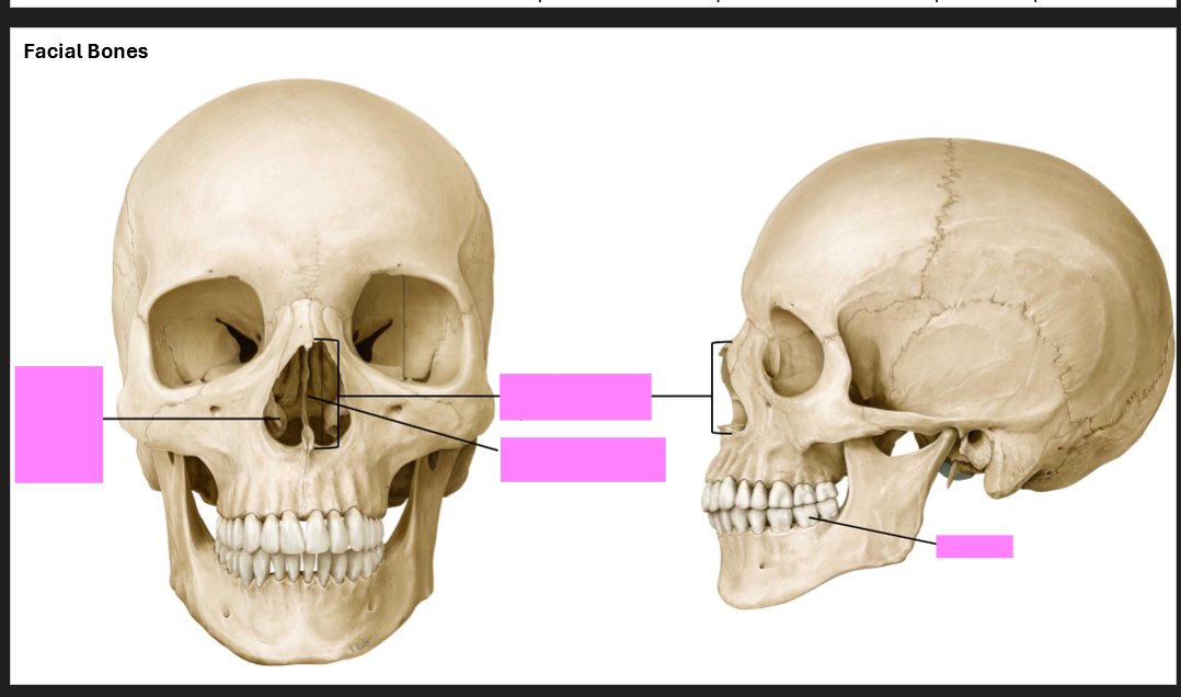
Label the facial bones
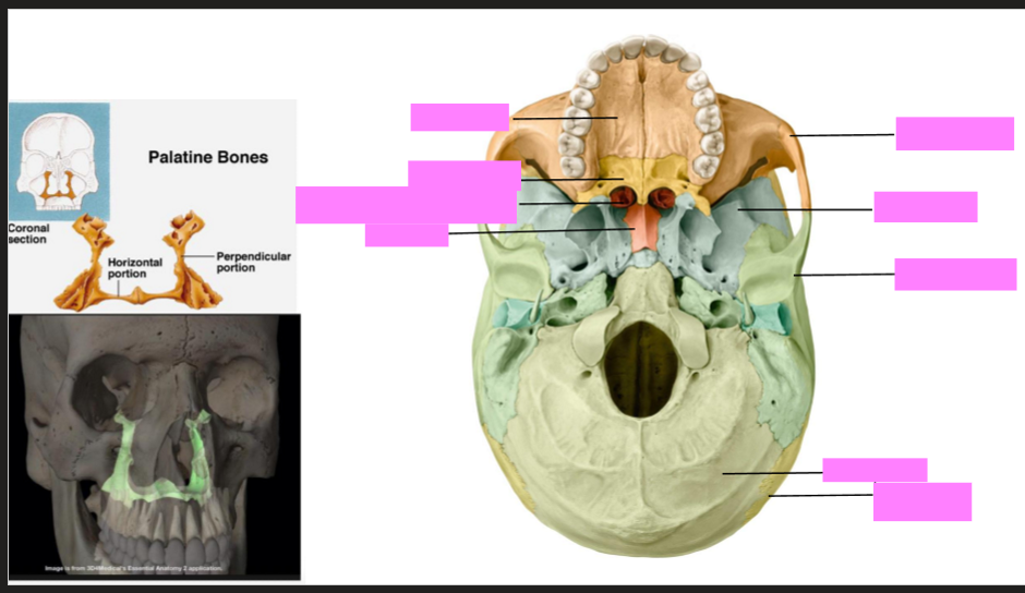
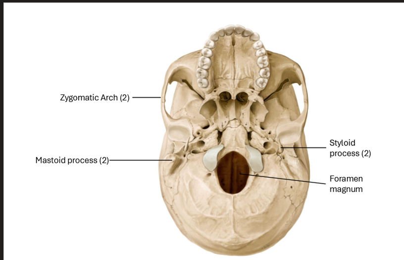
Label the skull
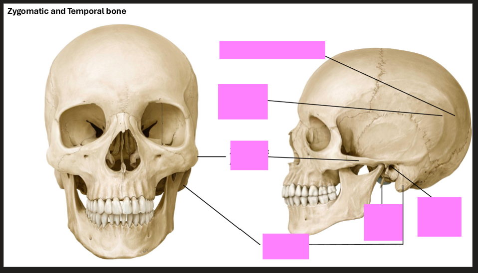
Label the skull
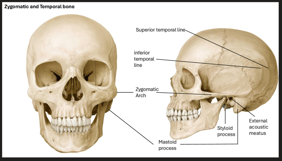
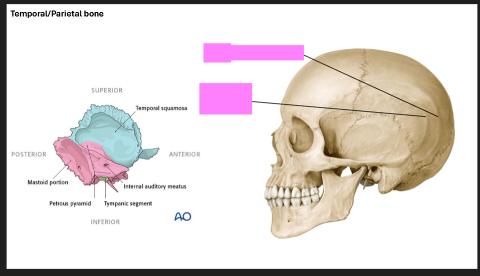
Label the skull
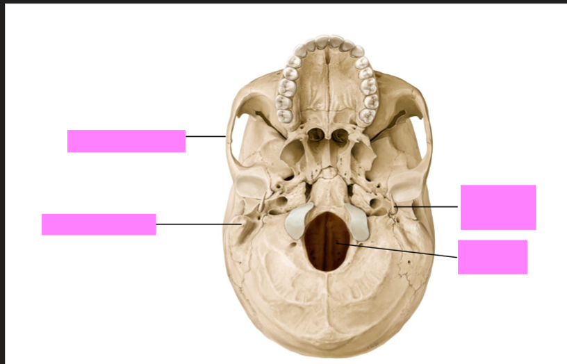
Label the skull
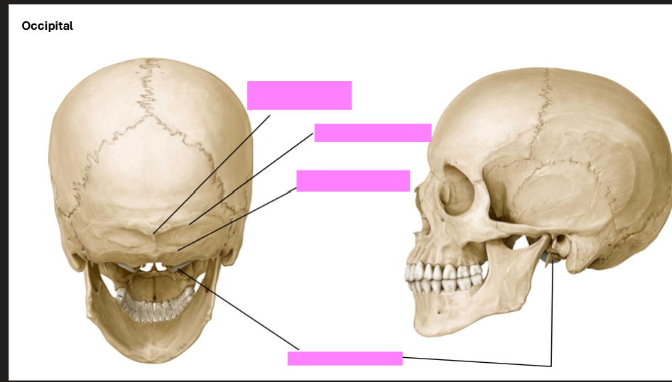
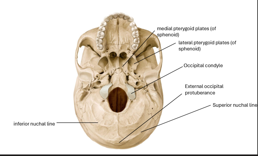
Label the Occipital Bone
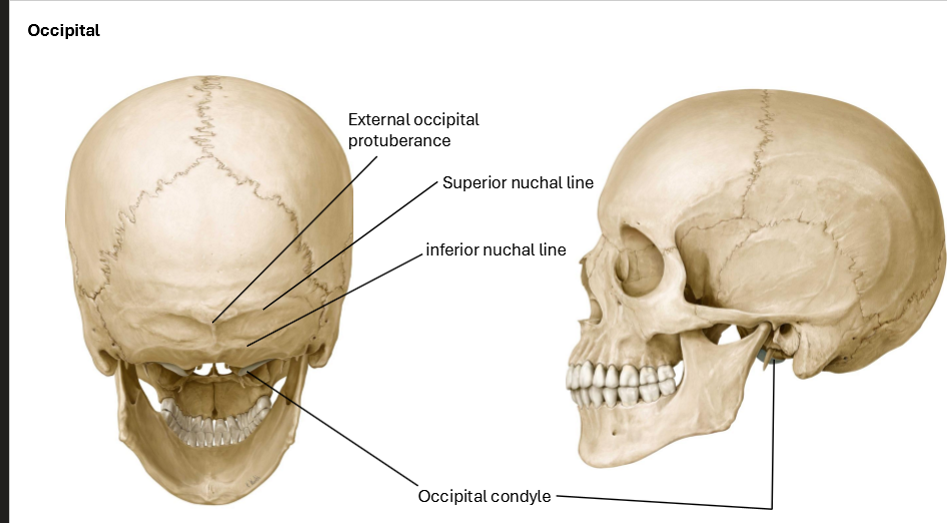
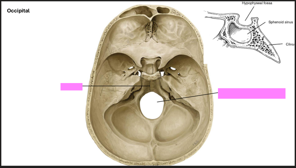
Label the Occipital Bone
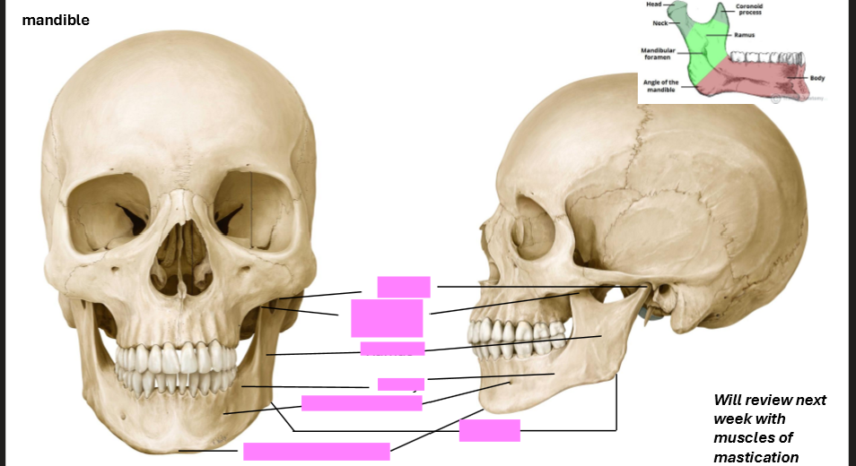
Label the Mandible
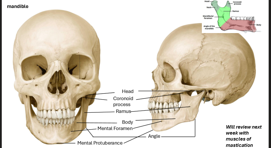

Label the Skull
Label these bones
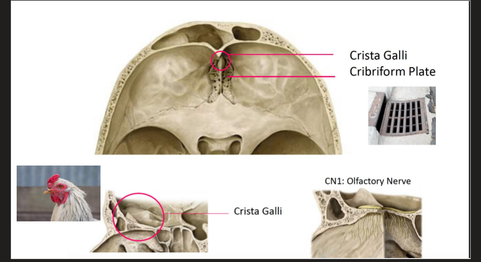
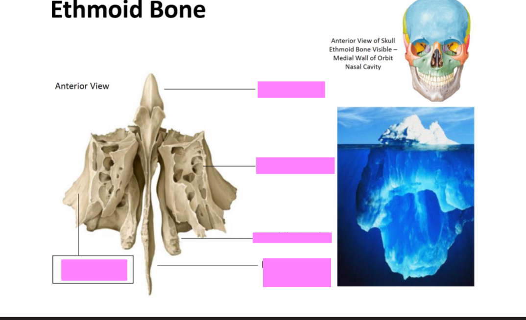
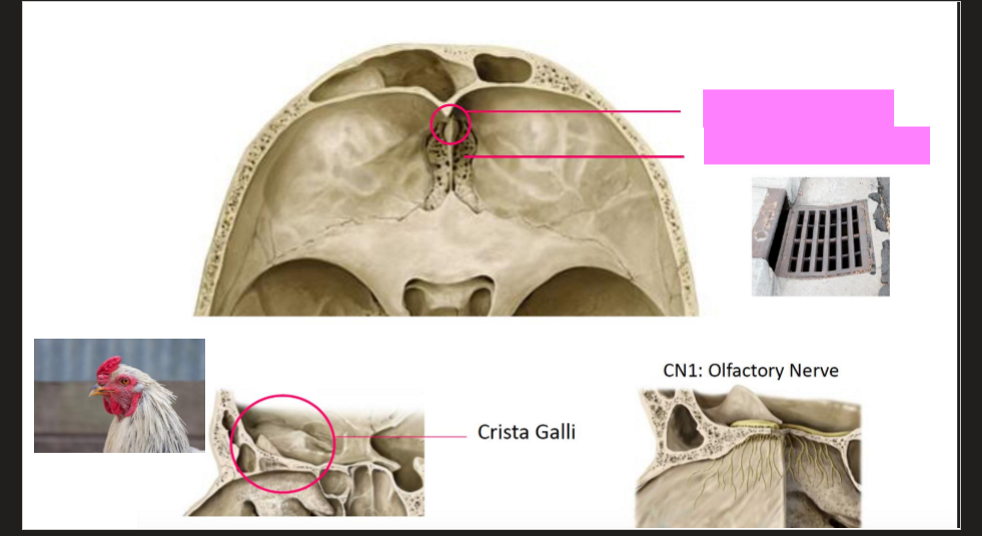
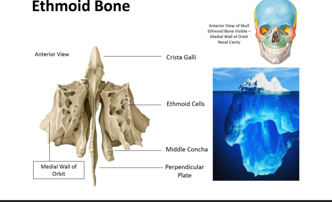
Label the Ethmoid bone
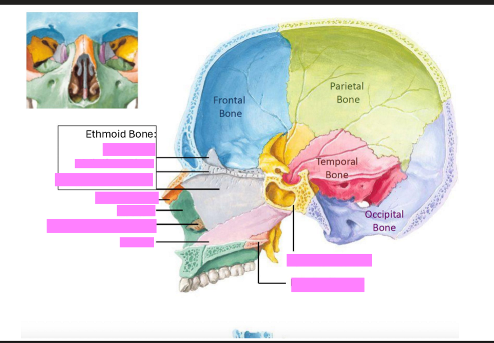
Label this diagram of the skull
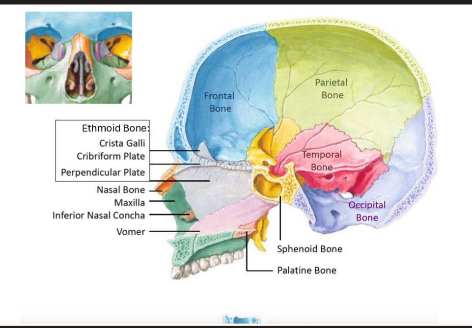
Label this diagram of the skull
Label this diagram of the skull
What does the Sphenoid Bone articulate with?
Occipital, Frontal, Ethmoid, Vomer, Tempral, Parietal, Zygomatic, Palantine
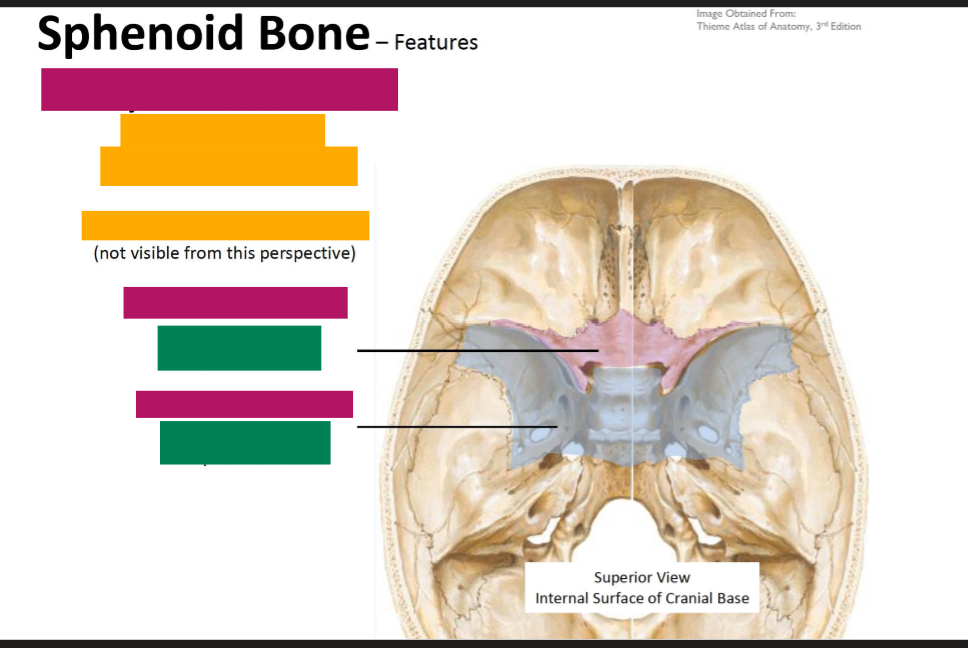
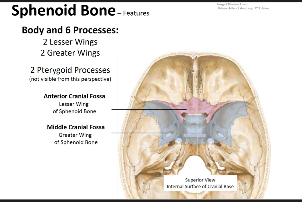
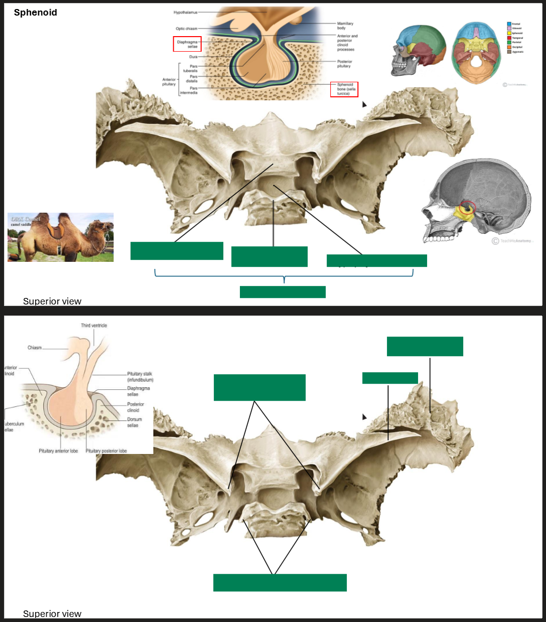
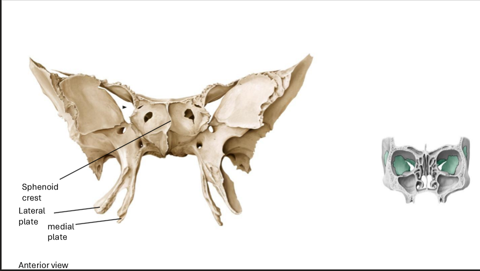
Describe the Sphenoid Bone
Has a body and 6 processes
2 greater and 2 lesser wings
2 Pterygoid Processes
Label the sphenoid Bone
Label the Sphenoid
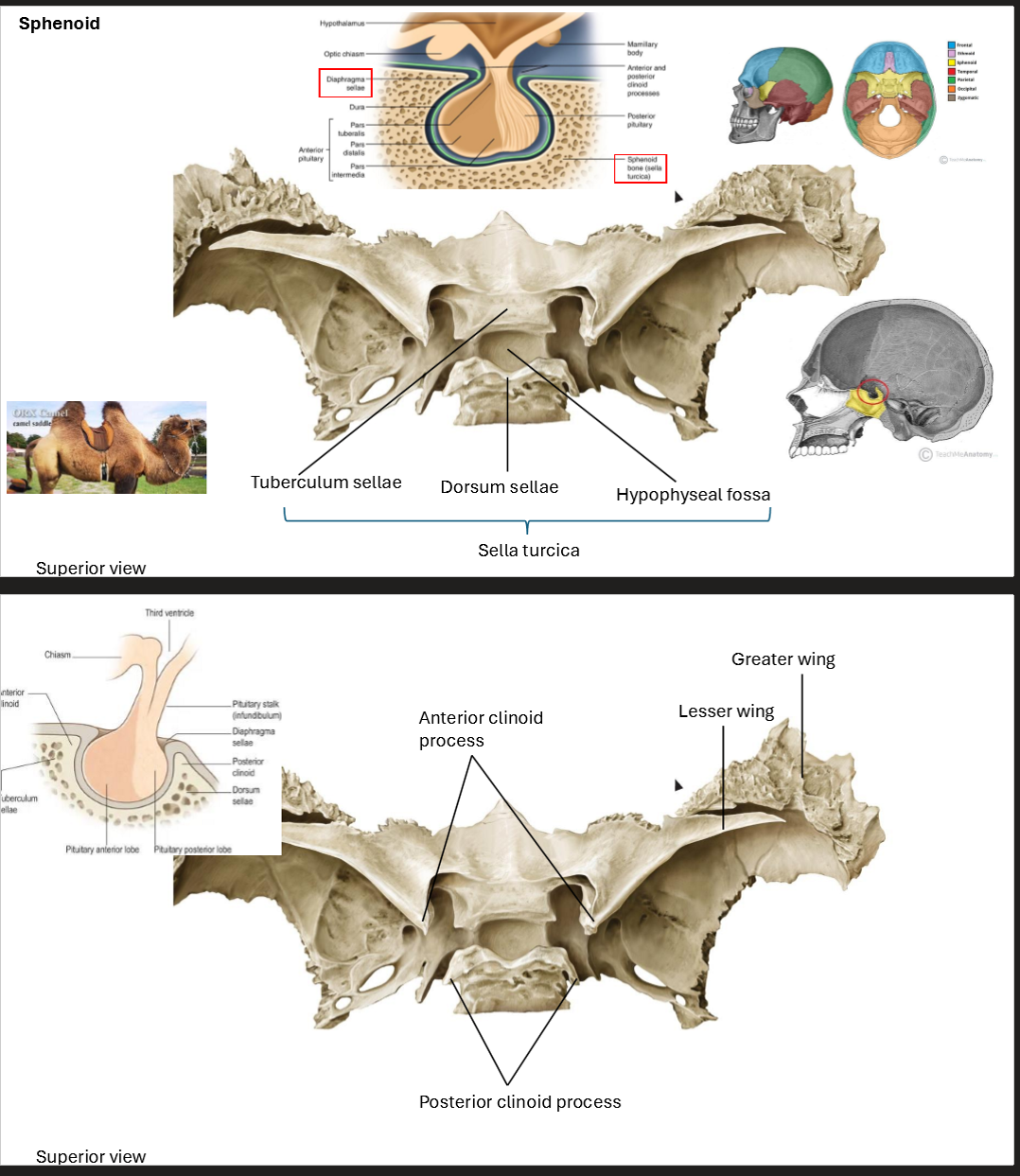
Label the Sphenoid
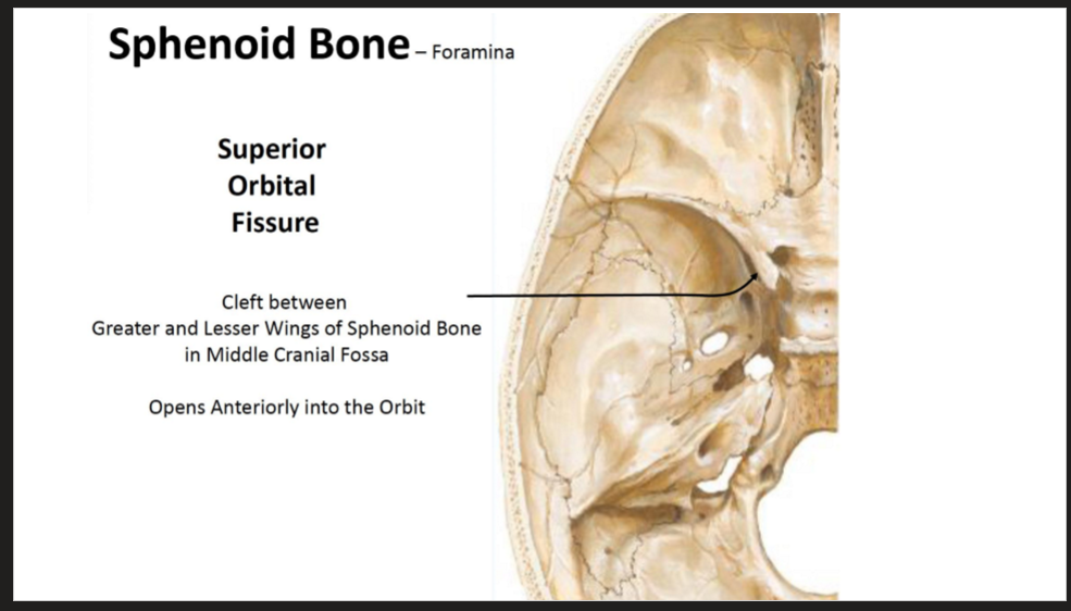
Describe the Superior Orbital Fissure
Cleft between Greater and Lesser Wings of Sphenoid Bone in Middle Cranial Fossa
Opens Anteriorly into the Orbit
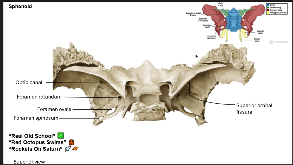
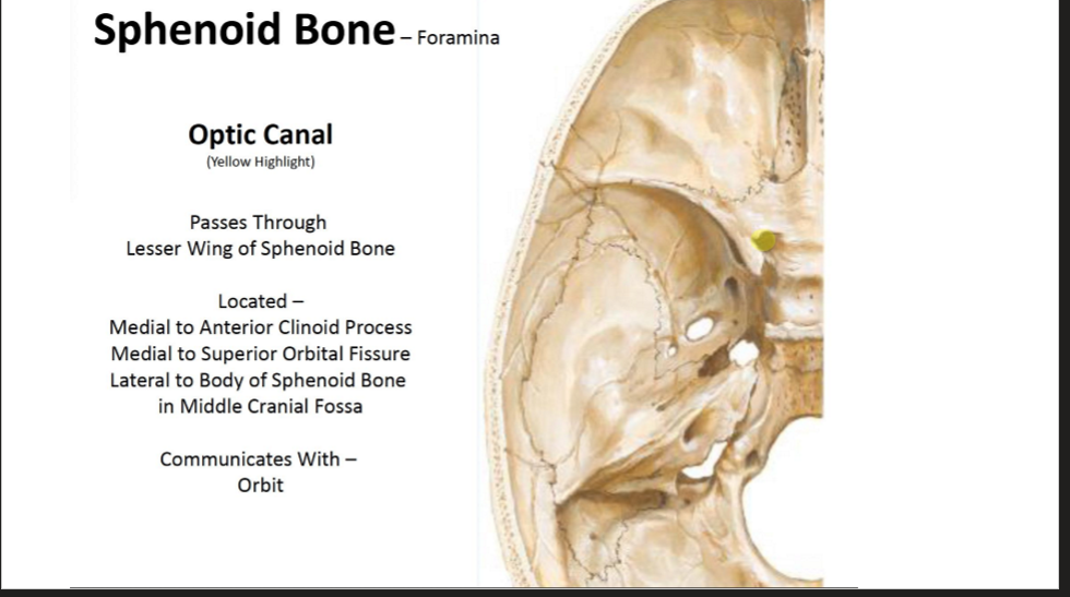
Describe the Optic Canal
Passes through the lesser wing of the Sphenoid Bone
Located -
Medial to Anterior Clinoid Process
Medial to Superior Orbital Fissure
Lateral to body of sphenoid bone in middle cranial fossa
Communicates with Orbit
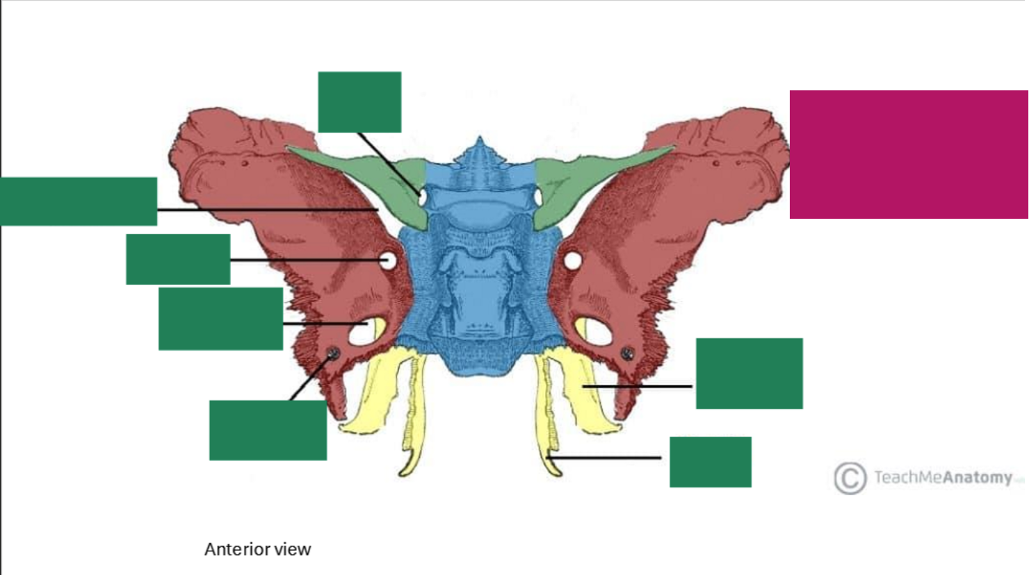
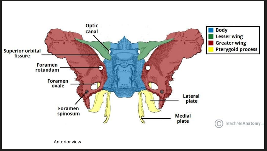
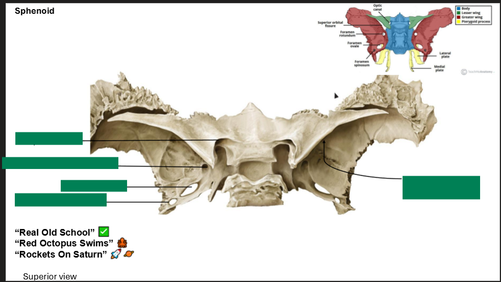
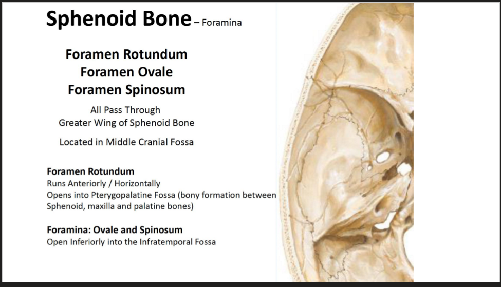
Describe the three foramina in the sphenoid bone (the foramens)
Foramen Rotundum, Oval, and Spinosum
All pass through the Greater wing of the sphenoid bone
Located in the middle Cranial Fossa
Foramen Rotundum
Runs anteriorly/Horizontally, opens into pteryogopalantine fossa (Bony formation between sphenoid, maxilla, and palantine bones)
The foramina Ovale and Spinosum
Opens inferiorly into the infratemporal fossa
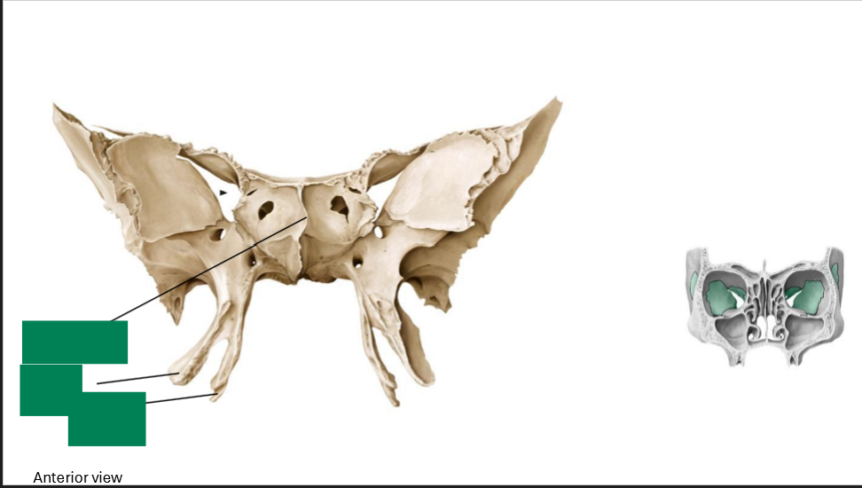
Label this diagram of the sphenoid
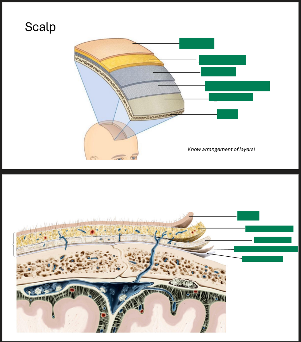
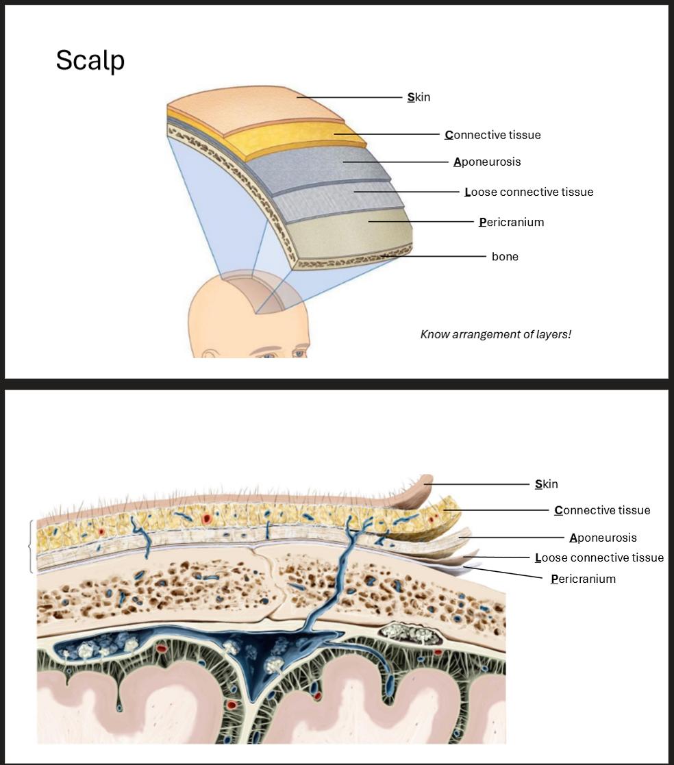
Label the scalp of these two diagrams
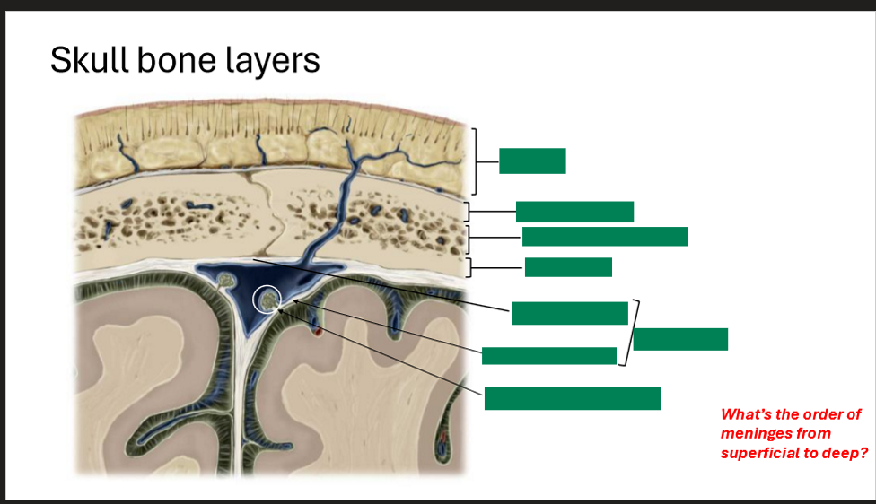
Label the skull bone layers
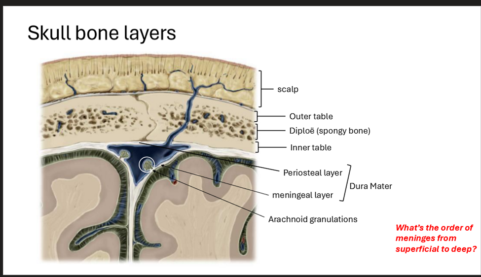
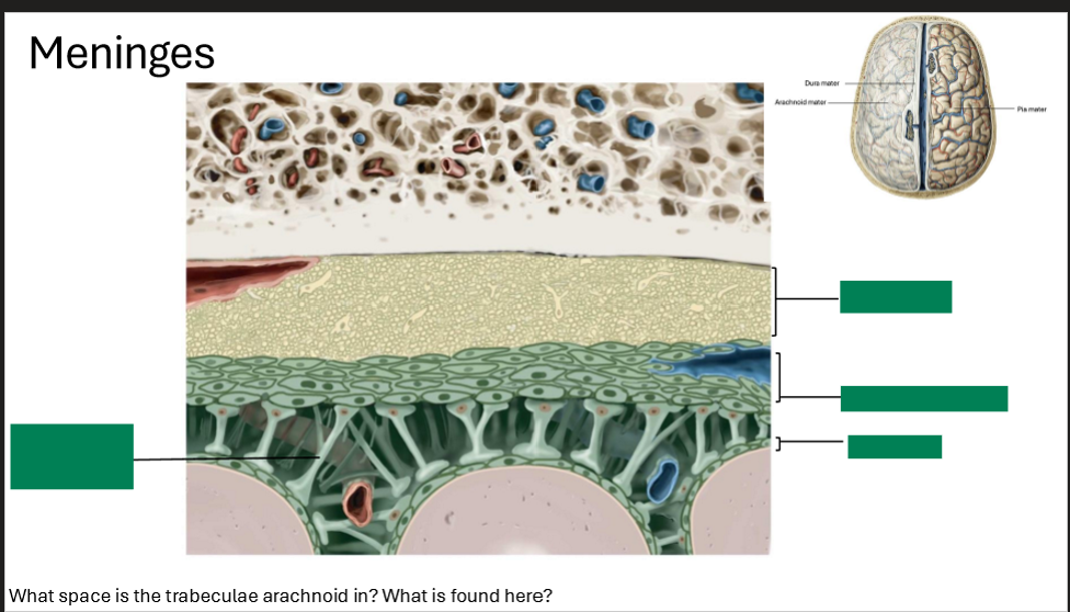
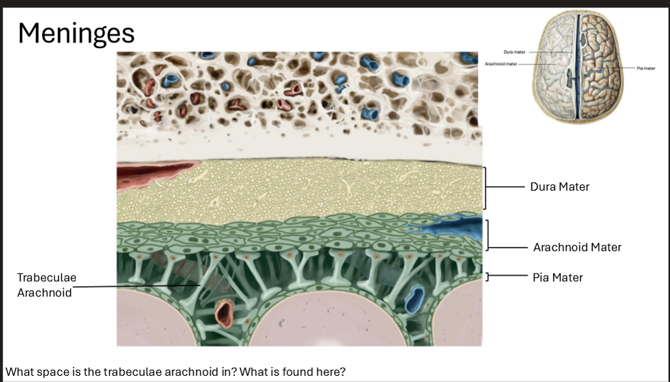
Label the meninges
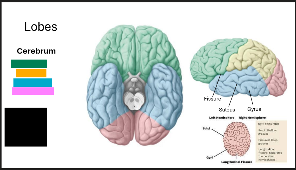
Label the lobes of the brain
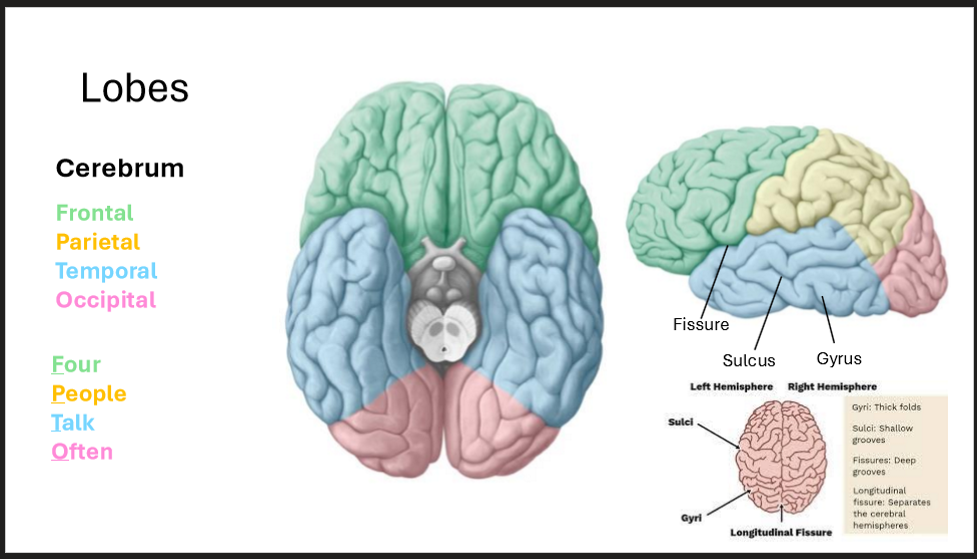
What does each lobe of the brain do?
Frontal → Planning, Decision-making, personality
Parietal → Senses and spatial awarness
Temporal → Hearing, Memory, Language
Occipital → Vision
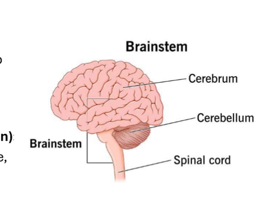
Describe the Brainstem and Cerebellum
BS → connects spinal cord to cerebrum
Cere. → Inferior to occipital lobe, helps with balance and coordination
What are the three parts of the brainstem?
Midbrain, Pons, Medulla Oblongota
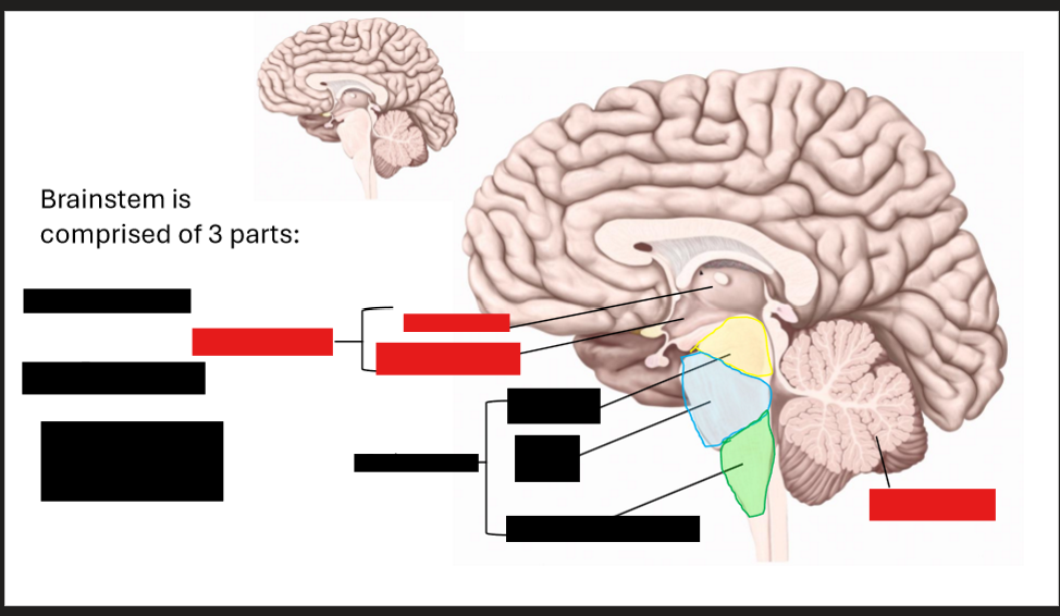
Label this diagram
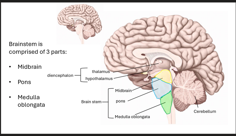
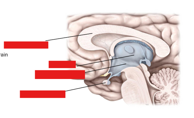
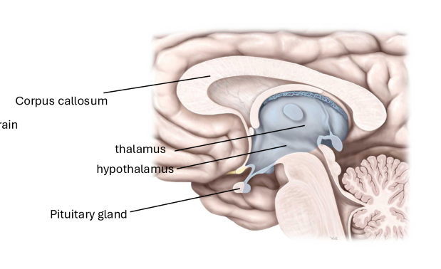
Label this diagram and describe each part of the diencephalon
Hypothalamus
Inferior to the thalamus, and superior to brainstem
Thalamus
Superior to the hypothalamus
Corpus Callosum
Spans the midline of the brain, connecting the right and left hemispheres
Pituitary Gland
Inferior to the hypothalamus
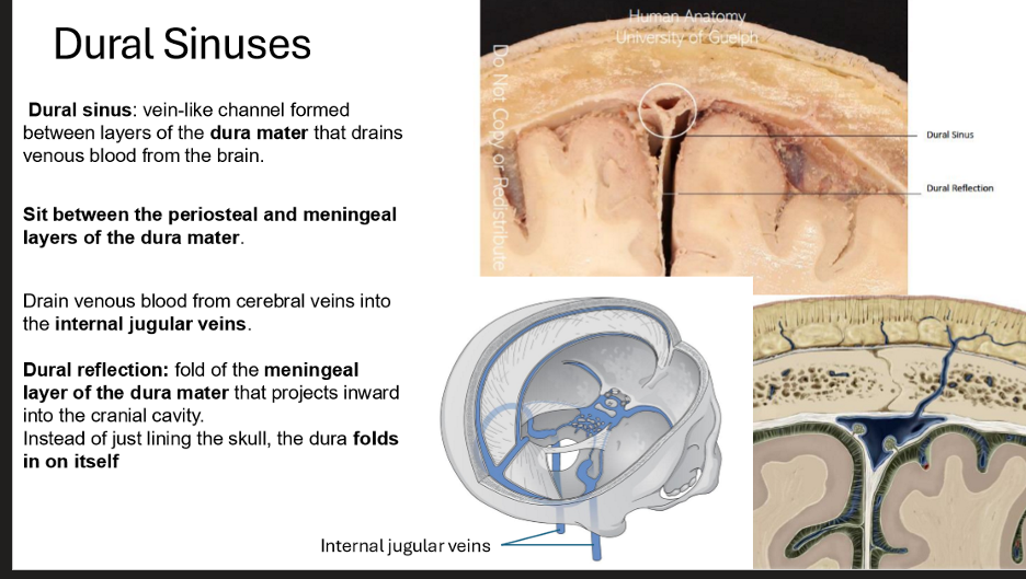
Describe the Dural Sinuses & Dural reflection
Vein-like channel formed between layers of the dura mater that drains venous blood from the brain
sits between the periosteal and meningeal layers of the dura mater
Drains venous bloodfrom the cerebral veins into the internal jugular veins
Dural reflection:
Fold of the meningeal layer of the dura mater that projects inward into the cranial cavity
Instead of just lining the skull, the dura folds in on itself
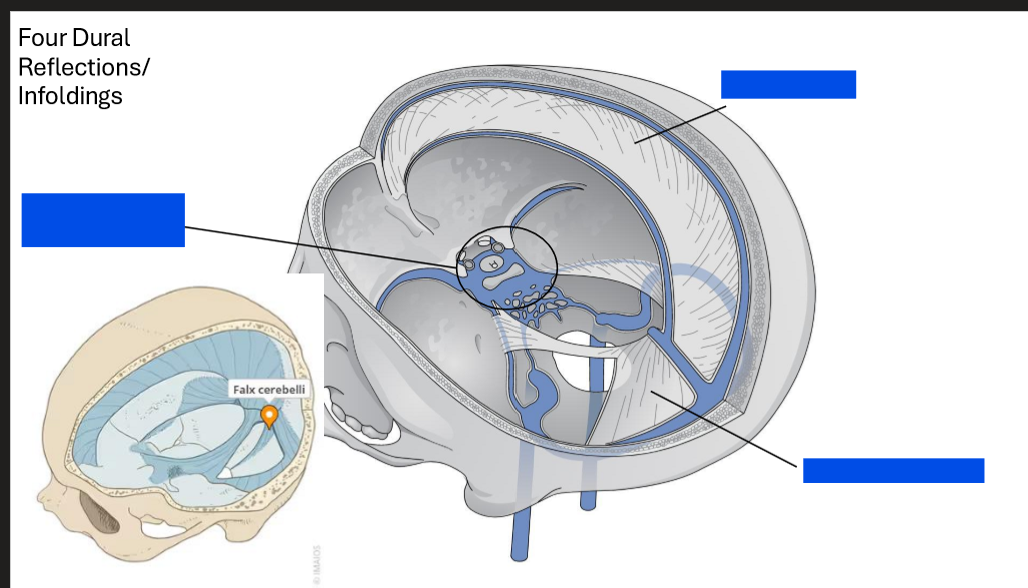
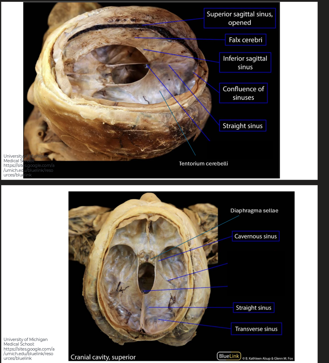
Label these dural reflections/infoldings
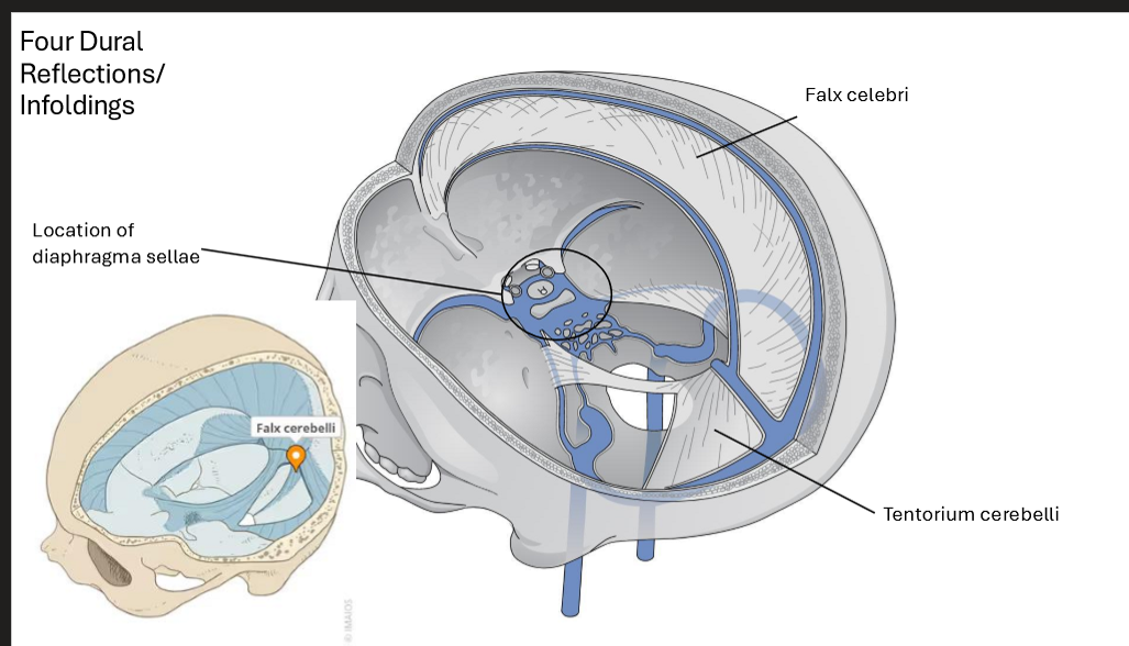
Describe Each of the Dural Reflections/Infoldings
Falx Cerebri
Seperates right and left cerebral hemispheres
From the crista gali and frontal crest to the internal aspect of the occipital bone
Tentorium Cerebelli
Seperates Occipital lobes and Cerebellum
Falx Cerebelli
Inferior to Tentorium Cerebelli
Seperates cerebellar hemispheres
Diaphragma Sellae
suspended between clinoid processes
forms a partial roof over sella turcica and covers pituitary gland
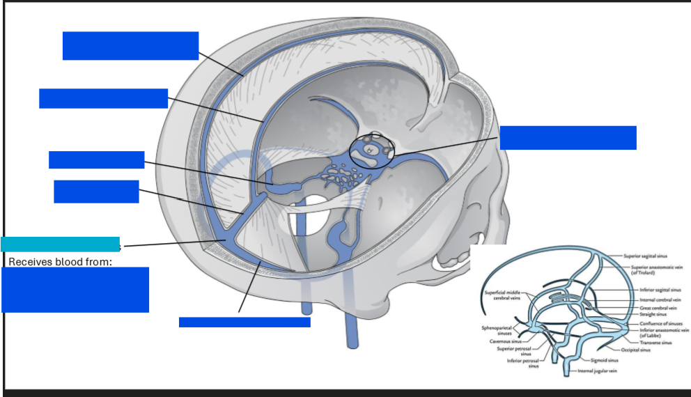
Label tis diagram of the veins
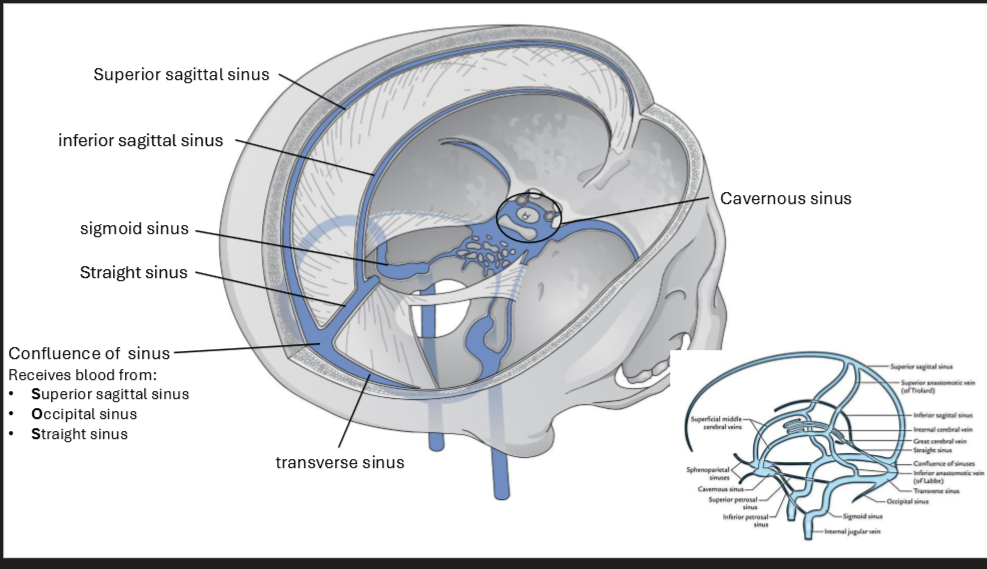
Describe the locations of each of the sinuses
Superior Sagittal Sinus
Superior border of falx cerebri
Inferior Sagittal Sinus
Inferior border of falx cerebri, ends in straight sinus
Straight Sinus
Runs along attachment of falx xerebri and tentorium cerebelli
Occipital Sinus
Posterior border of falx cerebelli
joins with confluence of sinuses
Transverse sinus
runs laterally from the confluence, continuous with sigmoid sinus
Sigmoid sinus
Continous with transverse sinus, s-shaped and continues as internal jugular veins
Cavernous Sinus
On sides of the sella turcica (or pituitary gland)
Describe the different relationships of the brain
Center → pituitary gland
Floor → sella turcica
Roof → Diaphragma sellae (dura)
Sides → Cavernous sinus (venous channels with cranial nerves + ICA)
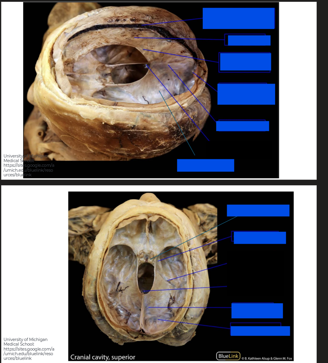
Label these diagrams of the cranial cavity
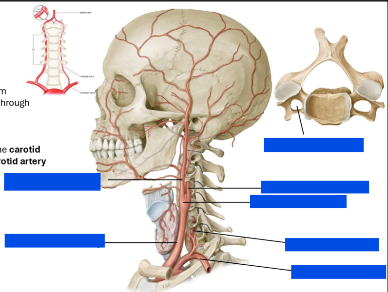
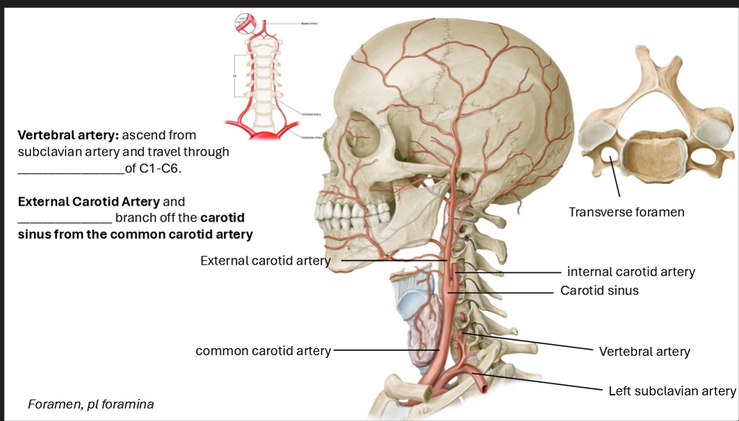
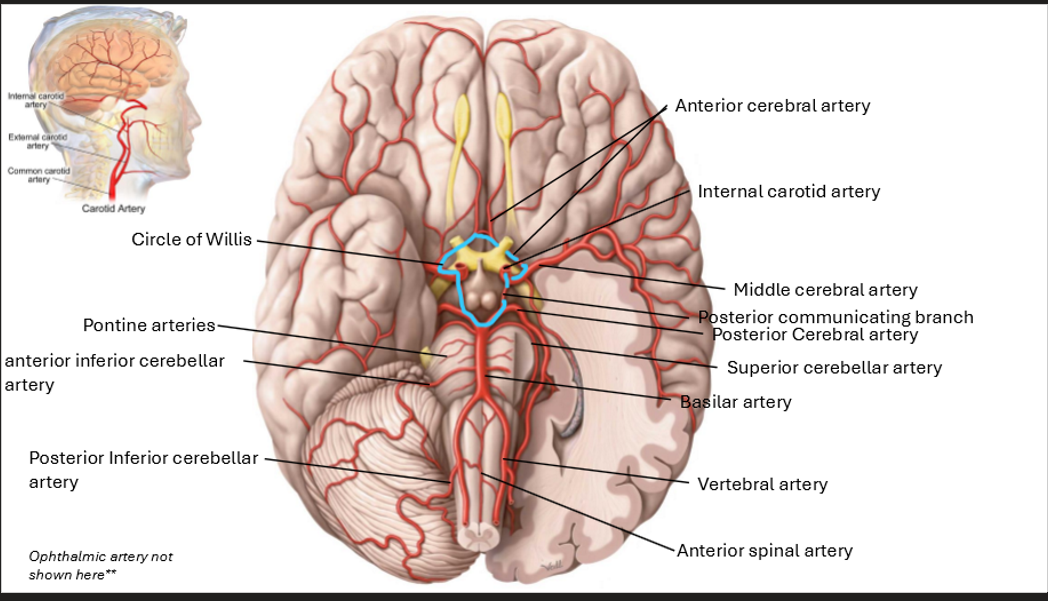
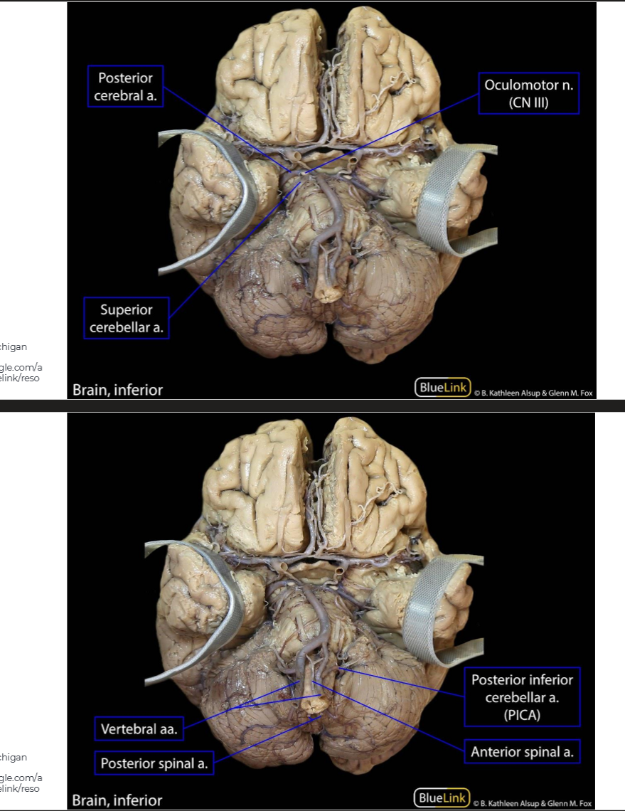
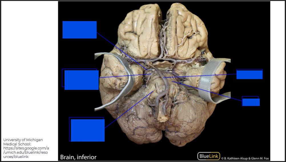
Label the diagram and describe the arteries that supply the brain
Vertebral Artery
Ascends from subclavian artery and travels through transverse foramen of C1-C6
External and Internal Carotid Artery
Branch off the carotid sinus from the common carotid artery
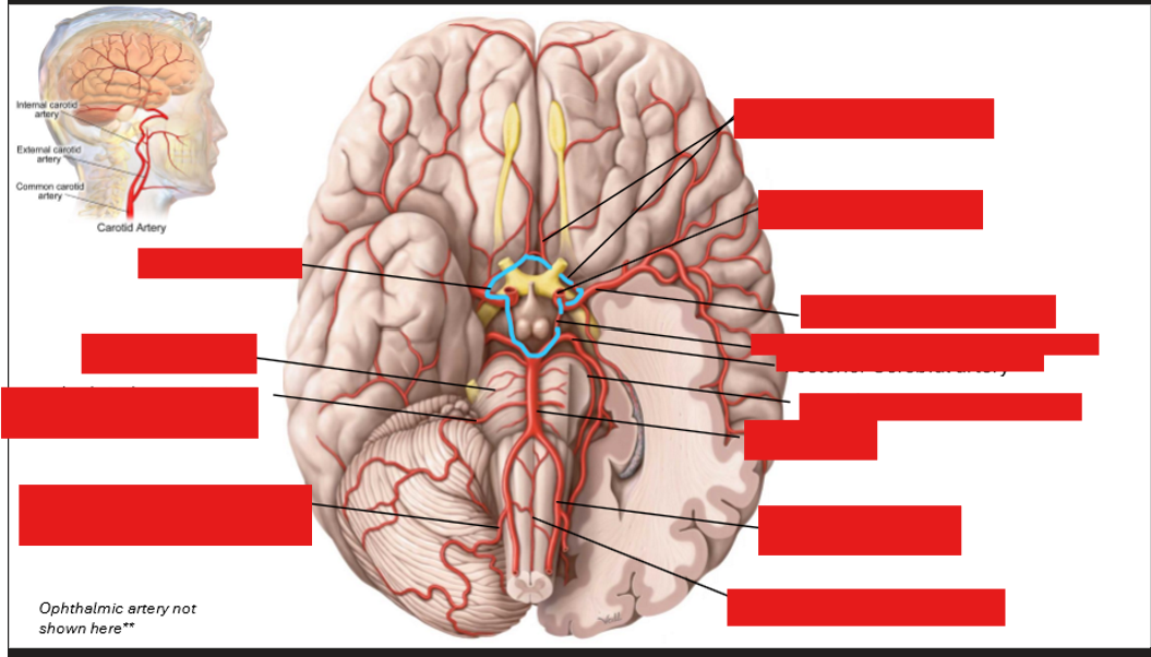
Label the arteries in the brain
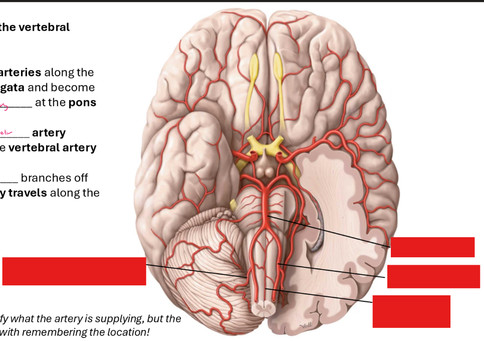
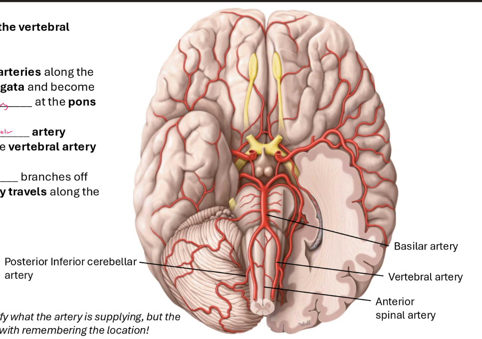
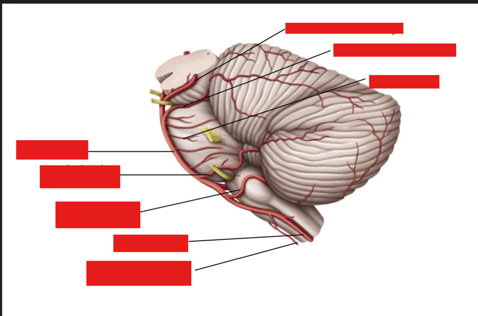
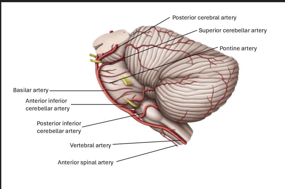
What are the three branches of the vertebral artery?
Two vertebral arteries along the medulla oblongata and becomes the 1. Basilar Artery at the pons
2. Posterior Inferior Cerebellar artery branches off the vertebral artery
3. Anterior Spinal artery branches off the vertebral artery, and travels along the spinal cord
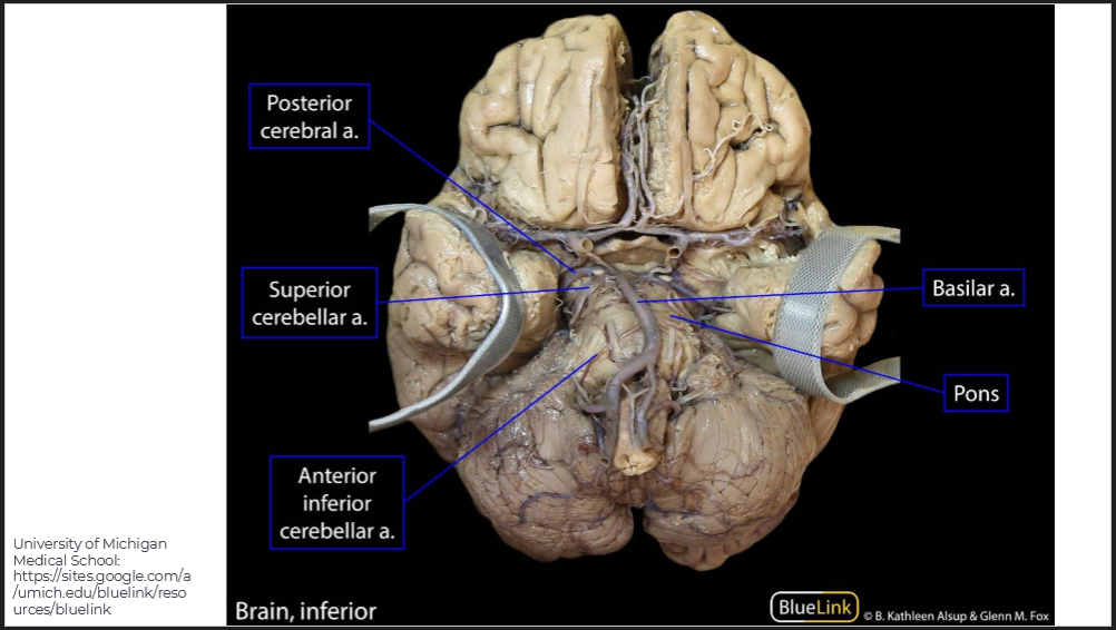
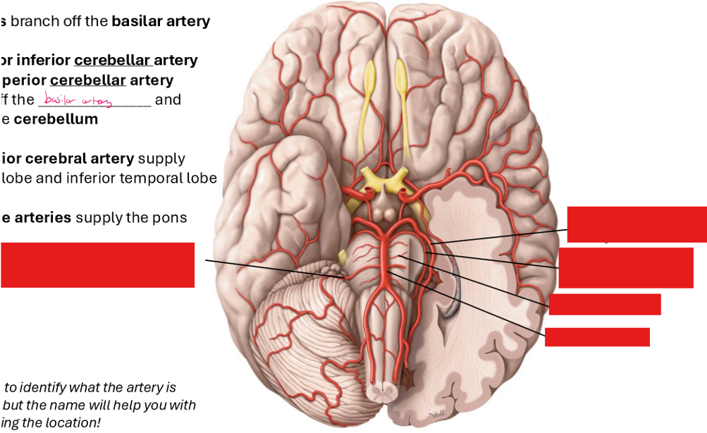
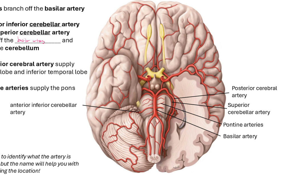
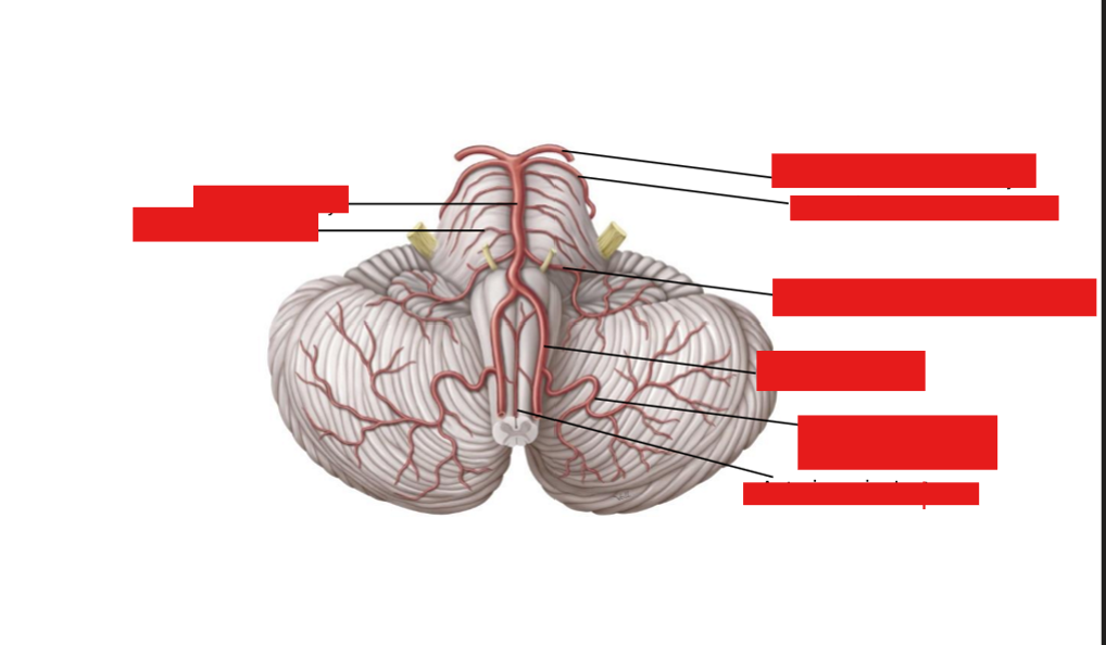
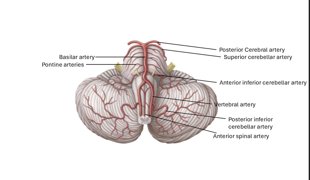
What are the arteries that branch off the basilar artery
Anterior Inferior Cerebellar artery
Superior Cerebellar artery
Both branch off the basilar artery and supply the cerebellum
Posterior Cerebral artery
Supply occipital lobe and inferior temporal lobe
Pontine arteries
Supply the pons
Label these arteries
Label these arteries
The basilar artery branches to give the —— which is connected to the internal carotid artery via ——
Posterior Cerebral Artery, Posterior Communicating artery
Label this diagram
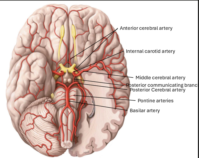
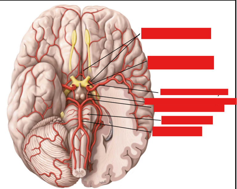
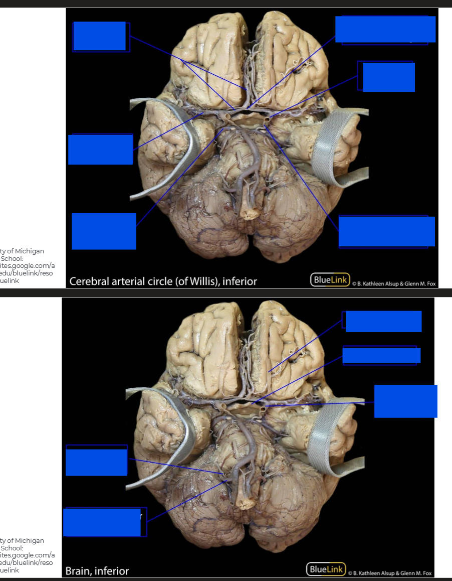
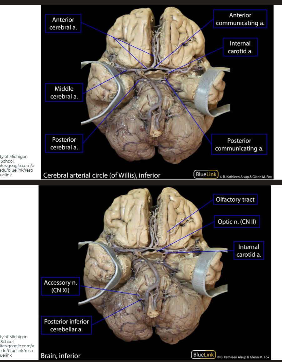
What are the branches of the internal carotid artery
Middle Cerebral Artery
Opthalmic Artery
Anterior Cerebral artery
Posterior Communicating branches
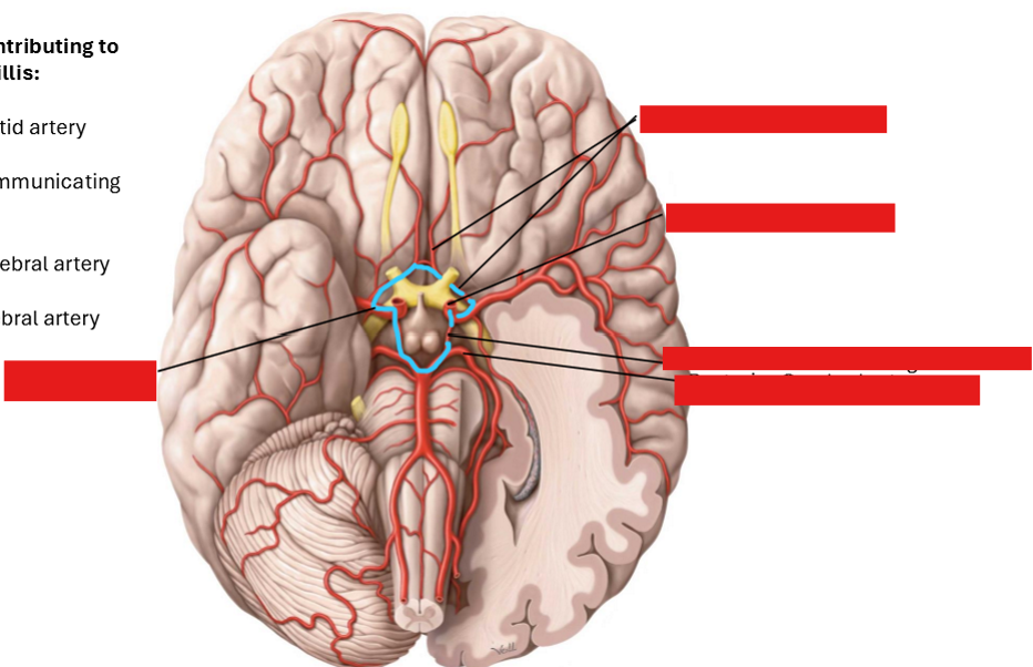
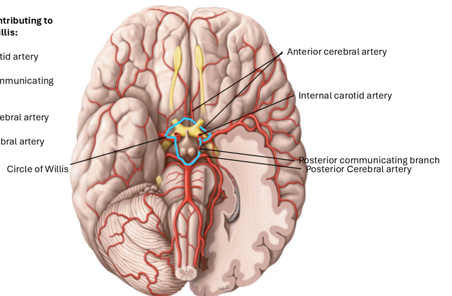
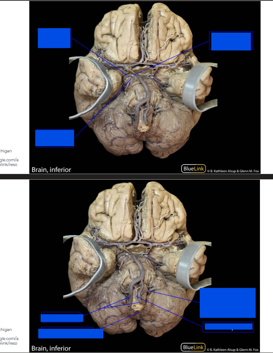
What are the arteries that contribute to the circle of willis
Internal carotid artery
posterior communicating branch
Posterior cerebral artery
Anterior cerebral artery
Label this diagram
Label the arteries in the cerebrum
Label these arteries of the cerebrum
Label the brain
Label the brain
Label the brain