Overview of the Nervous System
1/216
Earn XP
Description and Tags
These flashcards cover the key concepts related to the structure and function of the nervous system as discussed in the lecture.
Name | Mastery | Learn | Test | Matching | Spaced | Call with Kai |
|---|
No analytics yet
Send a link to your students to track their progress
217 Terms
Function of the Nervous System
The nervous system is designed to detect changes in the internal and external environment, evaluate, interpret that information, and then respond accordingly.
Central Nervous System (CNS)
Consists of the brain and spinal cord, where it receives, processes, and responds to information.
Peripheral Nervous System (PNS)
Includes all nerves outside the central nervous system; consists of arms, legs, and other nerves.
Afferent Neurons
Sensory neurons that carry signals toward the central nervous system.
Efferent Neurons
Motor neurons that carry signals away from the central nervous system to muscles and glands.
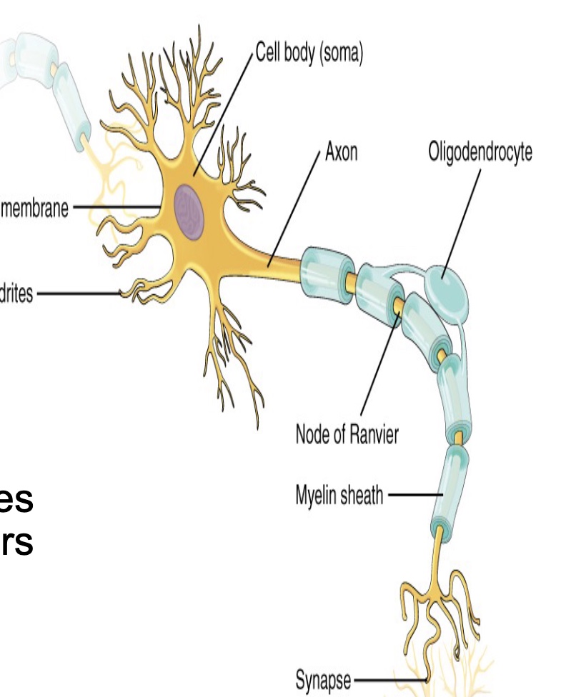
Myelin Sheath
A protective covering made of satellite cells in the peripheral nervous system and oligodendrocytes in the central nervous system.
Multipolar Neuron
A neuron with several dendrites and one axon; includes most neurons in the brain and spinal cord.
Bipolar Neuron
A neuron with two extensions; found in sensory areas such as the eye, ear, and nose.
Unipolar Neuron
A neuron with a single process that extends from the cell body; always sensory and conducts impulses toward the CNS.
Glial Cells
Helper cells in the nervous system, which include astrocytes, oligodendrocytes, microglia, and ependymal cells.
Astrocytes
The most abundant glial cells, forming the blood-brain barrier in the CNS.
Oligodendrocytes
Glial cells found in the CNS that myelinate axons.
Schwan Cells
Glial cells in the peripheral nervous system that also myelinate axons.
Interneuron
Neurons that connect other neurons within the central nervous system.
Nervous Tissue Components
Includes dendrites, cell body, and axon.
What is the overall structure of the nervous system?
Brain, Spinal Cord, Nerves
Which type of neuron has two extensions branching off of the cell body?
Multipolar
Anaxonal
Bipolar
Unipolar
Bipolar
Voltage-gated channels open in response to:
Only sodium ions
Any ion
A particular voltage
Any stimulus
A particular voltage
What are the three parts of a neuron?
Pole
Dendrite
Axon
Connective tissue
Cell body
Sarcomeres
Dendrite, Axon, Cell body
Unipolar neurons are always motor neurons, delivering information.
True
False
False
The two ions that are involved with entering and exiting the neuron plasma membrane down the length of an axon are:
Calcium and phosphorous
Potassium and sodium
Sodium and calcium
Calcium and potassium
Potassium and sodium
The mechanism for moving sodium and potassium ions across the neuron plasma membrane is:
Dialysis
Endo and Exocytosis
Active transport pumps
Simple diffusion
Active transport pumps
A neuron at rest has moved Potassium ions to the outside of the plasma membrane.
True
False
False
For the neuron to establish its resting membrane potential, energy use is required.
False
True
True
What is a membrane potential?
A measurement between 2 sides of the cell
the possibility of a neuron being able to work properly.
The number of ions outside the cell minus the number of ions inside the cell.
The percent difference between ions on the outside of the cell and ions on the inside of the cell.
A measurement between 2 sides of the cell
Functions of the nervous system
Control, communication, and integration
Communication of the nervous system
Sending messages, Cells in the NS send messages to cells in other systems
control of the nervous system
Regulation of body functions, The NS regulates a number of body functions (important in maintaining homeostasis)
integration of the nervous system
Unification of body functions, Allows the body to function as a unit

what is the yellow line (side)?
right cerebral hemisphere

What is the green line? (line down the middle)
longitudial fissure
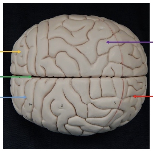
what is the blue line? (side)
left cerebral hemisphere

What is the purple line? (raised area)
gyri

what is the red line? (groove)
sulci
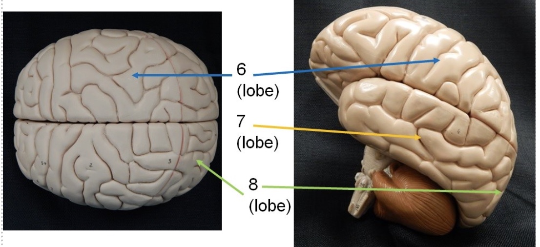
What is the blue line? (lobe)
parietal lobe

What is the yellow line? (lobe)
temporal lobe
Broca’s area
The area of the brain that is used for speech. Located on the left side of the brain
parietal
somatic (skeletal muscle movement) sensory info from skin and muscles arrives here, and its located in the parietal lobe
temporal lobe
auditory and olfactory area (smell)
what is the light green line?
olfactory nerve

what is the green line? (lobe)
occipital lobe
what is the blue line ? (lobe)
frontal lobe

what is the green line? (lobe)
temporal lobe

what is the red line?
cerebral cortex
what is the blue line?
fornix
Is the pituitary part of the brain?
Yes, it is part of the brains anatomy. It hangs below the brain which can be confusing.

What is the purple line?
corpus callosum

What is the green line?
septum pellucidum

what is the teal line? (white)
cerebral tracts

what is the blue line? (deep groove)
transverse fissure
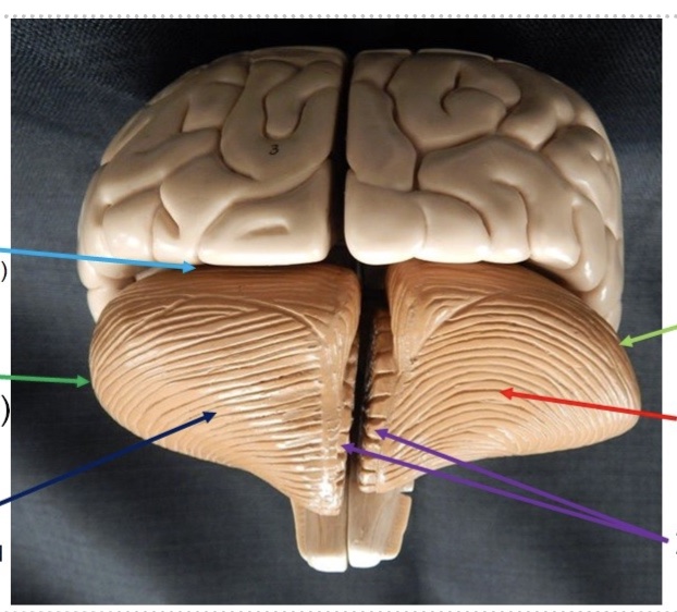
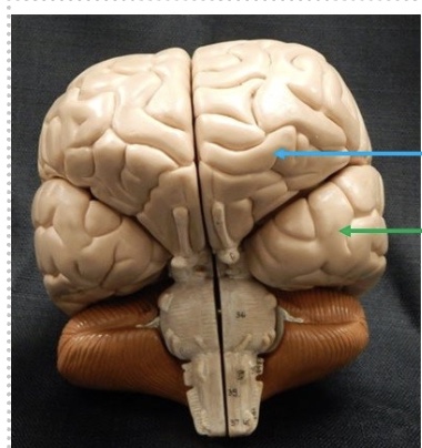
what is the dark green line? (specific side)
left cerebellar hemisphere

what is the dark blue line? (raised part)
convulsions

what is the light green line?(specific side)
right cerebellar hemisphere

what is the red line? (groove)
sulcus

what are the purple lines?
vermis

what is the blue line?
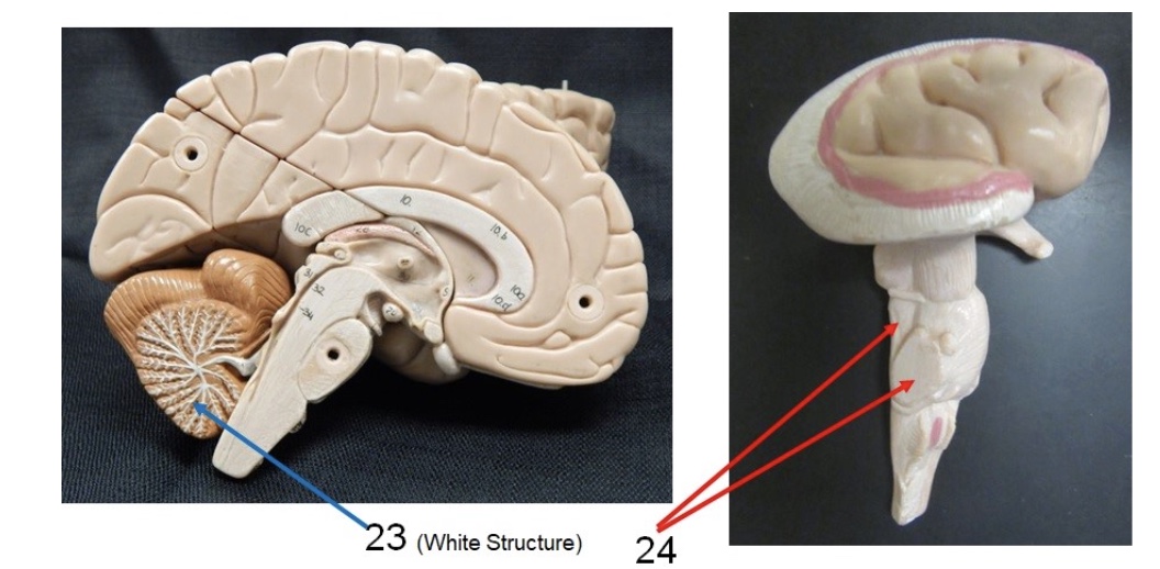
arbor vitae
what is the red lines?
cerebellar pundcles

what is the green line?
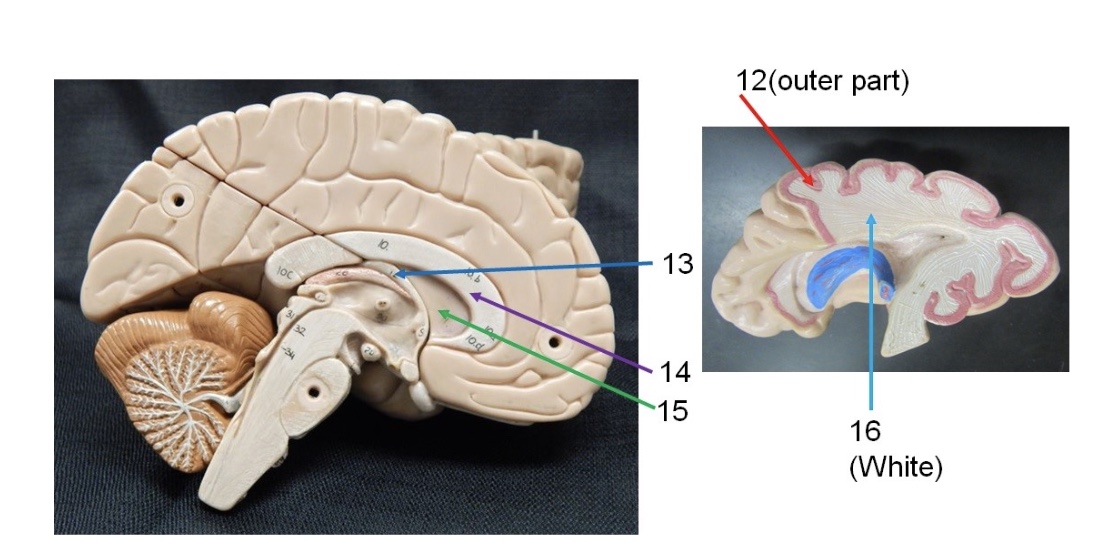
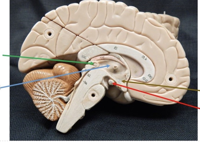
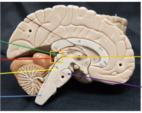
pineal body

what is the blue line?
thalamus
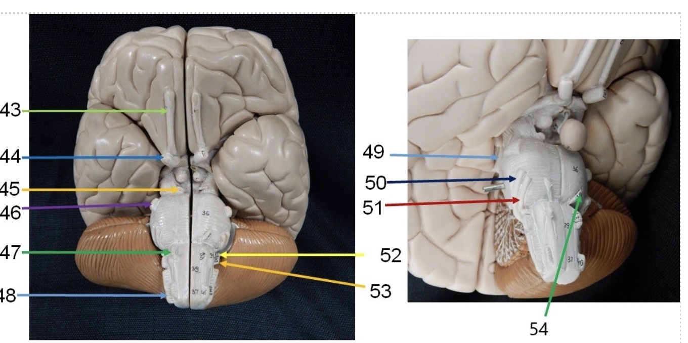
what part of the brain has vomiting?
medulla oblongota
what is the brown line?
hypothalamus

what is the red line?
mamillary body

what is the green line? (top part)
superior colliculi

what is the red line? (bottom part)
inferior colliculi
what is the yellow line? (division)
midbrain

what is the light blue line? (division)
medulla oblongota

what is the yellow line? (whole structure)
corpora quadregima

what is the purple line? (division)
pons

what is the light blue line? (space)
3rd ventricle

what is the green line? (space)
central aqueduct

what is the dark blue line? (space)
4th ventricle
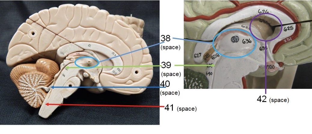
what is the red line? (space)
central canal

what is the purple line? (space)
lateral ventricles
what is the olfactory (I) nerve?
sensory nerve that detects smell
what is the optic (II) nerve?
sensory nerve that aids in vision
what is the oculomotor (III) nerve?
motor nerve that aids in motor movement of eyeballs
what is the trochlear (IV) nerve?
motor nerve that aids in eye movement of superior oblique
what is the trigeminal (V) nerve?
it is both a sensory and motor nerve, deals with facial sensations and mastication (chewing)
what is the abducens (VI) nerve?
motor nerve that moves the eye in the lateral rectus

what is the medium blue line? (44)
optic nerve

what is the yellow line?
oculomotor nerve

What is the purple line?
trigeminal nerve

what is the light blue line? (49)
trochlear nerve

what is the green line? (54)
abducens nerve
is the Somatic Sensory Division afferent or efferent?
afferent
is the Somatic Motor Division afferent or efferent?
efferent
is the Visceral Sensory Division afferent or efferent?
afferent
is the Sympathetic Division efferent or afferent?
efferent
is the Parasympathetic Division afferent or efferent
afferent
Endocrine and nervous system share systems:
they share Communication, Control, and Integration, they differ in how they communicate, control, and integrate: Nerve Impulses – Rapid, short-lasting, Hormones – Slow, long-lasting
neurons
Conduct nerve impulses (NI), We have about 100 Billion. Structure: Plasma membrane, Cytoplasm, Cytoskeleton, Microtubules, Microfilaments, Neurofibrils, Mitochondria, vesicles with neurotransmitters
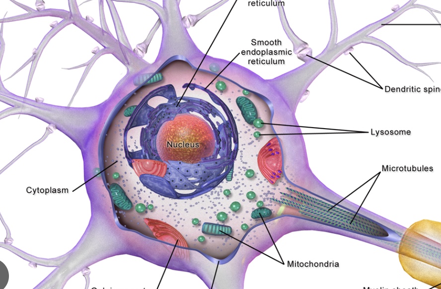
cell body of neuron
Largest part of neuron, Contains nucleus and typical organelles, Contains Nissl Bodies, Rough ER of neurons (protein synthesis)
Way to remember the nerves
oh once one takes the anatomy final very good vacations are heavenly
processes (nerve fibers)
Threadlike extensions from cell body, Two types: dendrites and axons

dendrites
One or more per neuron (short), Conduct NI toward cell bod
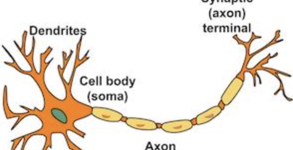
axon
One per neuron (Long) Conduct NI away from cell body
axon collateral
Side branches: Can have one or more, Divide into Telodendria, Telodendria (terminal branches) terminate into SYNAPTIC KNOBS (terminal ends-bulges)
impulse conduction pathway
The impulse is conducted one-way on a neuron. A nerve impulse is conducted from dendrites to the cell body to the axon and to the synaptic knobs
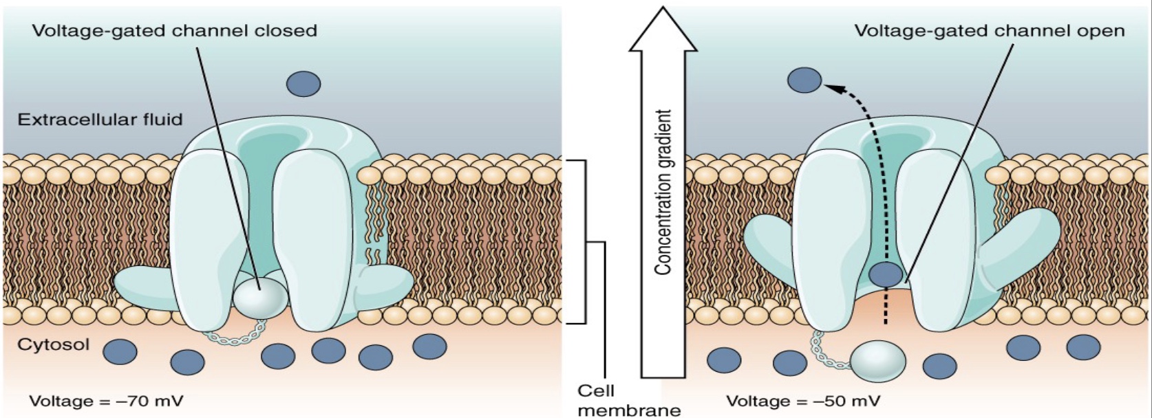
resting membrane potential RMP
Resting Membrane Potential (RMP) refers to the resting state of the neuron (when it is not conducting a nerve impulse) RMP for neurons: The outer surface of the plasma membrane contains a slight excess of positive ions compared to the inner surface, The membrane is said to be polarized, and more positive at
this point
factors of RMP
Factors contributing to the neuron’s RMP- Large negative proteins can’t cross the membrane – this makes inside negative, Most Na+ channels are closed, so Na+ can’t re-enter the cell, The Na+/K+ Pump is pumping 3 Na+ out, and 2 K+ in, K+ can diffuse back out, but Na+ can’t diffuse back in This maintains positive charge outside