13B Center-surround antagonism
1/43
There's no tags or description
Looks like no tags are added yet.
Name | Mastery | Learn | Test | Matching | Spaced | Call with Kai |
|---|
No analytics yet
Send a link to your students to track their progress
44 Terms
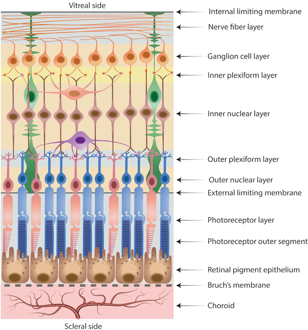
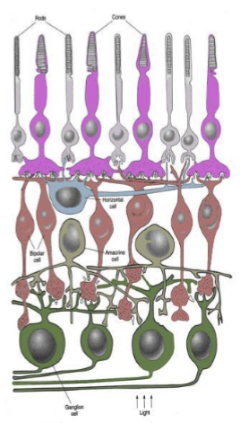
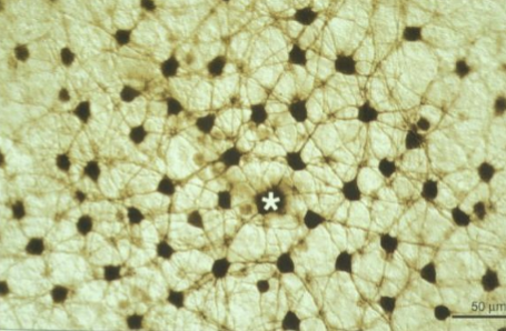
Where are horizontal cells located in retinal architecture, and what is their functional role?
Horizontal cells are located in the inner plexiform layer, where they lie between photoreceptors and bipolar cells.
They receive input from multiple photoreceptors and provide lateral inhibition, helping to enhance contrast and refine spatial signaling before information is passed to bipolar cells.
What is the main functional role of horizontal cells in visual processing?
Horizontal cells perform lateral inhibition, integrating information from multiple photoreceptors to:
Enhance contrast and edge detection
Regulate photoreceptor → bipolar cell signaling
Shape receptive fields for early visual processing
How do horizontal cells influence bipolar cell activity?
They adjust bipolar cell sensitivity by feeding back onto photoreceptors, controlling neurotransmitter release.
This helps create center-surround receptive fields, a key early step in spatial vision.

Where are horizontal cells located within the retina, and how are they arranged?
Horizontal cells are stratified horizontally in the outer margin of the inner nuclear layer.
They extend laterally across the retina, positioned to integrate signals from photoreceptors.

How are horizontal cells classified in primates?
Primates have 2-3 classes of horizontal cells, classified by the photoreceptors they predominantly contact:
Some preferentially contact cones
Others preferentially contact rods
This specialization helps shape wavelength‑specific and luminance‑specific processing.
How do horizontal cells differ from the typical neuronal structure?
Unlike typical neurons with distinct post‑synaptic dendrites and pre‑synaptic axon terminals, horizontal cells:
Have neurites that are post‑synaptic to photoreceptors
Lack a classic axon
Their neurites also behave functionally like pre‑synaptic terminals, feeding back onto photoreceptors

What is the functional significance of horizontal cells having neurites that act both post‑synaptically and pre‑synaptically?
This dual role allows horizontal cells to:
Receive signals from photoreceptors
Modulate photoreceptor output via feedback
Enable lateral inhibition, sharpening contrast and forming center–surround receptive fields

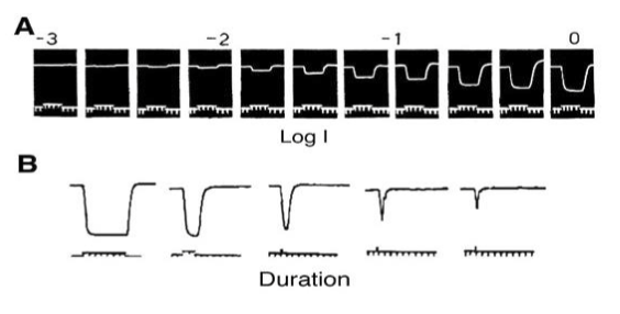
How do horizontal cells responds to light?
All horizontal cells hyperpolarize to light. They also exhibit graded potentials that are linked to the intensity and duration of the light stimulus.
How do photoreceptors signal to horizontal cells in dark vs light?
Dark: Photoreceptors continuously release glutamate
Light: Photoreceptor glutamate release decreases/stops
What type of glutamate receptors do horizontal cells express, and how is this similar to bipolar cells?
Horizontal cells express ionotropic kainate/AMPA-type glutamate receptors at their neurites.
This is similar to OFF bipolar cells, which also depolarize in response to glutamate.
What happens to horizontal cells in the dark at the level of ion flow and membrane potential?
In the dark, glutamate binds kainate/AMPA receptors →
Channels open
Cation influx (primarily Na⁺)
Horizontal cell becomes relatively depolarized
What is the electrical response of horizontal cells to light, and why?
In light, decreased glutamate →
Kainate/AMPA channels close
Reduced Na⁺ influx
Cell hyperpolarizes
This change enables horizontal cells to participate in contrast processing via lateral inhibition.
What is a receptive field? (Definition + retinal context)!
A receptive field is the area of sensory space over which stimulation produces a response in a specific neuron.
In the retina, it is the region of visual space in which light (or darkness) alters the neuron’s activity.
Do all visual neurons have receptive fields? What does this imply?
Yes, every neuron in the visual pathway has a receptive field.
This means each neuron encodes information about a specific region of visual space, with increasingly complex receptive field properties at higher levels.
How do physiologists experimentally map receptive fields?
By recording from a neuron and presenting small spots of light:
On the retina (isolated retina prep)
On a screen (anesthetized animal)
They observe where stimulation evokes a response.
What properties are determined when mapping a receptive field?
Physiologists determine:
Location in visual space that elicits responses
Extent/size of the receptive field
How much the stimulus can be moved before the response changes (spatial sensitivity)
What determines the size of a photoreceptor’s receptive field?
The width of the outer segment.
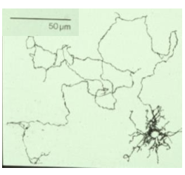
Is the receptive field of a horizontal cell limited to its dendritic field?
No. Horizontal cell receptive fields are much larger than their dendritic arborization.
They extend far beyond the region of direct photoreceptor contact.
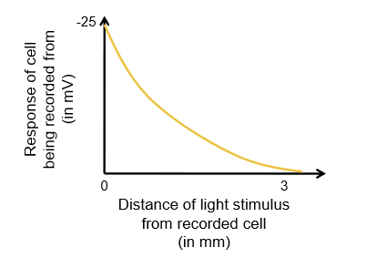
What is observed experimentally when mapping a horizontal cell receptive field with a moving light spot?
Maximum response when light is near the recording electrode
As light is moved away, response decreases in magnitude
However, responses remain detectable even millimeters away
What types of synapses are present in the retina?
Chemical synapses (neurotransmitter‑mediated)
Electrical synapses formed by gap junctions
How do gap junctions transmit signals compared to chemical synapses?
Gap junctions allow direct passage of ions and small molecules from one cell to another.
They do not use neurotransmitter release, enabling fast, bidirectional signal transmission.
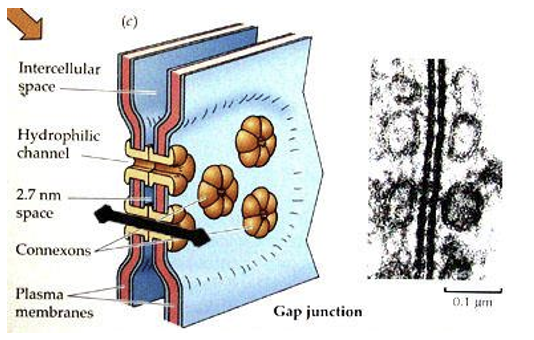
What explains the unusually large receptive fields of horizontal cells?
Horizontal cells are extensively electrically coupled (via gap junctions), allowing signals to spread laterally across many cells.
This electrical coupling explains why horizontal cell receptive fields extend far beyond their dendritic spread.
Why is describing bipolar cell receptive fields as simply ON vs OFF incomplete?
Bipolar cells have center-surround receptive fields, not uniform ON or OFF responses.
Their response depends on where within the receptive field the light stimulus is applied.
How does an OFF bipolar cell respond to light in the center of its receptive field?
Light in the center of an OFF bipolar cell’s receptive field →
↓ glutamate release from photoreceptors
Reduced activation of ionotropic glutamate receptors
Hyperpolarization of the OFF bipolar cell
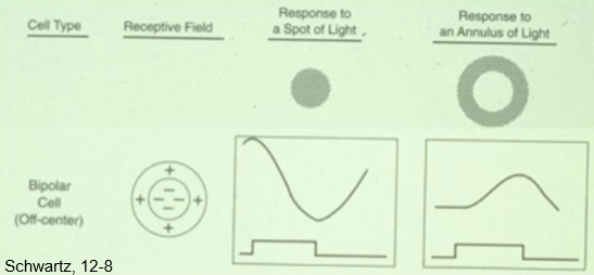
How does an OFF bipolar cell respond to light in the surround of its receptive field?
Light in the surround only →
Horizontal‑cell–mediated feedback onto center photoreceptors
Relative ↑ glutamate release from center photoreceptors
Depolarization of the OFF bipolar cell
→ Opposite effect of center stimulation

How does an ON bipolar cell respond to center vs surround illumination?
Center spot of light: Depolarizes
Surround (annulus) of light: Hyperpolarizes
→ Same center–surround structure as OFF cells, but polarity is reversed.
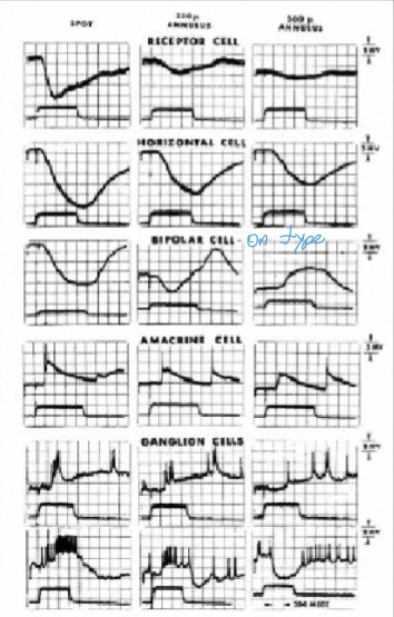
What circuit mechanism creates opposite center vs surround responses in bipolar cells?
Horizontal cell–mediated lateral inhibition:
Center response reflects direct photoreceptor input
Surround response is mediated indirectly via horizontal cell feedback
This produces center-surround antagonism.
What are the two most common inhibitory neurotransmitters in the CNSA?
Gamma-aminobutyric acid (GABA) and glycine
How much higher is the concentration of Cl outside the neurons vs inside?
20x higher
How many cells does every cone connect to near the fovea?
Every cone connects to two midget bipolars. One is an ON-center and the other is an OFF-center.
What is the polarization of an OFF-center bipolar cell?
Glutamate release from cone keeps it relatively depolarized.
What is the polarization of an ON-center bipolar cell?
Glutamate release from cone keeps it relatively hyperpolarized.
Since horizontal cells expresses kainate/AMPA-type glutamate receptors, what is its polarization in the dark?
Glutamate release in the dark causes it to be relatively depolarized.
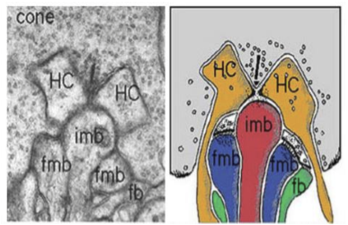
How do horizontal cell (HC) dendrites function at photoreceptor synapses?
HC dendrites act both post‑synaptically and pre‑synaptically.
While receiving input from photoreceptors, they also release an inhibitory transmitter back onto photoreceptor terminals.
What neurotransmitter do horizontal cells release, and what is its effect?
Horizontal cells release a GABA‑like inhibitory compound (exact identity debated).
This inhibitory signal causes the photoreceptor axon terminal to hyperpolarize, reducing glutamate release.
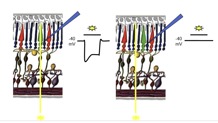
What is the state of horizontal cells and their transmitter release in the dark?
In the dark, horizontal cells are depolarized and actively release inhibitory (GABA‑like) transmitter onto photoreceptors, modulating their resting membrane potential.
How does horizontal cell feedback affect photoreceptor membrane potential in the dark?
With HC feedback intact: photoreceptor rests around –40 mV
Without HC feedback: photoreceptor is more depolarized (~–30 mV)
HC inhibition therefore hyperpolarizes photoreceptors relative to their intrinsic dark state.
What happens to a horizontal cell when a cone photoreceptor is stimulated by light?
Light causes the cone to hyperpolarize and reduce glutamate release.
Because the horizontal cell receives input from that cone, the horizontal cell also hyperpolarizes in response to light.
How does stimulation of surrounding cones affect inhibitory feedback onto a central cone?
When surrounding cones are stimulated, horizontal cells hyperpolarize and release less inhibitory feedback. This disinhibits the central cone terminal, causing increased glutamate release onto bipolar cells, contributing to center-surround antagonism.
What is meant by “disinhibition” of a cone photoreceptor, and what are its consequences?
Disinhibition refers to reduced inhibitory (GABAergic) feedback from horizontal cells onto a cone terminal.
As a result, the cone experiences less suppression of its synaptic output, leading to a relative increase in glutamate release onto bipolar cells, even without direct light stimulation of that cone.
How does increased glutamate release from a cone affect ON and OFF bipolar cells?
More glutamate causes:
OFF bipolar cells to further depolarize
ON bipolar cells to further hyperpolarize
This amplifies center–surround antagonism and contrast sensitivity.
Why was the classic model of direct GABA feedback from horizontal cells to photoreceptors questioned?
If horizontal cells directly caused center–surround antagonism in photoreceptors:
Photoreceptors themselves should show center–surround receptive fields, they do not.
Additionally, GABA receptors on photoreceptors have not been conclusively identified, weakening the original model.
What is the currently favored mechanism for horizontal cell feedback if not direct GABA action on photoreceptors?
Horizontal cells likely release GABA onto themselves, activating GABA receptors on HCs.
This leads to proton (H⁺) release into the synaptic cleft, altering pH, which modulates acid‑sensing channels on photoreceptors and bipolar cells.
Why is the classic GABA feedback model still useful for learning bipolar cell receptive fields?
Despite mechanistic revisions, the final effect on bipolar cell responses is the same:
Surround stimulation produces antagonistic effects
Center–surround contrast is enhanced