AP 1 Lecture Exam 3
1/111
There's no tags or description
Looks like no tags are added yet.
Name | Mastery | Learn | Test | Matching | Spaced | Call with Kai |
|---|
No analytics yet
Send a link to your students to track their progress
112 Terms
general functions of bones
support and protection
levers for movement
hematopoiesis
storage of mineral and energy reserves
hematopiesis definition
blood cell production, occurs in red bone marrow CT
what minerals are stored in the bone
calcium and phosphate
gross anatomy of bones
highly vascularized (spongy bone regions)
nutrient foramen
high sensory component
what is the nutrient foramen
small opening or hole in bone, artery entrance and exit
what is red bone marrow (myeloid)
soft connective tissue
hematpoietic
hematopoiesis definition
biological process by which body produces all of its blood cells
driven by hematopoietic stem cells, takes place primarily in the bone marrow
what is yellow bone marrow
product of red bone marrow degeneration as child matures
fatty substance
can convert back due to severe anemia
facilitates production of additional erythrocytes
what is bone connective tissue (osseous connective tissue) composed of
primary component of bone
composed of cells and extracellular matrix
4 types of cells in bone connective tissue
osteoprogenitor cells
osteoblasts
osteocytes
osteoclasts
what are osteoprogenitor cells
bone stem cells from mesenchyme
during cell division committed cell is produced that matures to become osteoblast
*stem cells are located in periosteum and endosteum
what are osteoblasts
immature bone cells
synthesize and secrete osteoids
*later undergo calcification
*produce new bone
what are osteocytes (hollow cells)
mature bone cells derived from osteoblasts (lost bone-forming ability)
become embedded within calcified osteoid (organelles are still functioning)
*maintain bone matrix and detect mechanical stress on a bone
what are osteoclasts (broken)
multinuclear cells
*derived from fused bone marrow cells
phagocytic cells involved in breaking down bone in bone resorption
composition of extracellular (bone) matrix
organic components
inorganic components
what are the organic components
osteoids
*gives tensile strength (resists stretching)
*bone flexibility
what are the inorganic components
salt crystals: calcium phosphate Ca3(PO4)2
*interacts with calcium hyroxide (forms crystals: hydroxyapatite Ca10(PO4)6(OH)2
*harden matrix and give rigidity of the bones
bone formation (ossification/osteogenesis) definition
formation and development of bone connective tissue
when does ossification begin (age), when does it end (age)
in embryo, continues after brith and as the skeleton grows during childhood and adolescence
ends 20-25 years old
*by 8th-12th weeks of embryonic development skeleton beings forming from either thickened condesations of mesenchyme or hyaline cartilage model of bone
what does bone formation begin with, what happens
starts with hyaline cartilage
then osteoblasts secrete osteoid
calcification (mineralization occurs) → deposition of hydroxyapatite crystals
what does bone matrix formation require
vitamin D: enhances calcium absorption from GI tract
vitamin C: collagen formation
calcium and phosphate for calcification
what happens in bone resorption
bone matrix is destroyed by substances released from osteoclasts
lysosomes realease:
proteolytic enzymes → digest organic matrix components
hydrochloric acid (HCl) → dissolves inorganic mineral parts of matrix
osetolysis
calcium and phosphate ions enter extracellular fluid of nearby tissues then blood
what is osteolysis
release of stored calcium and phosphate from bone matrix
when does bone resorption occur
occurs during bone remodeling
blood calcium levels are low
main funcitonal unit of bone
osteons
osteoids
collagen proteins, semisolid ground substance of proteoglycans and glycoproteins, semisolid, later calcifies
what happens in intramembranous ossification
ossification centers from within thickened regions of mesenchyme (8-12 weeks)
osteoid undergoes calcification (traps osteocytes)
woven bone and surrounding periosteum form
lamellar bone replaces woven bone, as compact and spongy bone form
what is intramembranous ossification (dermal ossification)
bone growth within a membrane
what is enchondral ossification
within cartilage
*begins with hayline cartilage model
*produces most bones of skeleton (upper and lower limbs, pelivs, vertebrae, ends of clavicle)
what happens in enchondral ossification
fetal hyaline cartilage model develops (8-12 weeks)
cartilage calcifies, and a periosteal bone collar forms around diaphysis (fetal period)
primary ossification center forms in the diaphysis (fetal period)
secondary ossification centers form in the epiphysis (newborn to child)
bone replaces almost all cartilage except the articular cartilage and epiphyseal plates
lengthwise growth continues until the epiphyseal plates ossify and form epiphyseal lines
when does the epiphyseal plate convert to epiphyseal line
between 10-25 years old
compact bone (cortical bone) characteristics
relatively rigid, dense
80% of bone mass
spongy bone (cancellous or trabecular bone) characteristics
appears porous (full of trabeculae)
internal to compact bone
20% of bone mass
has importance with weight
bone remodeling definition
constant dynamic process of continual addition of new bone tissue and removal of old bone tissue
what is bone remodeling based on
blood calcium levels
activities of osteoblasts, osteocytes, and osteoclasts
hormones and mechanical stress
hormones that promote bone growth
growth hormone, thyroid hormone, calcitonin, sex hormones
hormoes that inhibit bone growth (or increase bone resorption)
parathyroid hormone, calcitriol, glucocoricoids, serotonin
what does growth hormone (somatotropin) do
*produced by anterior pituitary gland
stimulates liver to produce hormone called insulin-like growth factor (IGF)
stimulate growth of cartilage in epiphyseal plate (bone elongation)
what does thyroid hormone do
*secreted by thyroid gland
stimulates bone growth by stimulating metabolic rate of osteoblasts
continues after epiphyseal plate and line
*regulates normal activity at epiphyseal plates
what do sex hormones (estrogen and testosterone) do
stimulate osteoblasts
promotes epiphyseal plate growth and closure
what do glucocorticoids do (blood → sugar levels)
*released form adrenal cortex
regulate blood glucose level
increase bone loss
*in children impair bone growth in response to chronically high levels of glucocorticoids
what does serotonin do
neurotransmitter and hormone
role in rate and regulation of normal bone remodeling
*inhibits osteoprogenitor cells from differentiating into osteoblasts when there is chronically high levels of serotonin
what are the 2 primary hormones that regulate blood calcium
calcitriol
parathyroid hormone
what does calcitonin do
in childhood promotes calcium deposition in bone and inhibits osteoclast activity
response to high blood calcium levels
*minimal effects in adulthood
what does calcitrol do
increases blood calcium levels by encouraging bone resorption by osteoclasts and stimulating absorption of calcium ions
*from small intestine into the blood
negative feedback loop of blood calcium levels
stimulus: decrease in blood calcium
receptors: parathyroid gland detects decrease
control center: parathyroid gland, releases parathyroid hormone
PTH and calcitriol interact with major organs
in bone: PTH and calcitriol act together to increase release of calcium from bone into the blood by increasing osteoclast activity → blood calcium levels increase
in kidneys: stimulate kidneys to excrete less calcium in urine with decrease in calcium loss in urine → maintain blood calcium levels
in small intestine: (unique to calcitriol) increase absorption of calcium from small intestine into blood → blood calcium levels increase if calcium is ingested as part of the diet and is available for absorption
net effect: elevating blood calcium → returning to preexisting levels
increase of blood calcium back to typical range inhibits further release of PTH
what happens in response to low blood calcium levels
PTH and calcitriol is released → increase blood calcium levels to preexisiting levels
what happens in response to high blood calcium levels
calcitonin is released (or stress from exercise) → decrease in blood calcium levels
rickets
disease caused by vitamin D deficiency in childhood
characterized by deficient calcification of osteoid tissue (bowlegged)
disturbances in growth
hypocalcemia (low blood calcium)
tetany (cramps and muscle twitches)
calcitonin talks to what bone cell
osteoblast
PTH and calcitriol talks to what bone cell
osteoclast
what is a fracture
breaks in bone
what are the types of fractures
stress fracture
pathologic fracture
simple fracture
compound fracture
stress fracture definition
thin break caused by increased physical activity
pathologic fracture definition
occurs in bone weakned by disease
simple fracture (closed)
broken bone not penetrating skin
compound fracture (open)
one or both ends of the bone pierce overlying skin
process of bone repair after fracture
fracture hematoma forms
fibrocartilaginous (soft) callus forms
hard (bony) callus forms
bone is remodeled
what are the structural classes of the joints
fibrous
cartilaginous
synovial
what is a fibrous joint
bones held together by dense conenctive tissue
no joint cavity
*immobile or slightly mobile
what is a cartilaginous joint
bones joined by cartilage (hyaline or fibrocartilage)
no joint cavity
*immobile or slightly mobile
what is a synovial joint (most complex)
bones joined by ligaments with fluid-filled joint cavity seperating bone surfaces
*most joints in the body
diarthroses (freely mobile)
what are the funcitonal calsses of joints (degree of movement)
synarthrosis
amphiarthroses
diarthroses
what is synarthrosis
immobile joints
can be fibrous or cartilaginous joints
what is amphiarthroses
slight mobile joints
can be fibrous or cartilaginous joints
what are diarthroses/synovial joints
freely mobile joints
all synovial joints
3 most common types of fibrous joints
gomphoses (synarthrosis)
sutures (synarthrosis)
syndesmoses (amphiarthroses)
what are synchondroses joints
bone joined by hyaline cartilage
immobile (synarthrosis)
what are symphyses joints
fibrocartilage between articulating bones
slight mobility (amphiarthroses)
basic features of synovial
articular capsule and joint cavity
synovial fluid
articular cartilage
ligaments, nerves, and blood vessels
classes by movement of synovial joints
uniaxial joint
biaxial joint
multiaxial
uniaxial joint
1 plane/axis
biaxial joint
2 planes/axes
multiaxial joint
multiple planes
classes of synovial joint surfaces (least → freely mobile)
uniaxial
plane joints
hinge joints
pivot joints
biaxial
condylar (lypsoid) joints
saddle joints
multiaxial
ball-and-socket joints
first class levers
fulcrum between effort and resistance
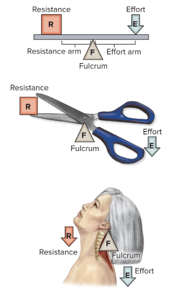
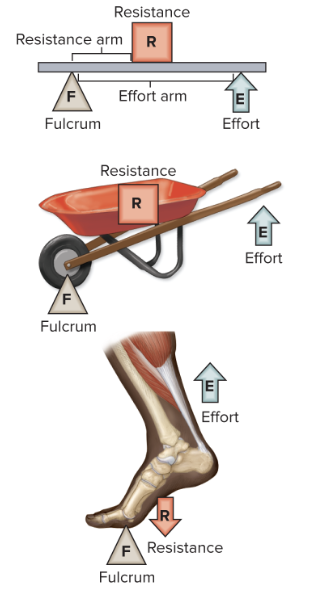
second class levers
resistance between fulcrum and effort
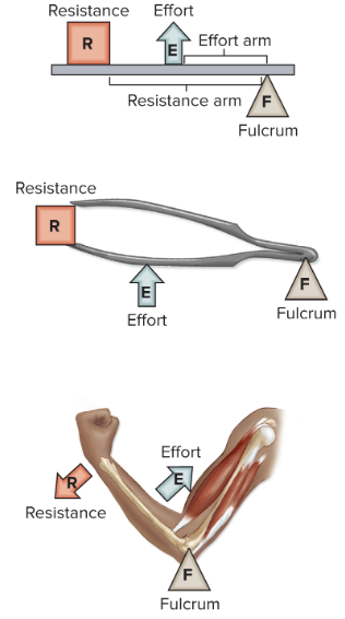
third class levers
effort applied between resistance and fulcrum
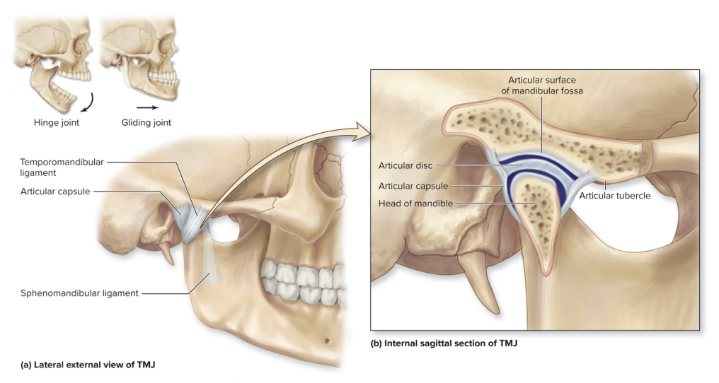
temporomandibular joint
articulation between head of mandible and temporal bone
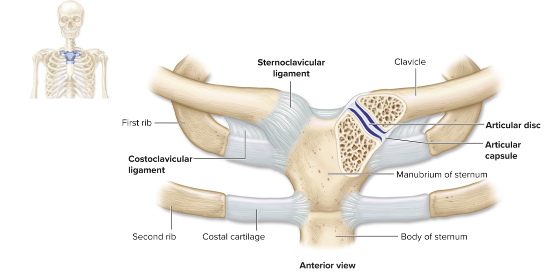
sternoclavicular
only joint where axial meets appendicular
*stabalize movements of entire shoulder
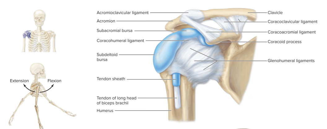
acromioclavicular joint
works with glenohumeral joint to give the upper limb full range of movement
knee joints
largest and most complex diathrosis (synovial)
primarily a hinge joint
tibiofemoral joint
patellofemoral joint
where is tibiofemoral joint
between condyles of femur and condyles of tibia
where is patellofemoral joint
between patella and patellar surface of femur
hierarchy of skeletal muscle
entire muscle → connective tissue epimysium
bundles of fasicles → epimysium
bundles of muscle fibers → perimysium
muscle cells → sarcolemma
myofibrils → sarcoplasm and sarcoplasmic reticulum
bundles of myofilaments → sarcoplasmic reticulum
protien filaments → sarcoplasm
what kind of channles are in the sarcolemma and t-tubules
voltage-gated Na+ channel
voltage-gated K+ channel
voltage sensitve Ca2+ channel
what are the regulatory proteins of the thin filament
tropomyosin
troponin
myoglobin of skeletal muscle
within cells allows for storage of oxygen used for aerobic ATP production (aerobic respiration → long term)
stored energy of skeletal muscle
glycogen
creatine phosphate
glycogen is used for what
when fuel is needed quickly (glycolysis → short term)
creatine phosphate is used for what
quickly give up its phosphate group to replenish ATP supply (immediate)
main functional unit of skeletal muscle
sarcomere
overview of events in skeletal muscle contraction
neurotransmitter junction: excitation of skeletal muscle fiber
sarcolemma, t-tubules, and sarcoplasmic reticulum: excitation-contraction coupling
sarcomere: crossbridge cycling
what do muscle fibers exhibit
RMP
*RMP of muscle cell is -90mV
RMP established by leak channels and Na+/K+ pumps (3 out/2 in)
what is sarcolemma
plasma membrane of a skeletal muscle fiber
what are t-tublues
deep invaginations of the sarcolemma
network of membranous tubules to the sarcoplasmic reticulum
where are the (2) voltage-gated channels (Na+and K+)
within membrane of sarcolemme (along its length)
t-tubules
thick filaments anatomy and physiology
assembled bundles of myosin protein molecules, each myosin has 2 strands: globular head and elongated tail
head contains binding site for actin of thin filaments and catalytic ATPase where ATP splits into ADP + P
2 tails of myosin intertwined
thin filaments anatomy and physiology
composed of 2 strands of actin protein twisted around each other
g-actin spherical molecules → myosin binding site
f-actin fibrous strand
tropomyosin anatomy and physiology
twisted filament protein
cover small regions of actin strands → myosin bidning sites