muscle and tendon
1/30
Earn XP
Description and Tags
Name | Mastery | Learn | Test | Matching | Spaced | Call with Kai |
|---|
No analytics yet
Send a link to your students to track their progress
31 Terms
aperneurosis
flat sheet of dense connective tissue connecting muscle to bones/ fascia
epimysium
dense irregular connective tissue surrounding each muscle protecting it from friction
fasicle
a bundle of muscle fibers
perimysium
connective tissue surrounding a bundle of muscle fibres
endomysium
surrounds individual muscle fibres
muscle belly
fleshy, thickest part of the muscle, is encased in the epimysium
skeletal muscle structure
multiple peripheral nuclei
voluntary
striated
regular parallel bundles
outermost layer surrounded by epimysium
cardiac muscle structure
striated
single central nucleus
involuntary
irregular arrangement
intercalated disks
intercalated disks have extensive gap junctions allowing cell to cell communication
smooth muscle contraction
not striated
single nucleus
involuntary
longer contractions
overlapping sheets of spindle shaped cells
microscopically appear homogenous
connected through end to end junctions called gap junctions creating a watertight seal
function of skeletal muscle
voluntary movement of skeleton controlled by somatic nervous system
maintain body position and posture
stabilise joints
support underlying organs and soft tissue
store nutrient reserves
maintain correc body temperature
smooth muscle function
involuntary contrction controlled by autonomic nervous system
lines inner wall of vasculature, hollow visceral organs, major bodily tracts
regulate blood pressure by altering systemic vascular resistance
peristalsis
regulate bodily secretion
lines respiratory tract
iris controls light entering
hair follicles
what is muscle architecture
the arrangement of muscle fibres relative to the axis of force
maximum force developed by muscle is proportional to the number of sarcoeres hence fibre length
pennate muscle
short fibres at an angle to internal tendon/aperneurosis
increases PCSA
PSCA is directly proportional to force
short fibres mean less contraction distance so it is economical
however trade off with speed
parallel muscles
fibres run parallel to line of pull of muscle
more sarcomeres in series mean more total muscle fibre shortening so more work
work= force x distance
moves joints through a large range of motion
speed= distance/time
roles of tendon
minimise distal limb mass
join muscle to bone
store elastic energy
conserve energy
power amplification: stretched tendons recoil faster than muscle shortens so more power. only a small amount of work is done but in a shorter time so power output is higher
power= rate of doing work
tendon structure
tenoblasts and tenocytes
chondrocytes, synovial cells and vascular cells
tendon collagen fibres are in a crimped pattern
collagen fibrils- collagen fibres- fascicles (surrounded by endotenon)
fascicles are bound together by the endotenon a dense irregular connective tissue sheath to form the tendon
type i
slow oxidative
low myosin ATPase activity
high oxidative capacity
smaller diameter
fatigue resistant
less force production
steady fatigue curve
type iia
fast oxidative glycolytic
high myosin ATPase activity
high oxidative AND glycolytic capacity
type iib
fast glycolytic
high myosin ATPase activity
high glycolytic capacity
larger diameter (stronger) fatigue easily
what can fibre types be influenced by
genetics
training
age
lifestyle
diet
isometric contraction
muscle contracts but does not change length
produces force but it is equal to resistance
eg holding something
concentric contraction
shortens as it generates force
force greater than resistance
movement
eccentric contraction
muscle lengthens under tension
lowering
high force and low energy
eg control or resist flexion of the elbow caused by the ground reaction force during landing impact
lactertus fibrosis
horse
Biceps stretches during stance when carpus locked in extension
– LF stores ELASTIC ENERGY
– When carpus buckles in late stance this rapidly releases the stored energy
– The leg swings forward
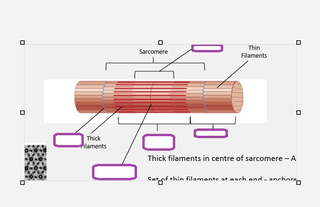
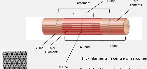
myosin
thick filaments
polypeptide chains
2 globular heads and a long tail
heads are the site of myosin ATP enzyme
thin filaments
2 intertwned chains of actin molecules plus
tropononin- small globular protein bound to actin and tropomyosin
tropomyosin- rod shaped, located end to end along thin filament
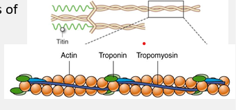
sliding filament theory
tropononin controls position of tropomyosin on the thin filament. myosin cant bind with tropomyosin on its binding site
acetylcholine diffuses from neuron and ca ions are released into sarcoplasm and bind to troponin
calcium acts on tropomyosin and the myosin binding site is exposed
myosin head binds to actin at newly exposed site. pi and adp are released
thin filament moves in the direction of its negative end because the myosin head is firmly attached to the thin filament during its power stroke
the two heads of each myosin molecule work independently. only one head attaches to actin at a given time
myosin head and atp bind. myosin head detaches from the thin filament
atp is hhydrilysed
events during a muscle contraction
resting state- troponin controls the position of tropomyosin on the thin filament- here tropomyosin blocks the myosin binding site on the actin molecules
excitation contraction coupling- calcium ions bind to troponin which changes shape. this moves tropomyosin on the thin filament away from the myosin binding site
myosin heeads bind to actin on the thin filament. causes detachment of adp and phosphate molecules
power stroke: myosin heads move performin a power stroke which drags the thin filament towards the centre of the sarcomere
detachment- ATP binds to myosin causing it to lose affinity for actin and to detach from the actin binding site
atp is hydrolysed into adp and phosphate which reenergises myosin head to return to prevous poition
no calcim- resting state
if calcium is oresent then return to stage 3
what are the 3 functions of ATP in skeletal muscle contraction
energy released from atp hydrolysis re enrges the myosin head providing enery for cross bride movement and force generation
binding of atp to myosin causes the release of the myosin head from actin allowing repeated contractions
in the sacoplasmic reticulum ca-atpase hydrolyses atp in order to take ca ions back to the sr to end a muscle contraction