Cellular Signal Trasnduction
 Call Kai
 Learn
 Practice Test
 Spaced Repetition
 Match
 Flashcards
 Knowt Play
1/136

Earn XP
Description and Tags
For Professor Leibowitz's CST/Advanced Cell Biology course
Name | Mastery | Learn | Test | Matching | Spaced | Call with Kai | Chat |
|---|
No analytics yet
Send a link to your students to track their progress
137 Terms
Types of Cellular Signaling
Pheromones: Between organisms
Endocrine: Long distance signaling
Paracrine: Local release/response
Autocrine: Cell talks to itself
Membrane-Attached: Cell A has signal that attaches to Cell B physically
Signal (Hormone) Categories
Small lipophilic molecules
Hydrophilic molecules
Lipophilic molecules
Gas molecules
Processing the message with…
Specificity
Regulation
Flexibility
Kinetics/Dynamics: How fast you respond to signal/amount of signal
Amplification: Increasing signal
General Signal Pathway
Receptor → Adaptor → Second Messenger → Effector
Receptor-Ligand Interactions
Affinity: Strength of interaction
R + L ⇆ RL
Kd: Dissociation constant
Molecular Switches
Conformation
Dimerization: Multimerization
Phosphorylation/Dephosphorylation
Post-Translational Modifications
Recruitment/Subcellular Localization
Stopping/Attenuating a Signal
Degradation of hormone, very slow
Internal mechanisms cause an immediate halt
Points of Potential Drug Intervention
How it works:
Inhibit function by directly blocking target receptor
Receptors can have other functions/downstream effectors
Block entire pathway (signal transduction)
Could work/could not, need empirical evidence
General Drug Research and Development Scheme
Target Identification
Target Validation
Drug ID
Drug Validation
Animal Testing
IND → Phase I-III Clinical Trials → NDA
Target Identification
Don’t need a specific target, just need to know it works
Starting from nothing: Comparative biology between sick and healthy
Starting from something: Drug/modulator
Target Validation Methods
Knockout/Knock-in mutants
Cell-based assay with modulator
Rational Drug Design
Understand molecular details of drugs so it binds properly
Structure Determination: Lots of AI used to synthesize compounds
NMR Spectroscopy
X-Ray Crystallography (very expensive/time consuming)
Modeling
Structure Activity Relationships
Random Drug Screening
Three types:
Assay Development: In well plate
Library: Can have millions of compounds
Combinatorial chemistry
Natural product
Screening
Semi-Rational Drug Discovery
Know as much as you possibly can about the target so you can have a library of potential compounds (combine rational and random approaches
Validation
Secondary Screening: Use animal models
Move proof up a notch
Preclinical Testing
Drug design systems: How fast drug is absorbed, does drug work
Diagnostics
Diagnosis is key
Importance: Arguably 50% of treatment
Types of Diagnostic samples: Biopsies, bodily secretions, scans
Diagnostic Tools: Analyte Measurement, Antibodies, genomic DNA/SNP
GPCR Activation
GTPases linked to receptors
Heterotrimeric G Protein Pathway: Induce guanine nucleotide exchange
Receptor binds ligand → Conformational change in G protein
Gα releases GDP to bind GTP → Gα dissociates from Gy
Gα and Gβy interact with different effectors
GTP is hydrolyzed to GDP + Pi
Gα associates with GDP again and Gβy
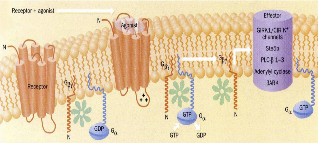
G-Proteins
Gα: GTPase activity, Receptor and Effector Interactions
Gαs: Stimulates adenylyl cyclase
Gβy: Effector interactions
Gβ: Interface that interacts with Gα has key residues for effector interactions
GPCR Structure
Integral membrane protein
Seven transmembrane α-helices
Extracellular loops: Ligand binding site outside, G protein binding site inside
Three Classes of GPCR
Class I: Rhodopsin-like
Class II: Glucagon-like
Class III: Metabotropic Glutamate-like
Molecular Switch: GPCR
Conformational change w/ ligand binding
Ligand-GPCR complex binds to G-protein
Ligand binds to receptor and changes structural conformation
GPCRs Regulate Ion Channels
Direct regulation via G protein
Activates a signal pathway up-regulating a kinase or phosphatase to open/close it

Second Messengers
cAMP: Activate proteins inside the cell (protein kinases, ion channels, other enzymes, transcription)
cAMP
Formation: Adenylyl Cyclase + ATP → cAMP + Pi
Utilization: cAMP Dependent Protein Kinase (PKA)
Degradation: Use phosphodiesterase to hydrolyze it to AMP
GRKs
Serine-threonine kinases recognize and phosphorylate receptors in their agonist-stimulated form
DLEESSSSD: Intracellular sequence exposed when ligand-bound
GRKs are alternatively regulated/expressed in different cells, different conditions
GPCR Regulation: GTP
For G-proteins/GTP: Built in self-timer (shut-off mechanism)
RGS Proteins: Accelerate GTP hydrolysis by binding Gα (higher catalytic rate)
GPCR Regulation: Receptor Phosphorylation
PKA: Doesn’t matter if ligand is still bound, receptor is turned off due to dissociation from G Protein
GRK (G Protein-Coupled Receptor Kinase): Targets ligand directly, desensitization
GPCR Regulation: cAMP
Bind to regulatory subunit in a cooperative fashion, first ligand makes second ligand bind better
Regulatory domain has pseudosubstrate that binds active site, inhibit catalytic domain
cAMP binding causes regulatory and catalytic domains to separate, allow phosphorylation of substrate
GPCR Specificity
Link specific GPCR to specific G-Protein via
Effector specificity
Differential cell expression
Toxins
Coupling Promiscuity
Many GPCRs can couple to more than one G protein subtype
Isoforms of cellular components can promote signal integration by having partially overlapping connectivity
Adrenergic Receptors
Three Types:
Gs-coupled: More camp, stimulate adenylyl cyclase
Gq-coupled: α1
Gi-coupled: α2 for smooth muscular lining blood cells, inhibit Adenylyl Cyclase
Gs vs. Gi
Gs stimulates adenylyl cyclase
Cholera: ADP ribosylates Gαs to lock it in a GTP-bound form → No GTP hydroysis → Overproduce cAMP
Gi inhibits adenylyl cyclase
Pertussis: Stuck in GDP-bound/inactive form → Inhibit adenylyl cyclase → Overproduce cAMP
Crosstalk | GPCR and RTK Pathway
Gα+GTP dissociates and activates Phospholipase C
Phosphoinositol pathway activated by RTKs produce PIP2
Phospholipase C hydrolyzes PIP2 → IP3 + DAG
IP3 associates with voltage-gated Calcium ion channels to release CA2+
DAG + CA2+ → Release Protein Kinase C which can modulate RTK pathway proteins

FULL GPCR PATHWAY
Ligand binds GPCR, triggering a conformational change in the G-protein subunits
Gα loses GDP for GTP binding, dissociates from Gβy
Gαs + GTP bind to adenylyl cyclase, cause conversion of ATP to cAMP
cAMP binds to inactive PKA complex
Two catalytic domains dissociate from two regulatory domains, so PKA is now active
PKA travels to the nucleus and phosphorylates CREB TF, binds to DNA

Receptor Tyrosine Kinases
Signal Transduction receptors that can form homodimers or heterodimers w/ many combinations depending on the cell and varying numbers based on expression
Structure:
Extracellular Ligand Binding Domain
Single Transmembrane Helix
Cytosolic Domain including a protein Tyrosine Kinase
Molecular Switch: RTK
Dimerization: Activates all RTKs
Ligand binding locks dimers in active conformation when they were once inactive
Tyrosine Phosphorylation
Ligand forces kinase domains to interact → Chains phosphorylate each other making open kinase domains for even more phosphorylation
Structure on its own inhibits range of interaction/action, need ligand to alter conformation permanently

RTK Amplification: Adaptor Proteins
Has binding site for phosphotyrosine, binds to exposed phosphate on tyrosine chain
Can branch off and link tyrosine to multiple pathways
Membrane interactions
Enzymatic activity domain
RTK Regulation | Desensitization
Limited number of cell surface receptors, so regardless of the concentration of ligand, prevent prolonged receptor activity
Specificity | Phosphotyrosine Interacting Domains
Recognize phosphotyrosine directly on chains to recruit adaptor proteins
SH2: Src Homology 2 domains
Looks for 1-6 AAs on C-terminal side of chain, if there’s recognition, binding happen
P-tyr Binding Domain: Recognizes phosphotyrosine and 3-5 AAs in N-Terminal
Specificity | Protein-Protein Interactions
SH3: Ser Homology 3 domains
Bind to Proline Rich Domains (PXXP); bind in multiple places for stronger overall interaction
WW domains: PXPX
PD2: Bind to hydrophobic AA at C-Terminus of protein
Specificity | Membrane-Interacting Domains
Stick to surface of membrane, interact with head groups of phospholipids
Pleckstrin Homology (PH) domain: Bind PIP3
FYVE domain: Bind PI-3-P
Docking proteins
RTK Effectors
Ras-GEF: MAP Kinase
PLCy
PI-3 Kinase: Kinase phosphorylates PI
How to Activate Effector Proteins | Membrane Translocation
Protein in cytosol
Ligand binds receptor → Translocation from membrane to cytosol to find substrate
Convert PIP2 → PIP3
How to Activate Effector Proteins | Conformational Change
Two molecular switches
Membrane translocation then conformational change
How to Activate Effector Proteins | Tyrosine Phosphorylation
Inactive Phospholipase C recruited to membrane, kinase of receptor phosphorylates Phospholipase C to activate it
Getting Your RAS in Gear
RAS: Guanine Nucleotide Binding Protein (GTPase)
Nucleotide exchange needs GEF (Guanine Exchange Factor: Interacts with small G protein with only one subunit)
Inactive: GDP + GEF → Active: GTP until GTP hydrolyzes
Hydrolysis needs GAPs: Change conformation of RAS to increase hydrolysis capabilities
RAS lives near membrane, undergoes palmitoylation (palmitoyl group is covalently attached, stuck in membrane)
Identifying RAS as an RTK Downstream Inhibitor w/ VEGF
Remove RAS with anti-RAS Abs
No cell activation because VEGF is a downstream growth factor
Have mutant RAS that is constitutively active
Out of control cell growth, regardless of presence of VEGF
Linking RAS and RTK: Bug Eyes
Sevenless: RTK (named after mutated eye cell in drosophila)
Bride of Sevenloss (BOS): RTK ligand/agonist that causes dimerization after binding
Grb2: Adaptor protein (SH2/SH3 Homolog) that grabs receptor w/ SH2 domain and recruits SOS with SH3 domains in cytosol
Son of Sevenless (SOS): GEF that interacts with and activates RAS via conformational change
Ras+GDP + SOS → RAS+GTP
Amplification | MAP-Kinase Pathway
RAF: A MAP-KKK activated by RAS+GTP, further activated by RAS+GDP
MEK: MAP-KK phosphorylated by RAF
ERK: MAPK phosphorylated by MEK
ELK-1: TF in nucleus is phosphorylated by ERK, leads to cell cycle progression
Other MAPKs: Jun, Myc, Fos, ATF-2
Crosstalk | Phosphoinositol Pathways I
Phospholipase Cγ: Inositol or PI can be further phosphorylated to get PIP, PIP2, or PIP3
PIP2 cleaved by Phospholipase C → DAG + IP3
GPCRs activate Phospholipase Cβ!
Phosphoinositol Pathways II
PI3 Kinase: Activates PKB
AKT/PTEN: pH domain interacts with PIP3; phosphorylation by PDK1,2
PTEN: Phosphatase and Tensin homolog, promotes apoptosis
BAD: Induces apoptosis, inhibited when phosphorylated by PKB
p27: Regulator, inhibitor of inhibitor of cell cycle
RTK Regulation | Attentuation/Termination
Antagonistic Ligands
Heteroligomerization with receptor mutants
Inhibiting the RTK:
Ser/Thr phosphorylation: Turn on receptors
Protein Tyrosine Phosphatase: Remove phosphate, turn receptors off
Receptor Endocytosis/Degradation
RTKs in Pathophysiology | Cancer Treatments
Herceptin: Erbitux mAb - binds to receptor outside of cell
Improved by releasing toxin when degraded and killing cancer cells
Tarceva: Small molecule tyrosine kinase inhibitor
RTK | Role of EGFR in Cancer
Stimulating motility factor of cells (metastasis) and angiogenic factors
Binding of specific ligands, leads to different outcomes
Regulates tumor cell division, repair, survival
Block pathway → Block phenotypes
FULL RTK PATHWAY
RTK: Sevenless
Ligand, Bride of Sevenless (BOS) binds to RTK
Tyrosine Kinase domains phosphorylate one another, bind Grb2 adaptor protein
Grb2 recruits SOS via SH3 domain, which activates RAS
RAS exchanges GDP for GTP, and activates RAF
RAF activates MEK with ATP exchange
MEK activates ERK with ATP exchange
ERK translocates to nucleus and activates TF, ELK-1 to allow cell cycle progression
Other TFs: jun, myc, fos

How to Control Transcription?
Get region of DNA to extend and not be condensed AND recruit DNA Pol
Use transcription factors
Steroid Hormone Receptors
TFs recruited by sequence-specific TFs, and general TFs with RNA Pol II to start transcription
Molecular Switch: Intracellular Receptors
Subcellular localization with AhR
Intracellular/Nuclear Receptors
Ligands: Small lipophilic molecules, thyroid hormones
Receptor Activity:
Ligand binding
DNA binding
Protein-Protein Interactions
Nuclear Receptor Superfamily
Steroid Receptors: Binds palindromic sequences
RxR Heterodimers
Dimeric Orphan Receptors: Binding a direct repeat
Monomeric Orphan Receptors
DNA Binding Domain
Zn² Fingers: Coordinated by cysteine residues
P Box: DNA recognition
D Box: Dimerization
Recognition Helix: Binds major grroove because it needs to bind to specific sequences
C-Terminal Extension: T/A Boxes
Hormone Translocation
Hormone enters cell and binds to complex
If in cytosol, translocates to nucleus
Bind to promoter region of gene in DNA, start transcription to allow genes to be expressed (produce mRNA)
DNA Binding Domain binds DNA at major groove
Receptor Structure

Domain Structure of Nuclear Hormone Receptors
NLS: Nuclear Localization Sequence in between C and D
Absence of ligand: NLS is hidden
Hormone Response Elements (HRE)
Hormone response element is where TF binds
Consensus Motif: 6 bp core element
Steroid Class II: AGAACA
The rest: AGG/TTCA (/ represents G or T)
Dimeric HRE Recognition
Receptors may recognize singular motif but as dimers, there are two consensus motifs that are recognized
Direct repeats, palindromes
Inverted Palindrome: ACTT/GGA or ACAAGA
DNA Binding Sites of Receptors
DR5: Consensus motifs, in between are spacers
DBD: Only binds when it recognizes a core sequence, but it still looks at nucleotides nearby
Dimerization
Monomers: When binding as monomer, binds to DNA elements with 5’ A/T-rich sequence before the consensus sequence
Homodimers: Competitive binding, first one binds making second bind easier
Binding palindromes
Heterodimers
RXR as promiscuous partner (Pal., DR, Inverted Pal.)
Non-Permissive: Ligand binds RAR first activating transcription ALWAYS
Permissive: Ligand binds either PPAR or RXR activating transcription
2nd ligand binding increases activity
Low level transcription at first, then high level for the second
Two signal pathways, crosstalking
Intracellular Nuclear Receptors | Regulation
A/B Domain: Size/length varies between TFs
AF-1: Autonomous transcriptional activation factor, upregulate/downregulate activity of TFs via phosphorylation with protein kinase
AF-2: Ligand dependent; can only phosphorylate region if ligand is bound
Crosstalk | Intracellular Receptor and RTK
Phosphorylation of Steroid Nuclear Receptors
Use MAPK and AKTin A/B region of AR Basal
Direct phosphorylation of TFs
Phosphorylation of ELK-1 leads to conformational change → Stronger DNA binding → Transcriptional repression of ELK-1
Controlling Chromatin Structure (KNOW FOR EXAM)
Histone Acetylation/Deacetylation
HDAC: Removes acetyl groups from histones to condense DNA (deacetylation)
HAT: Add acetyl groups
TRAP/DRIP Complex: Holoenzyme recruitment
Recruit RNA Pol II and initiate RNA transcription
Recruitment Patterns
Sequential: One after the other is recruited
Combinatorial: Work together at same time
Parallel: Sort of combinatorial but two promoters near each other are bound by RNA Pol II, one recruits HAT, the other TRAP/DRIP
Crosstalk | Intracellular Receptors and GPCRs
GPCR/cAMP: CREB (TF)
Phosphorylation of CREB by Protein Kinase A due to increase in cAMP → Binds DNA with DBD
Binding of CREB to CRE → GPCRs can directly control transcription
Coactivator CBP/p300
Intracellular Receptors | Estrogen Receptor
In cytosol, released from HSPs, goes into nucleus; recruits co-activates to open DNA, recruit RNA Pol.
Estrogen..
Affects heart, brain, bone, breast, uterus, and liver due to estrogen receptors
Produces proteins to decrease LDL (low density lipids) to protect good cholesterol
Stimulates uterine wall/breast cells to proliferate → Increase cancer risk
Antiestrogens
Block estrogen’s growth stiulation → Bind ER, no coactivator interaction, no gene activation
SERMs
Selective Estrogen Receptor Modulator
Stops breast cell proliferation, not uteruine cell proliferation
Tamoxifen
Blocks binding at receptor
First: Surgery
Second: Check if ER+, use Tamoxifen
Even without recurrence, risk of uterine cancer increases
Need better SERMs
Raloxifene
Prevents osteoporosis; at menopause, lose estrogen → Decrease bone strength, blood clots
Less breast/uterine cancers compared to tamoxifen
Progesterone
Birth control
HRT
Abortion
HDAC Inhibitors
Zolinza: HDAC inhibitor for cutaneous T-Cell lymphoma
Allow continued expression of proteins that decrease cell growth
FULL INTRACELLULAR (ESTROGEN) RECEPTOR PATHWAY

Receptor Ser/Thre Kinases | Ligands
TGF-β: Dimer found by ligands
BMP: Bone morphogenetic protein
GDF: Growth and differentiation factor
Proteolytic cleavage → Activation
All homodimers
Receptor Ser/Thre Kinases | Regulation of TGF-β
TGF-β Bioavailability
Pre-pro-TGF-β: Made signal protein and have it secreted via secretory pathway
Synthesize part of protein, ribosome binds to ER to continue synthesis, then go to Golgi Apparatus and release a secretory vesicle
Signal peptide cleaved in ER, (that’s the “Pre”): Left with pro-TGF-β
TGF-β binds to surface of receiving cell, controlling when and how signal is made
Receptor Ser/Thre Kinases | Type I Receptor
Intrinsic Ser/Thr kinase, GS-domain, single transmembrane helix
Self inhibitory w/ GS domain
Receptor Ser/Thre Kinases | Type II Receptor
Same features as Type I but..
Phosphorylation/activation of Type I receptor: Ser in GS domain, removing inhibitory effect
Brought together by ligand
Receptor Ser/Thre Kinases | Type III Receptor
Regulatory proteoglycan for receptor activity
Receptor Activation
Ligand binding increases receptor hetero-tetramerization
Type II phosphorylates Type I → Activation (completely inactive unless interacting)
Receptor Ser/Thr Kinases | Flexibility
How does heterotetramerization increase flexibility?
Conformation can vary, lead to multiple downstream pathways being affected
Receptor Ser/Thr Kinases | Amplification
Direct link to nucleus: TF that is phosphorylated via signaling cascade, goes to nucleus
Receptor Ser/Thr Kinases | How to Find a Protein Downstream of the Receptor?
Type I phosphorylates something
Take a cell with TGF-β receptor and add to a well, solubilize, stain with antibody serine, add lots fo protein + 1 phosphorylated
Use gel to find protein
SMADs
TFs, three types to know:
R-SMADs: Receptor regulated, phosphorylated
Co-SMADs: Work with phosphorylated R-SMADs
Antagonistic SMADs: Regulate R-SMADs with non-productive interactions
Crosstalk: R-SMADs and MAPK (Receptor Ser/Thr Kinases and RTK)
MH1 + MH2 Domains
MH1: Recognizes DNA sequence CAGAC
MH2: Transcritpional activation via regulation of interactions with..
Co-SMADs (work as trimer)
Other TFs
Co-activators
Crosstalk with MAPK pathway to recruit new co-repressors (ERK-1 blocks SMAD complexes)
Receptor Ser/Thr Kinases | Specificity
R-SMADs: Differential interactions with receptors
Receptor Ser/Thr Kinases | Regulation
SARA: SMAD anchor for receptor activation
SARA presents R-SMAD to receptor → R-SMAD can only interact with Co-SMAD after dropping SARA, becoming a trimer → Enter nucleus
NES keeps Co-SMAD out of the nucleus
R-SMAD has a hidden NLS, revealed by SARA
SMAD4 links different downstream effectors and expressions
TGF-β and Cancer
Acts as a tumor suppressor
Mutations to pathway → Cancer (mainly pancreatic and colon)
TGF-β Signaling
Epithelial cells:
Leads to adhesion, death, cell cycle arrest
Cancer-inhibiting
Endothelial cells:
Motility, morphogenesis, growth
More migration and invasiveness
Cancer-promoting
Crosstalk | Receptor Ser/Thr Kinase and Intracellular Receptor
SMAD complex (2 R-SMADs + Co-SMAD) associate with CBP/p300 intracellular nuclear co-activators
R-SMADs: SMAD2/3
Increase transcription
FULL RECEPTOR SER/THR KINASE PATHWAY
