All labs 1-6 ap2
1/534
There's no tags or description
Looks like no tags are added yet.
Name | Mastery | Learn | Test | Matching | Spaced | Call with Kai |
|---|
No analytics yet
Send a link to your students to track their progress
535 Terms

What is A
lateral canthus

what is B
eye lashes
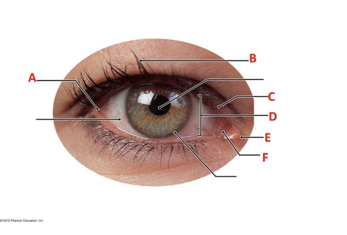
what is C
palpebrae

What is D
palpebrae fissure

What is E
medial canthus

What is F
lacrimal caruncle
What is the function of the levator palpebrae superioris muscle?
opens the eye
What actions are performed by the inferior oblique eye muscle?
eye rolls, looks superiorly and laterally
What actions are performed by the superior oblique eye muscle?
eye rolls, looks inferiorly and laterally
Which two bactericidal proteins are produced by tears?
lysozyme and IgA
What action is performed when the palpebrae are opened then closed in succession?
blinking
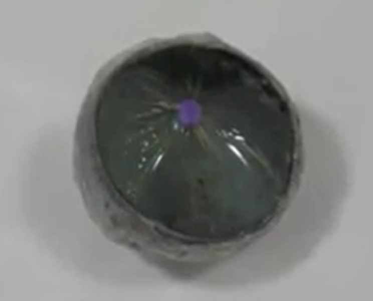
What eye structure is a non-vascularized, transparent, fibrous covering of the middle eye's outer surface which protects the eye and acts as an initial lens for focusing light into the eye?
cornea
What eye structure is the central opening that allows differing amounts of light into the eye depending upon its size?
pupil
Which muscles control enlarging pupil size in response to lower light levels?
pupillary dilator muscles
Which muscles control decreasing pupil size in response to brighter light levels?
pupillary constrictor muscles
Which pupillary reflex is assessed when light is shined into the eye to see how the pupil responds?
direct response
Which pupillary reflex assesses how the eye not being tested responds to differing amounts of light shined into the eye being tested?
consensual response
True or False? A normal pupillary reflex direct response to light shined into the eye is pupil dilation and pupil constriction occurs when light is stopped from shining into the eye.
false
What structures comprise the fibrous layer of the eye?
sclera and cornea
Which eye layer contains the iris, choroid, and ciliary body?
vascular layer
Which eye layer is the most deep?
retinal layer
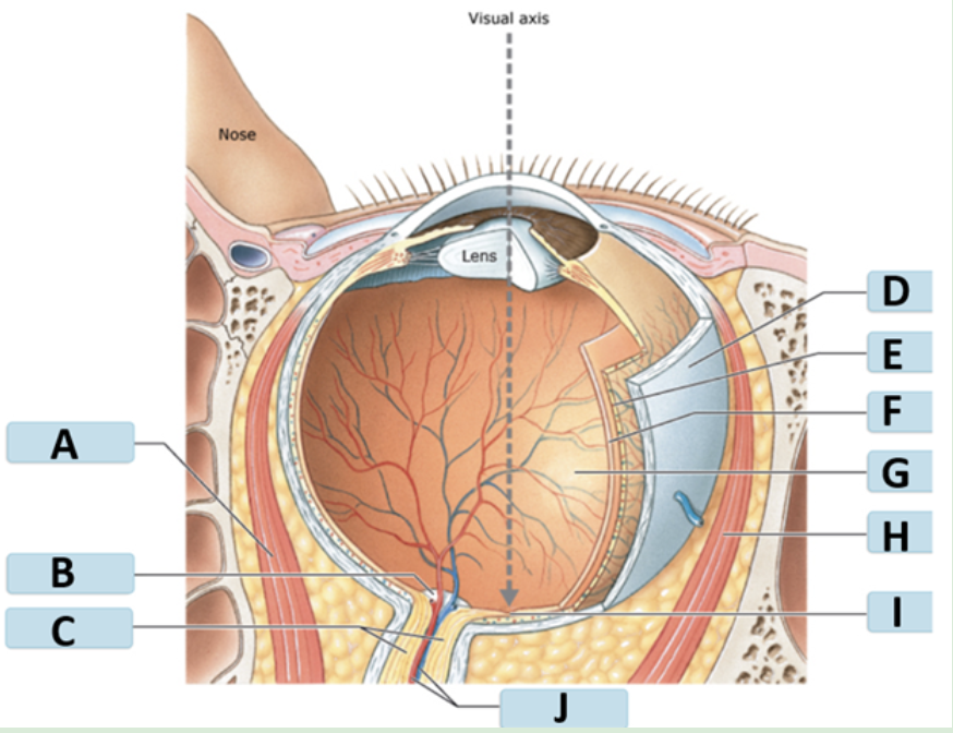
what is A
medial rectus

what is B
optic disc

what is C
optic nerve

what is D
sclera

what is E
choroid

what is F
retina

what is G
posterior cavity

what is H
lateral rectus

what is I
fovea centralis

what is J
central artery vien

what is A
amacrine cell

what is B
horizontal cell

what is C
cone

what is D
rod

what is E
pigmented layer of the retina
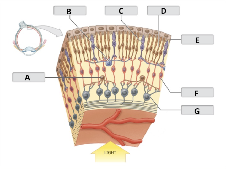
what is F
bipolar cell

what is G
ganglion cell
What area within the macula contains the highest concentration of cones?
fovea centralis
Name the area where the optic nerve originates.
optic disc
In addition to vitreous humor, what is vitreous body composed of?
collagen fibers and proteoglycans
Where would you find vitreous body?
posterior cavity of the eye
Where would you find aqueous humor?
anterior cavity of the eye
How do you check for pupillary accommodation?
put an object (penlight or finger) in front of the patient and slowly move it toward the patients face
Describe how an abnormal direct pupillary response appears when the light is shined into the eye.
absence or lesser constriction of pupil
Describe how to detect the consensual response of the pupils.
shine light in one eye and then see if the other eye responds the same way
Describe how to detect the direct response of the pupil.
shine light into one eye to see how that pupil responds
Name the photoreceptor cells.
rods and cones
Name three types of cones.
blue, green, and red
Light information is transmitted from photoreceptors to ____________.
bipolar cells
Axons of which cells make up the optic nerve?
ganglion cells
Which photoreceptors are mainly used in low light conditions?
rods
ID highlighted region
optic nerve
ID highlighted region
sclera
ID highlighted region
cornea
ID highlighted region
iris
ID highlighted region
pupil
ID highlighted region
ciliary body
ID highlighted region
lens
ID highlighted region
vitreous body
ID highlighted region
retina
ID highlighted region
optic disk
Thyroid Stimulating Hormone
TSH
Luteinizing Hormone
LH
anti-diuretic hormone
ADH
thyroxine
T4
melanocyte stimulating hormone
MSH
Triiodothyronine
T3
Oxytocin
OXT
Calcitonin
CT
Parathyroid hormone
PTH
Follicle-stimulating hormone
FSH
Growth hormone
GH
Adrenocorticotropic hormone
ACTH
erythropoietin
EPO
prolactin
PRL
atrial natriuretic peptide
ANP
epinephrine
E
brain natriuretic peptide
BNP
norepinephrine
NE
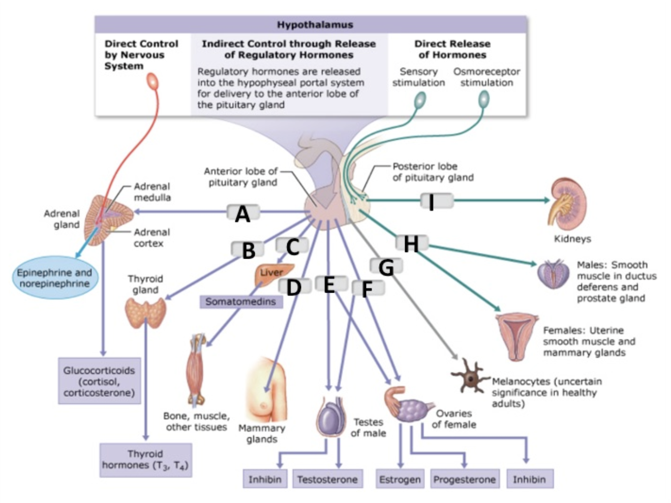
ACTH
A

LH
F

FSH
E

TSH
B

ADH
I

OXT
H

GH
C

inhibin (male)
E

somatomedins
B

inhibin (femal)
G

glucocorticoids (cortisol, cortisone)
C

estrogen
F

testosterone
H
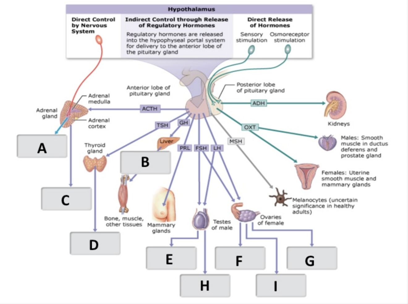
E, NE
A

T3, T4
D
The hormone ADH is produced by the ________ and is released from the ________.
hypothalamus; posterior pituitary
TSH is produced by the ____________, and ACTH is produced by the ____________.
anterior pituitary/anterior pituitary
The hormone ADH affects the target organ, the ___________, while the hormone ACTH affects the target organ, the _________.
kidney; adrenal cortex
The hypothalamus regulates the release of anterior pituitary hormones by releasing certain _________ and __________ hormones.
Inhibiting; releasing
When autonomic centers in the hypothalamus are stimulated by the sympathetic division of the ANS, it directly signals the ____________ to produce epinephrine and norepinephrine.
adrenal medulla