Skeletal Anatomy
1/54
There's no tags or description
Looks like no tags are added yet.
Name | Mastery | Learn | Test | Matching | Spaced | Call with Kai |
|---|
No analytics yet
Send a link to your students to track their progress
55 Terms
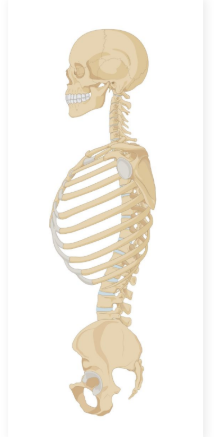
What is the skeleton
Skeleton: A structural framework providing support and integrity to the bodies of some animals
Can be made of different materials based on the type of skeleton
Exoskeleton
Exoskeleton: An external skeleton
Utilized by many invertebrate animals and all Arthropods
Composed of two protein layers
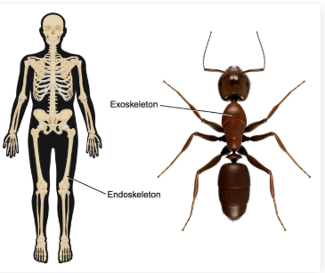
Endoskeleton
An internal skeleton
Utilized by vertebrate animals
Composed of bone and cartilage

Endoskeleton parts
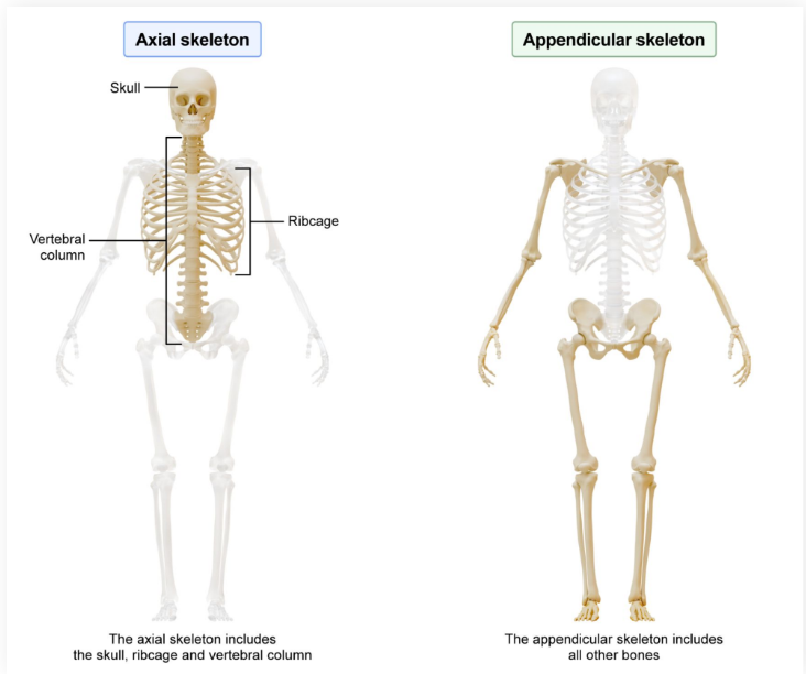
The endoskeleton can be broken up into the axial and appendicular skeletons

Axial skeleton
The “central bones” of the endoskeleton
Includes the skull, vertebral column, and rib cage

Appendicular Skeleton
The bones of the appendages
Long Bones
Long bones: Cylindrical bones of the appendicular skeleton
Longer than wide
Important roles include support, movement, and hematopoiesis

Hematopoiesis
Blood cell production

Epiphysis
The end of a long bone that forms joints with other bones
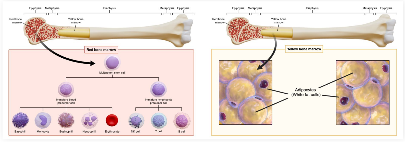
Contains red bone marrow

Diaphysis
Long, hollow shaft in the center of the bone
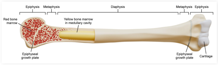
Medullary Cavity
Located within the diaphysis
Contains red and yellow bone marrow

Red Bone Marrow
Produces stem cells capable of generating red and white blood cells

Yellow Bone Marrow
Produces stem cells capable of generating fat, bone, cartilage, and muscle. Fat can be stored in addition to its production
Metaphysis
Found between the medullary cavity and epiphyseal plate
● Similar to the epiphysis
Epiphyseal Plate
Epiphyseal plate:
Capable of lengthening the diaphysis through ossification
What are long bones made of?
Pockets of cancellous bone
google def: “honeycombed tissue found at ends of long bones”
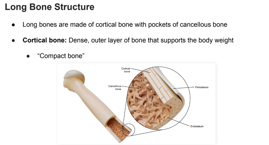
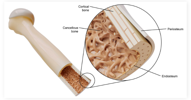
Cortical bone
Dense, outer layer of bone that supports the body weight
A.K.A: “Compact bone”
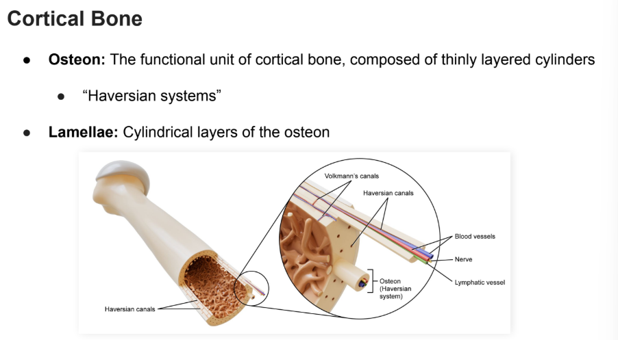
Osteon
The functional unit of cortical bone, composed of thinly layered cylinders
“Haversian systems”
Google def: “the fundamental functional and structural unit of compact bone. It acts as a microscopic transport and support network, giving dense bone its incredible strength and ability to heal.”
Lamellae
Cylindrical layers of the osteon

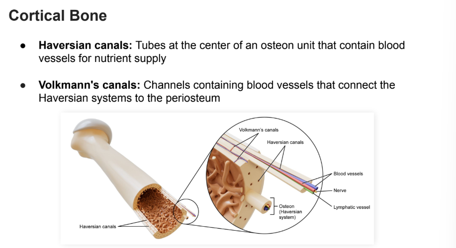
Haversian Canals
Tubes at the center of an osteon unit that contain blood vessels for nutrient supply

Volkmann’s Canals
Channels containing blood vessels that connect the Haversian systems to the periosteum.

Periosteum
An external sheath surrounding the cortical bone that provides nutrients in order to promote growth and healing
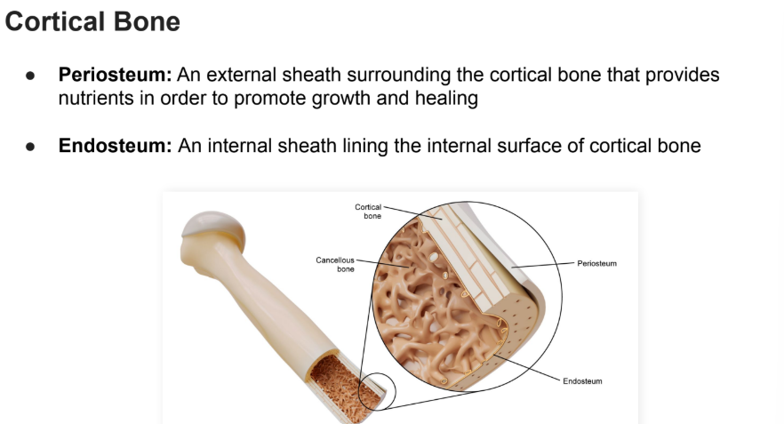
Endosteum
An internal sheath lining the internal surface of cortical bone

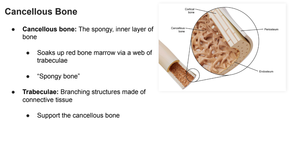
Cancellous Bone
The spongy, inner layer of bone
Soaks up red bone marrow via a web of trabeculae
“Spongy bone”

Trabeculae
Branching structures made of connective tissue
Support the cancellous bone
Bone Remodeling
● Bone remodelling: The process of alternating between ossification (bone formation) and resorption (bone loss)
● Old bone can be replaced with new bone
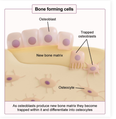
Osteoblasts
Bone building cells
Secrete proteins and utilize calcium from the blood
Mature into osteocytes

Osteocytes
Mature osteoblasts that have been trapped within the bony matrix
● Live within osteons
● Maintain bone
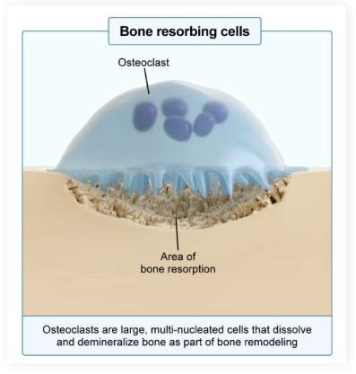
Osteoclasts
Bone Degrading cells
Eat and resorb bone by releasing enzymes and reducing pH (increasing acidity)
Release calcium and phosphate back into the blood
Bone Composition
What is bone made of?
Osteoid
Hydroxyapatite
Osteoid
Osteoid: Bone’s organic component
Contains many proteins, including collagen fibers
Provides tensile strength to bone (pulling or stretching without deforming)
Hydroxyapatite
Bone’s inorganic component
Mineralized material embedded within the osteoid
Contributes to bone density and strength (durability)
Bone remodelling and calcium homeostasis
Bone remodeling is involved in maintaining the homeostatic balance of the molecules used to generate bone
Key molecule is calcium
Increased bone production requires calcium usage, decreasing systemic blood calcium
Increased bone resorption frees up calcium from the bone, increasing systemic blood calcium
Calcium Homeostasis Hormones
Hormones can manipulate the homeostatic balance of calcium
PTH (parathyroid hormone)
Parathyroid hormone (PTH): Increases calcium in the bloodstream
Stimulates osteoclasts, increasing bone resorption and releasing calcium
Increase reabsorption of calcium in the kidney, decreasing calcium excretion in the urine
Calcitonin
Calcitonin: Decreases calcium in the bloodstream
Inhibits osteoclast activity, decreasing bone resorption, allowing calcium to remain in bone
Decreases reabsorption of calcium in the kidney, increasing calcium excretion in the urine
Mnemonic: Calcitonin “tones down” the calcium in the blood
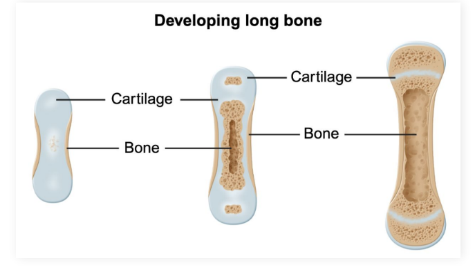
Embryonic Ossification
Embryonic ossification: Bone formation during fetal development
Models of embryonic ossification:
Intramembranous ossification
Endochondral ossification
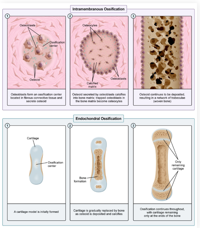
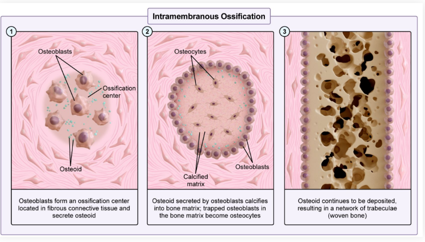
Intramembranous Ossification
● Intramembranous ossification: Direct bone formation within sheets of embryonic connective tissue
Osteoblasts deposit osteoid
Osteoid calcifies and forms cortical bone
Occurs mostly in flat bone
Include bones of the skull, etc.
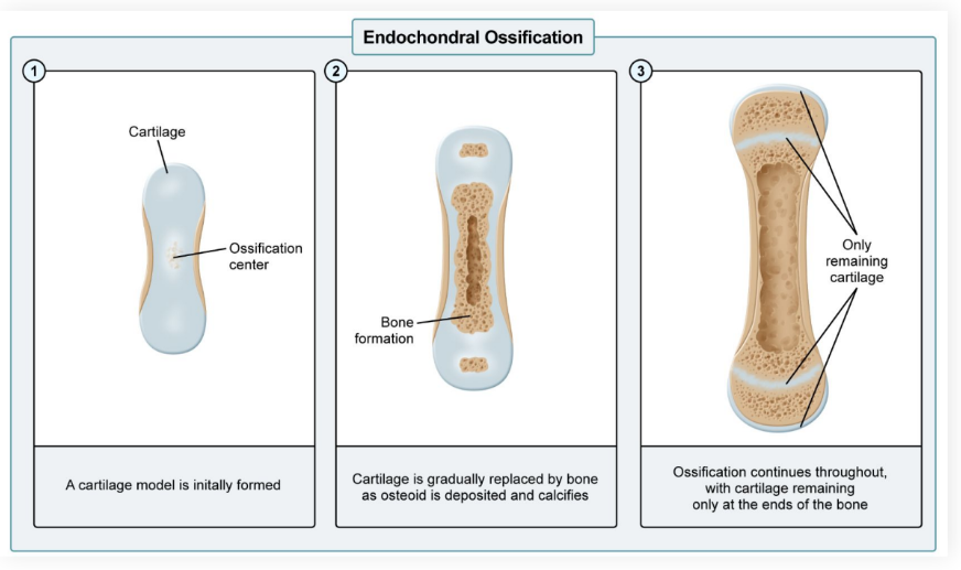
Endochondral Ossification
Endochondral ossification: Indirect bone formation requiring cartilage replacement with bone
An initial cartilage model forms first
Osteoid is deposited, gradually replacing the cartilage
The osteoid calcifies and forms cortical bone
Most bones in the body use this model, including long bones and the ribs
Connective tissue (CT)
A supportive matrix that can be composed of cells, fibers, and a gel-like filler substance
Fibroblasts
Fibroblasts: CT resident cells which secrete the constituents of the fibrous connective tissue, maintaining and remodeling the matrix
Fibrous Connective Tissue
CT that contains a high concentration of fibers
Fibrous Connective Tissue Functions
Fibrous connective tissue offers different function throughout the body

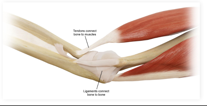
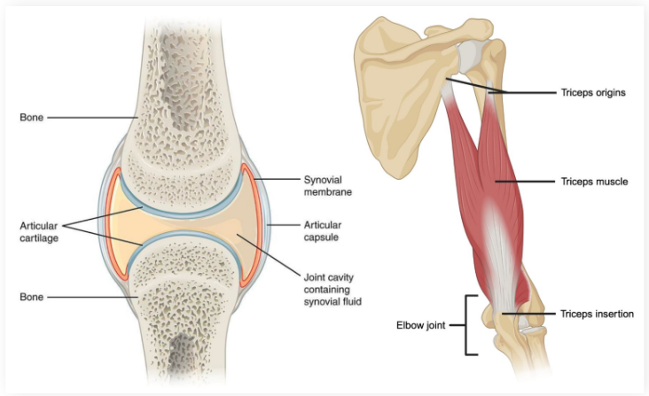
Tendons
Tendons: Attach muscle to bone
Two types (Muscle to Bone)

Ligaments
Ligaments: Attach bones to other bones
(Like to Like)

Periosteum
External sheath surrounding cortical bone
Provides nutrients and innervation to the bone
Endosteum
Internal sheath that exists between the cortical and cancellous bone

Cartilage
Cartilage: Fibrous connective tissue that does not possess blood vasculature or nerve innervation
In comparison, bone is highly vascularized and innervated
Joints
Meeting points between two or more bones
Vascularized, innervated, and contains connective tissue
Muscles can generate movement at joints
Muscles originate on one side of a joint, insert onto the bone(s) on the opposite side of the joint, and contract to generate movement
What is in charge of secreting calcitonin?
Thyroid Gland
When does the epiphyseal plate ossify?
It ossifies at puberty, turning into bone and preventing further growth.
What is the epiphyseal plate made of?
Hyaline cartilage
What does the epiphseal growth plate lengthen?
The diaphysis
Where does hematopoeisis occur in the bone?
The epiphysis
What do osteoclasts get derived from?
Monocytes, with their primary function being to resorb bone.