SAM3.4-3.5: Third eyelid and Nasolacrimal system + Cornea/Sclera
1/103
There's no tags or description
Looks like no tags are added yet.
Name | Mastery | Learn | Test | Matching | Spaced | Call with Kai |
|---|
No analytics yet
Send a link to your students to track their progress
104 Terms
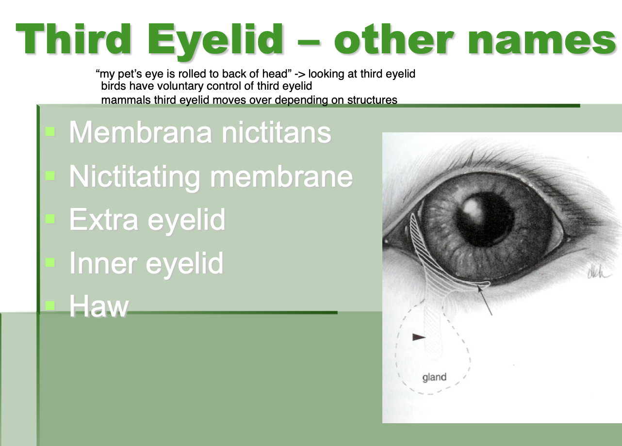
Other names for third eyelid
membrana nictitans.
nictating membrane.
extra eyelid.
inner eyelid.
haw.
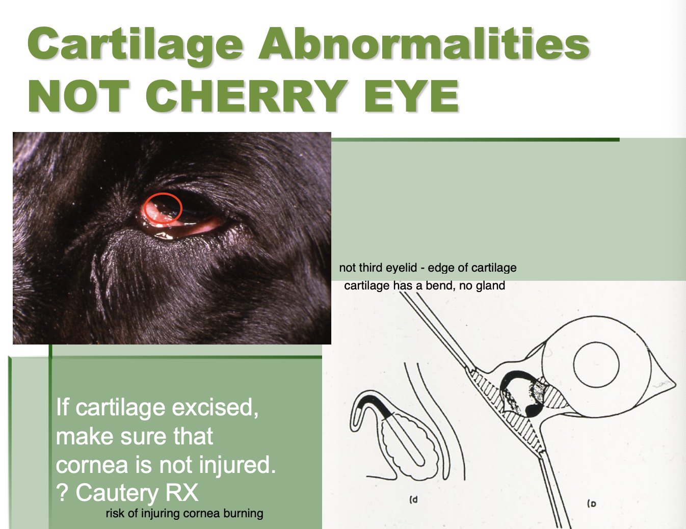
Everted cartilage of the 3rd eyelid (3)
Is not cherry eye.
tend to see in larger breed dogs.
Normal at birth then over the course of a few months the 3rd eyelid will begin to evert.
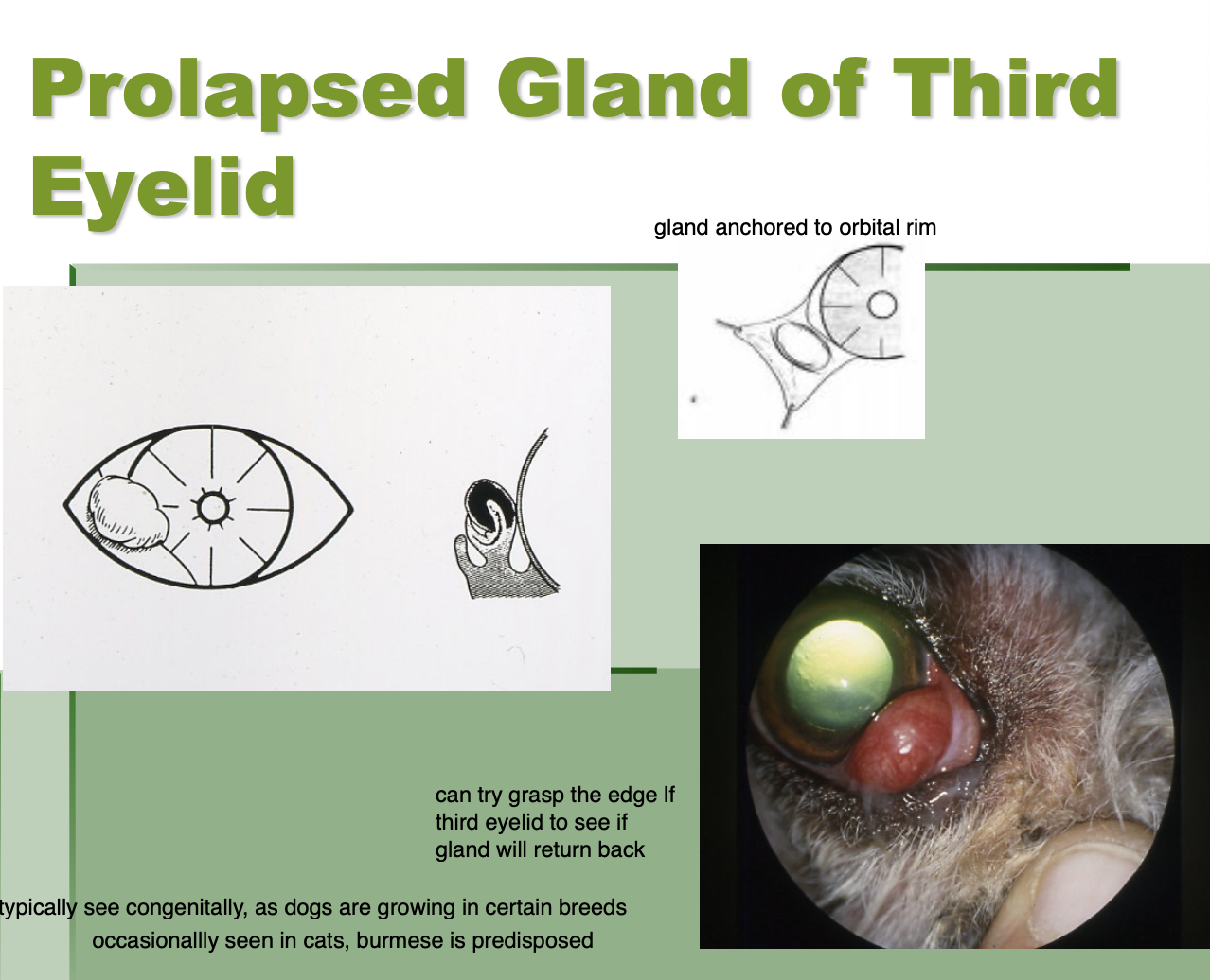
Prolapsed gland of the 3rd eyelid (cherry eye)
Seen in younger animals (certain dog breeds, burmese cats)
Base of the gland prolapses externally.

How do you tell the difference between a cherry eye and an everted 3rd eyelid
grasp the leading edge of the 3rd eyelid.
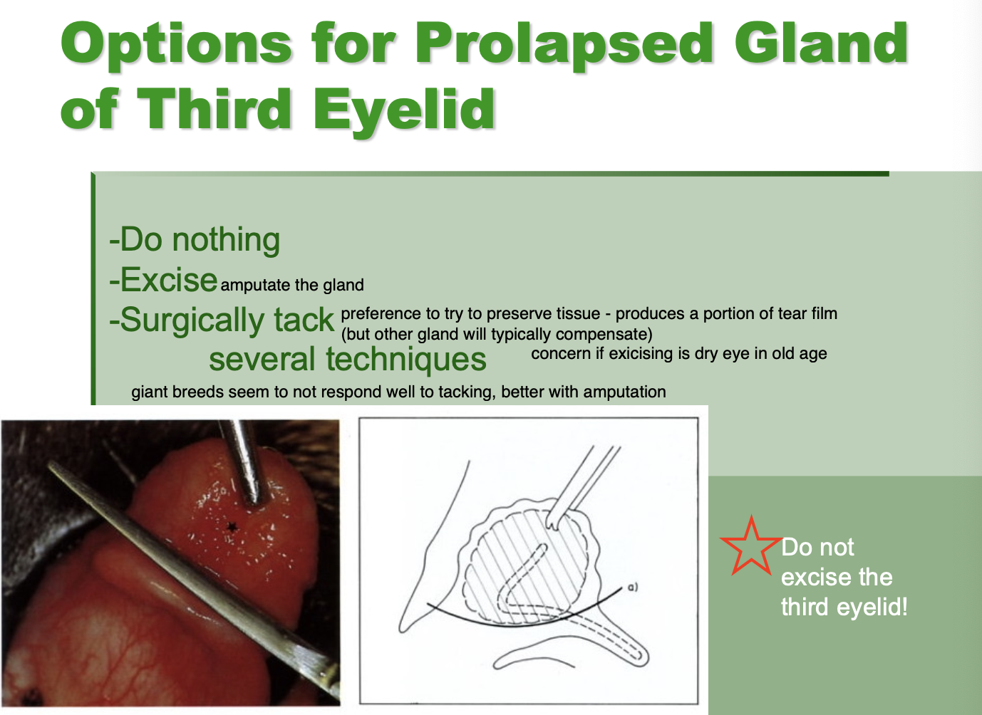
Options for Cherry eye (3)
Do nothing.
Excise gland.
Surgical technique - several techniques

T or F? A tx for prolapsed gland of third eyelid is excision of the third eyelid.
False. Can amputate GLAND but do NOT excise third eyelid.

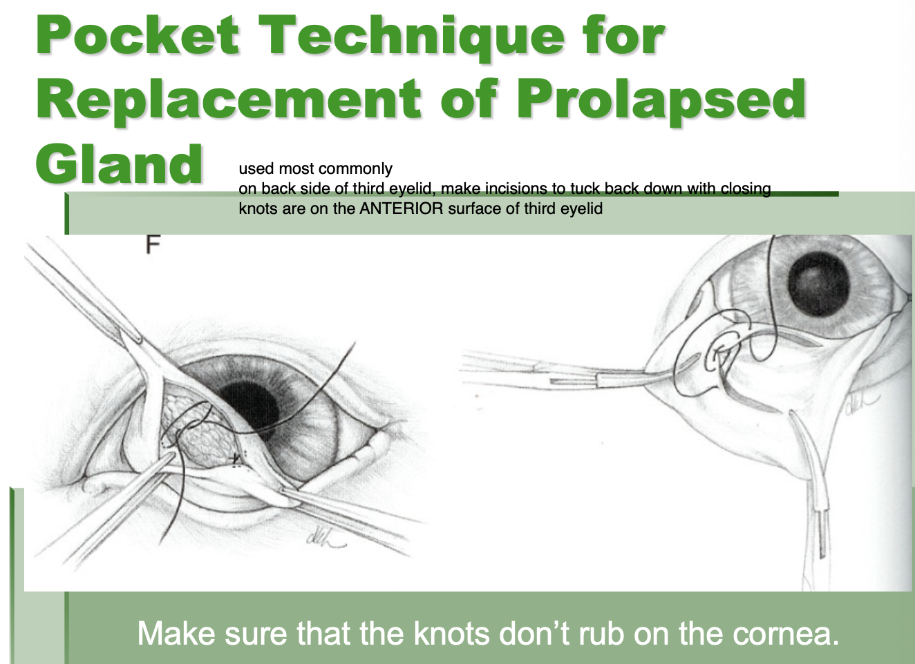
Surgical techniques for tx of prolapsed gland of third eyelid
tack gland of third eyelid to orbital rim
pocket technique for replacement (used most commonly)
Why do we try to preserve the gland of the third eyelid
removal increases risk of dry eye.
Elevation of the 3rd eyelid etiologies (10)
Primary protrusion.
Pain.
Micropthalmos.
Haws syndrome.
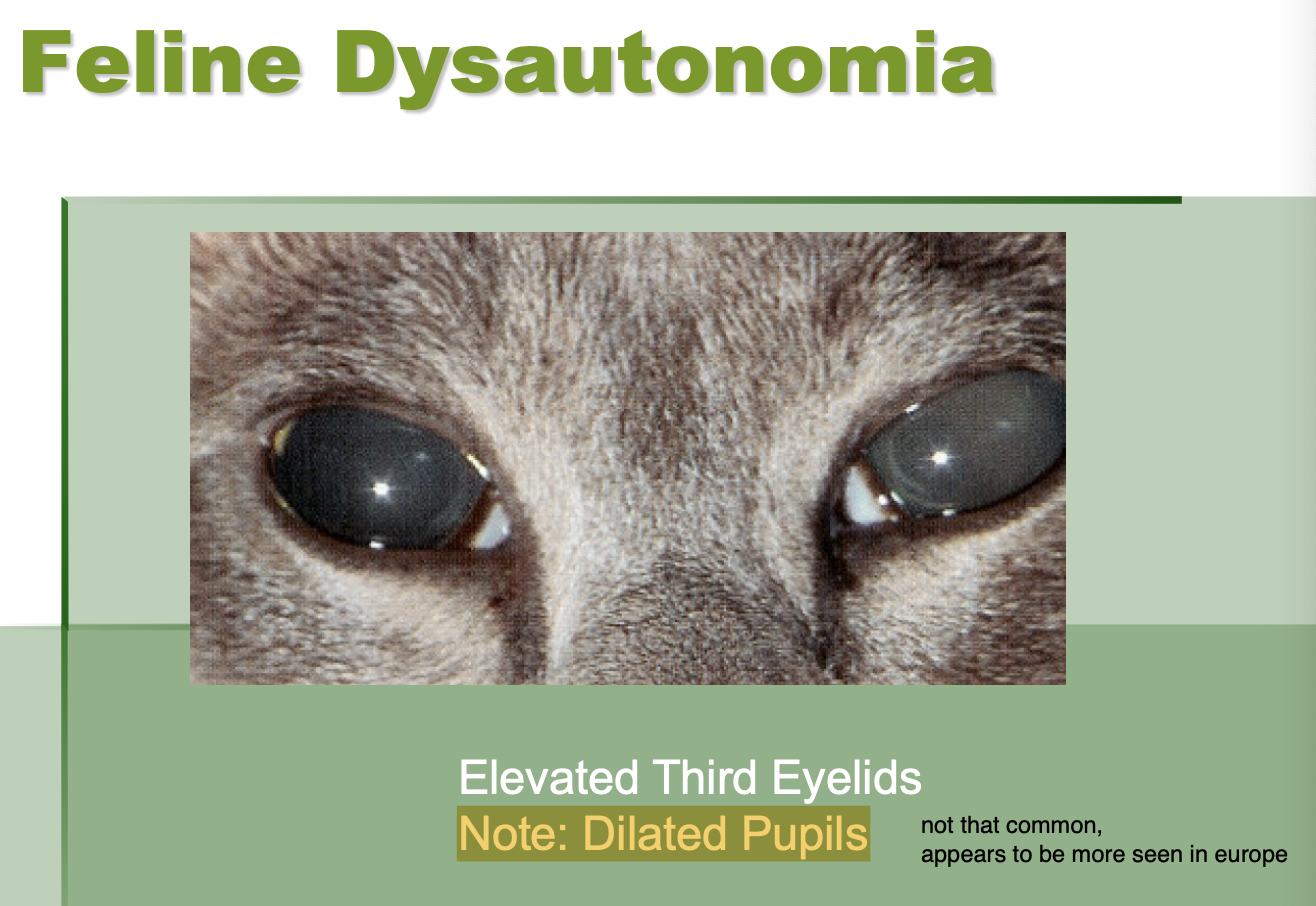
Feline dysautonomia.
Horner's syndrome
Enophthalmos - dehydration.
Cannibus toxicity.
Tetanus.
Rabies.
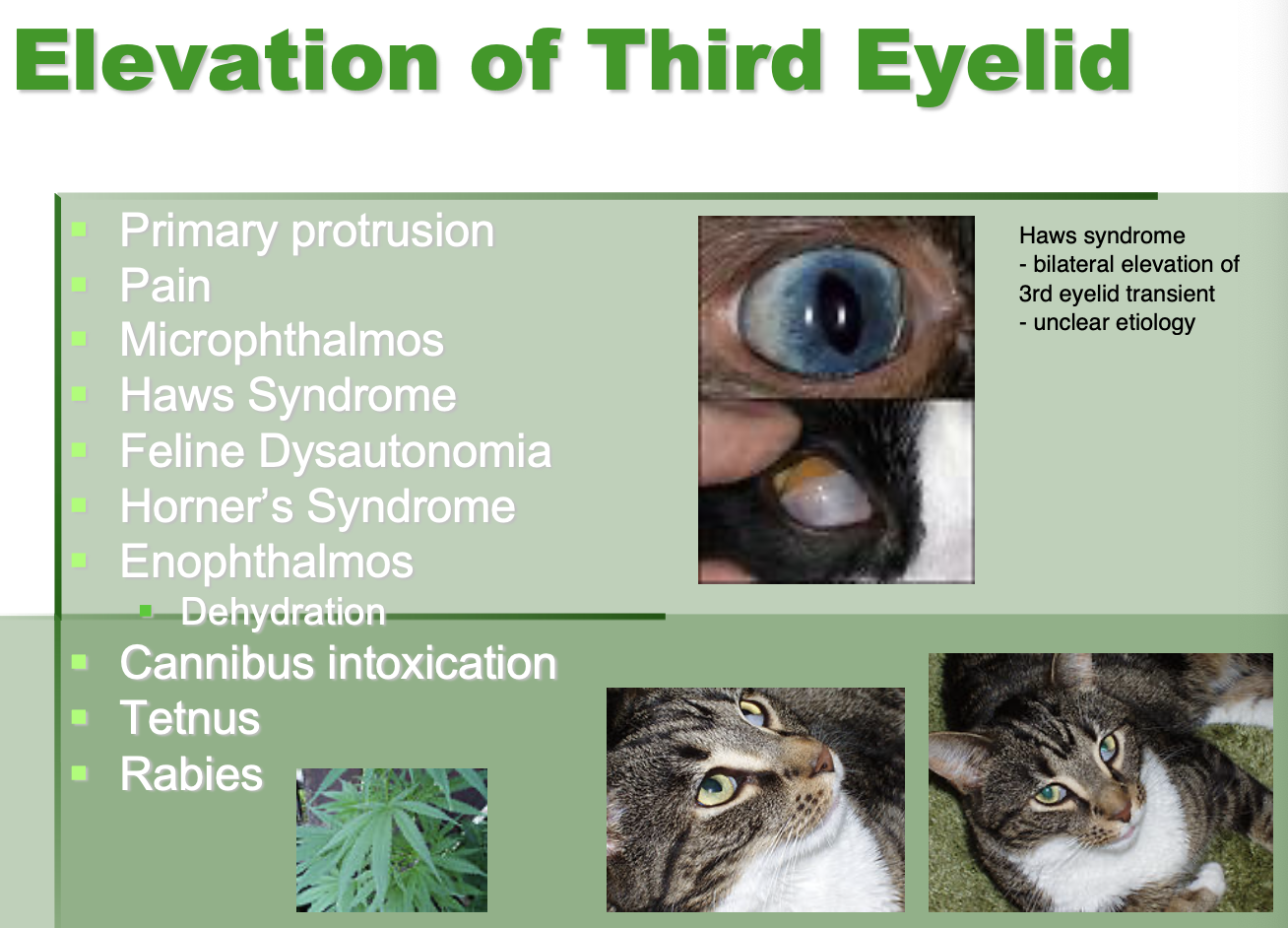
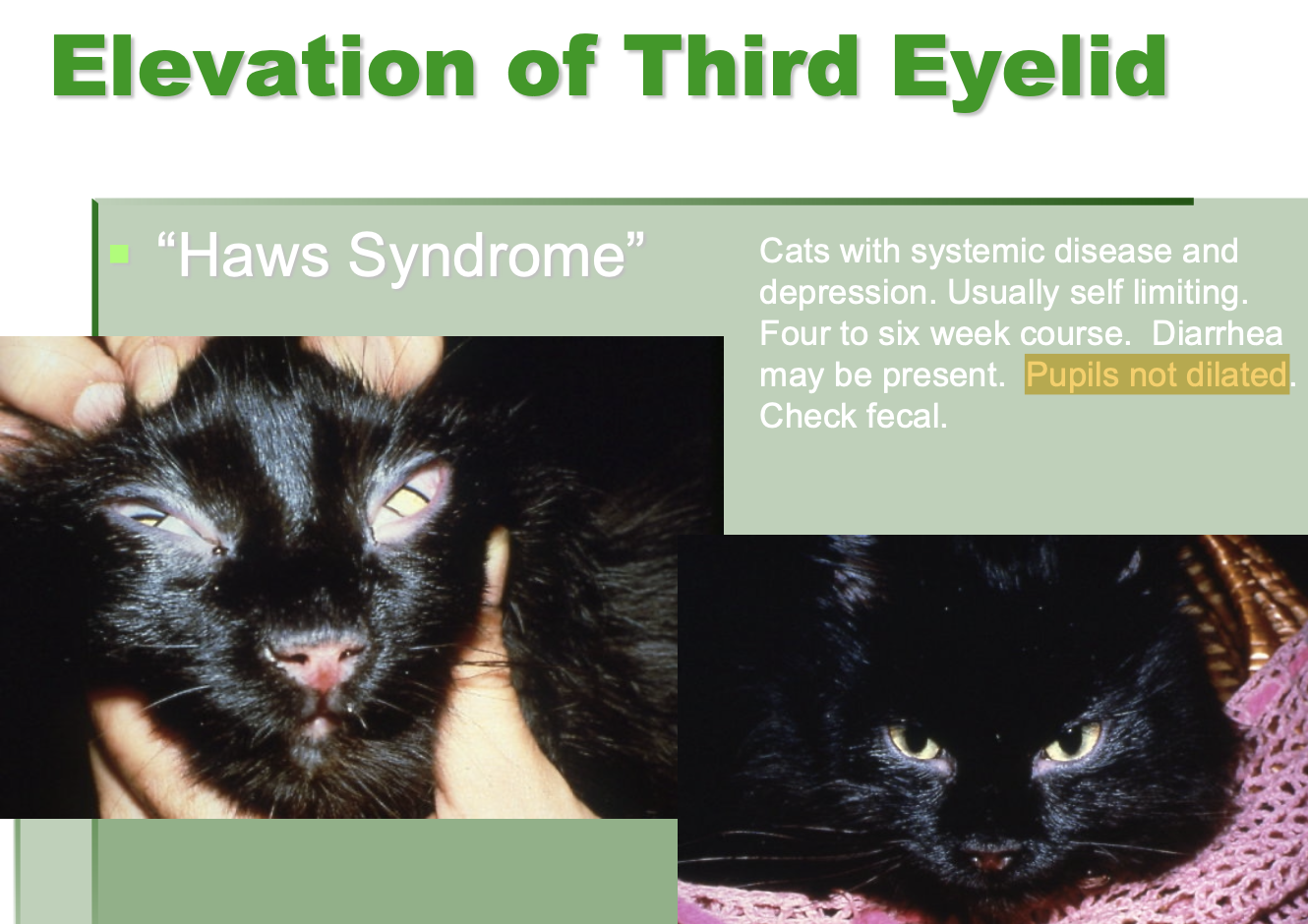
Haws syndrome is when
cats w/ systemic dz and depression have 3rd eyelid elevation.

Haws syndrome course of syndrome (4)
usually self-limiting.
4-6wk course.
D. may be present.
Pupils not dilated.
Feline dysautonomia - eye presentation (2)
elevated 3rd eyelid.
dilated pupils (difference between this and Haws).
Atypical pannus of 3rd eyelid (aka plasmoma or plasmacytic lymphocytic conjunctivitis) appearance (4)
plasma cell infiltrates.
pigment loss.
cobblestone appearance.
thick and hyperemic.
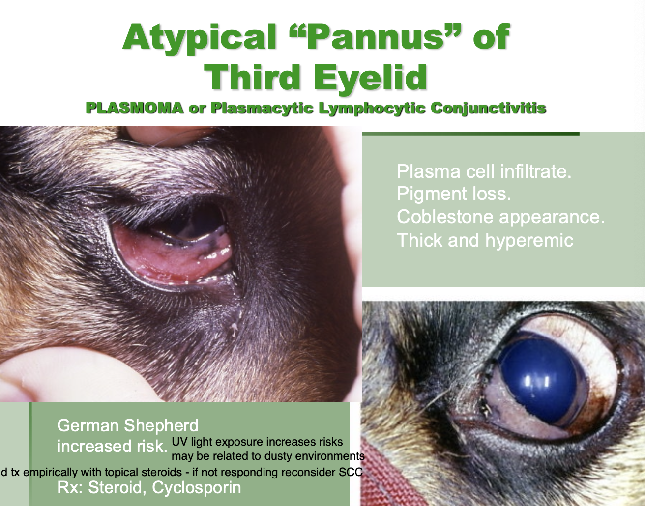
Atypical pannus of 3rd eyelid Tx
Steroids, cyclosporin

Signalment for plasmoma, plasmacytic lymphocytic conjunctivitis, atypical “pannus” of third eyelid
GSD increased risk

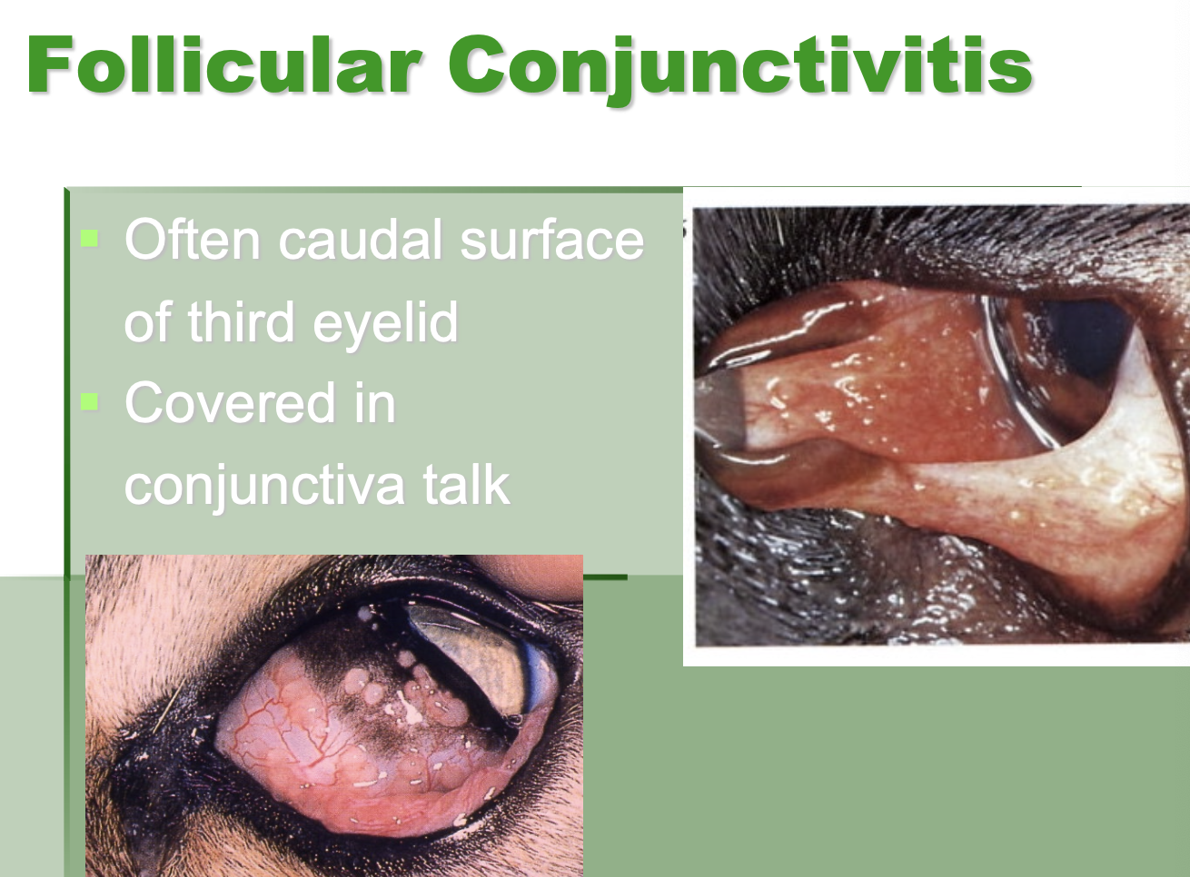
Follicular conjunctivitis often affects
Ca. surface of 3rd eyelid.
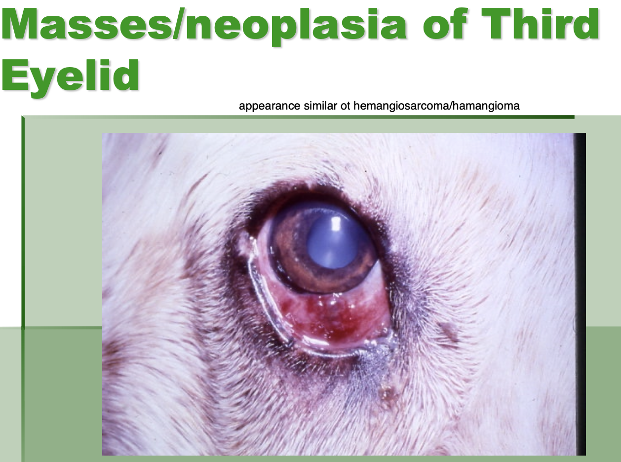
3rd eyelid neoplasia (3)
SCC.
Adenocarcinoma.
Lymphosarcoma.
(LSA, HSA, adenocarcinoma more common in SA)
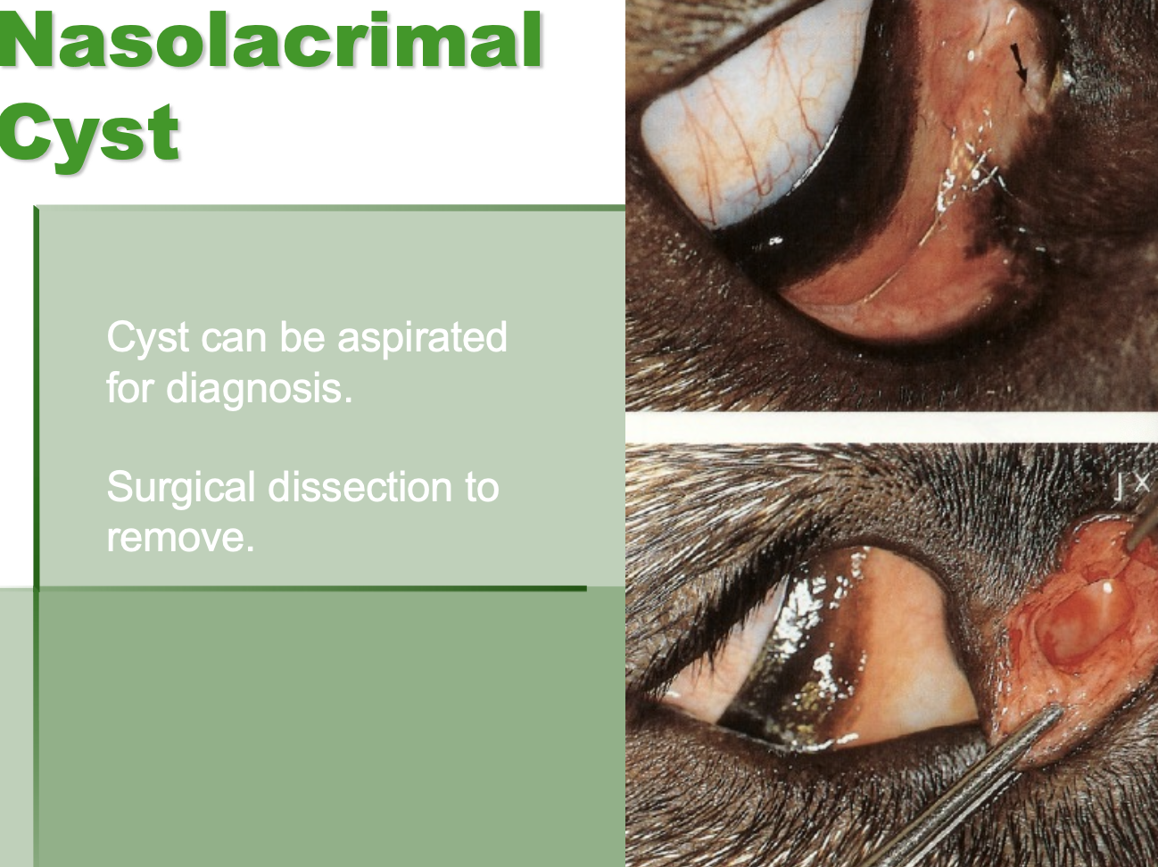
Nasolacrimal cysts (2)
can be aspirated for dx.
Surgical dissection to remove.
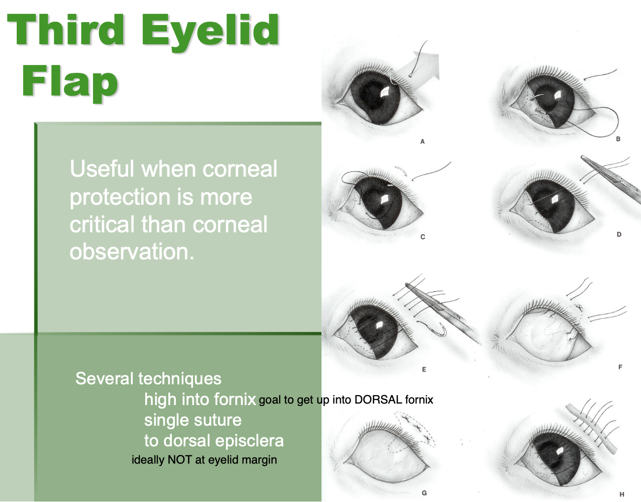
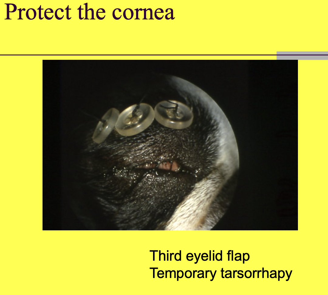
Third eyelid flap is useful when
corneal protection is more critical than corneal observation.
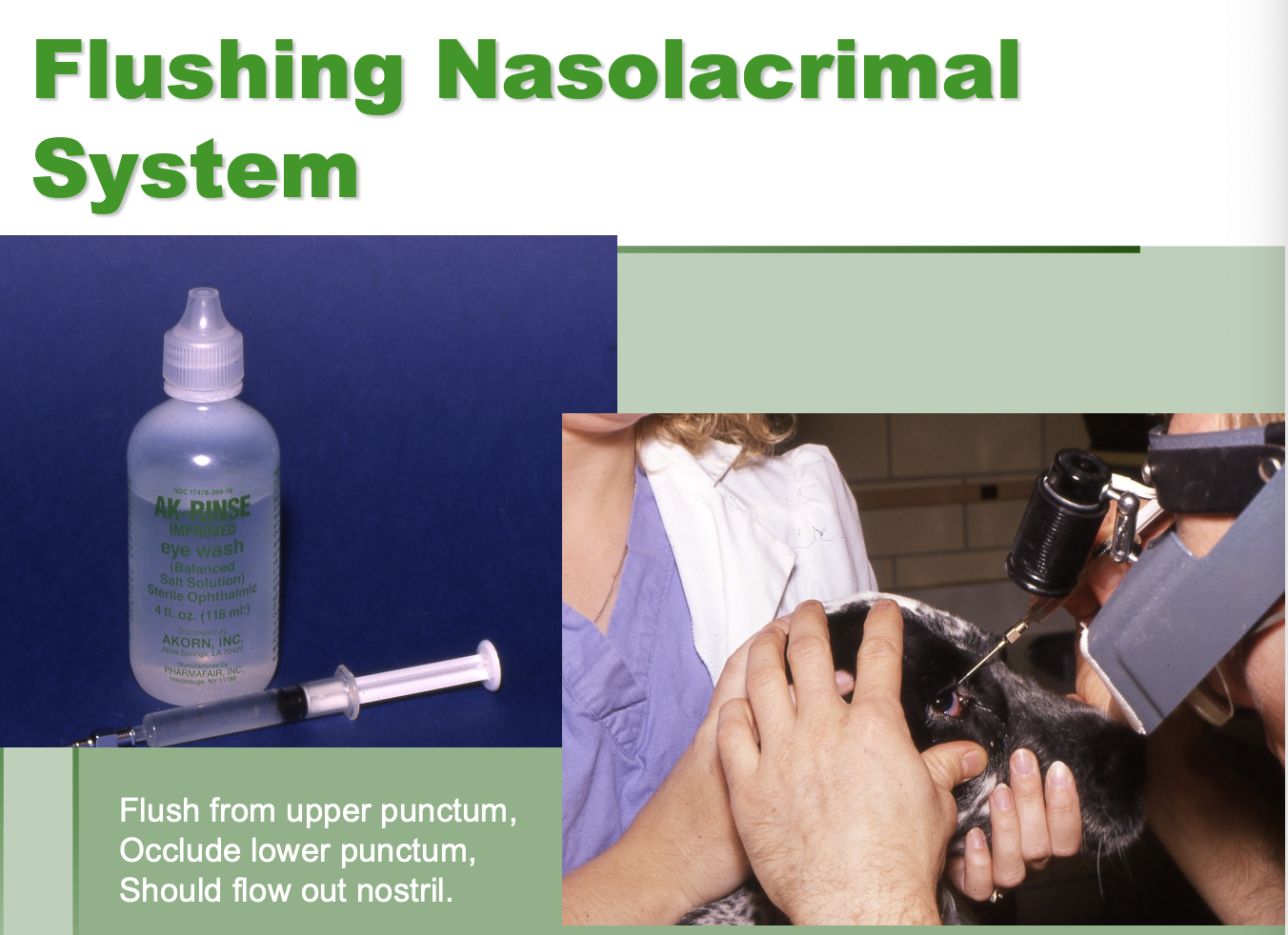
Flushing nasolacrimal system
Flush from upper punctum while occluding lower punctum.
Should flow out nostril.
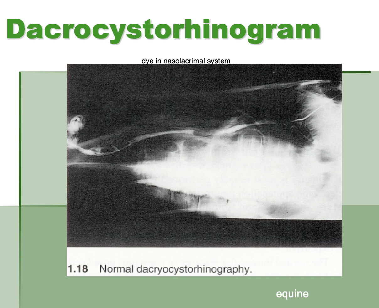
Dacrocystophinogram is
putting dye in the nasolacrimal system
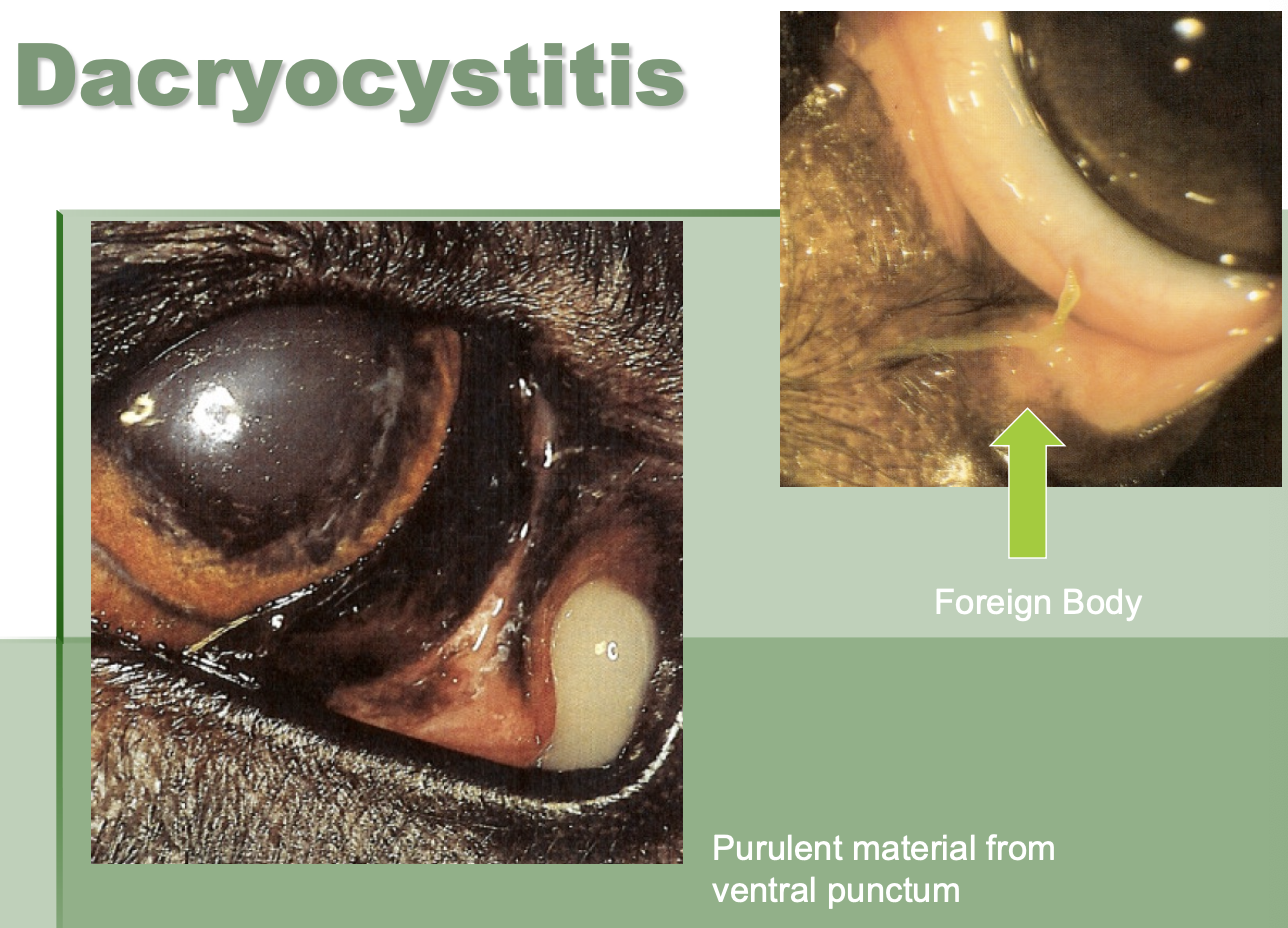
Dacrocystitis
inflammation of the tear duct often associated w/ FB.
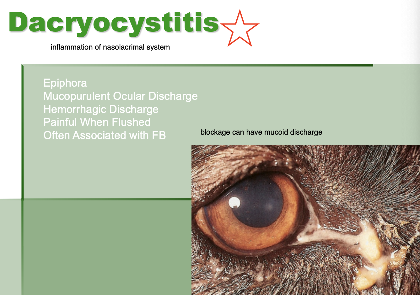
Dacryocystitis CS (4)
Epiphora.
Mucopurulent ocular discharge.
Hemorrhagic discharge.
Pain when flushed.

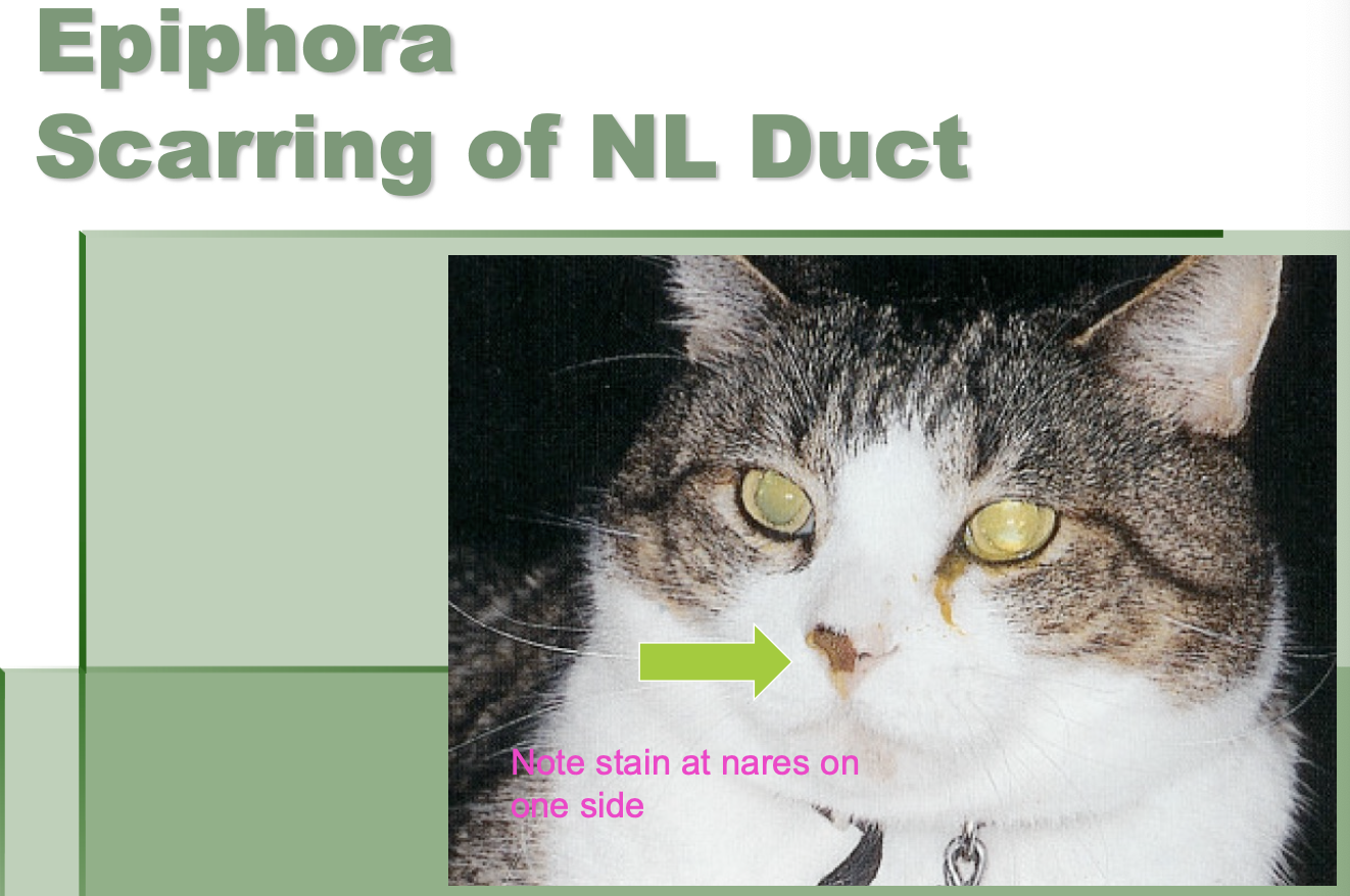
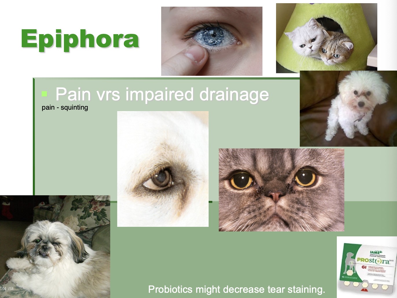
Epiphora def
excessive tearing or drainage

Etiology of epiphora - general
pain v. impaired drainage.
What can be tried to decrease tear staining
probiotics.
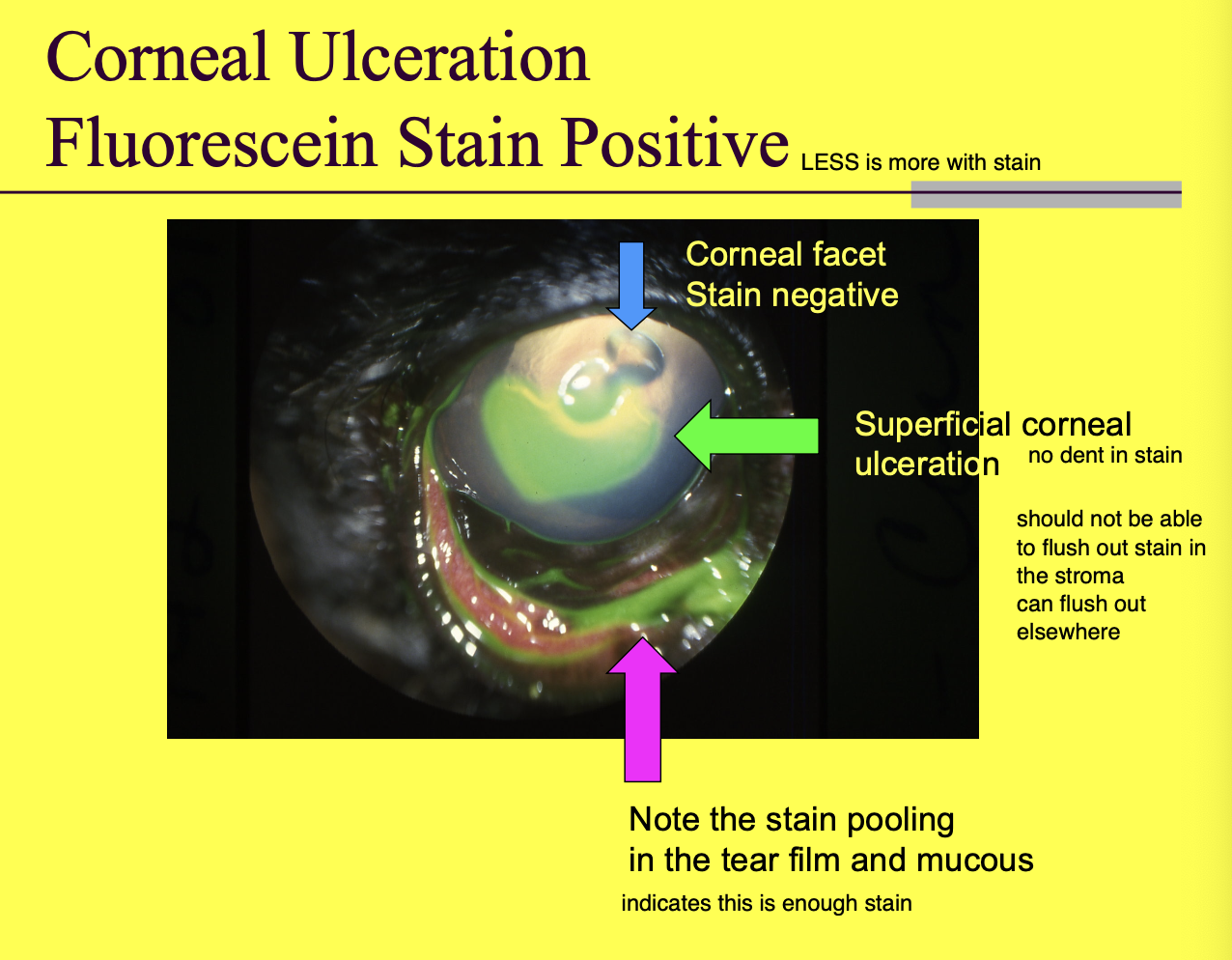
Corneal ulceration dx
positive fluorescein stain
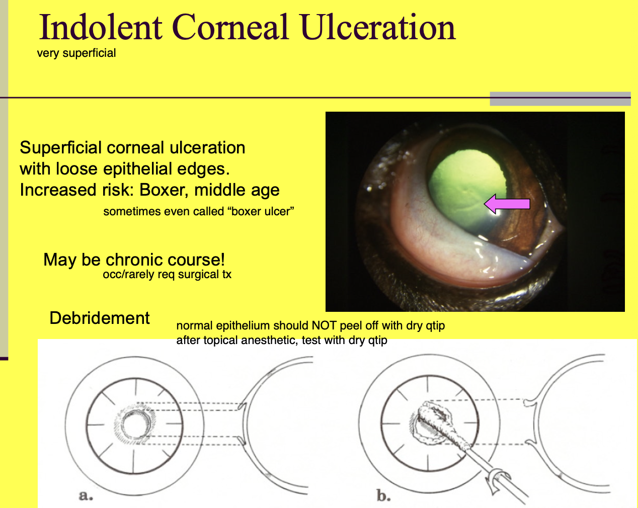
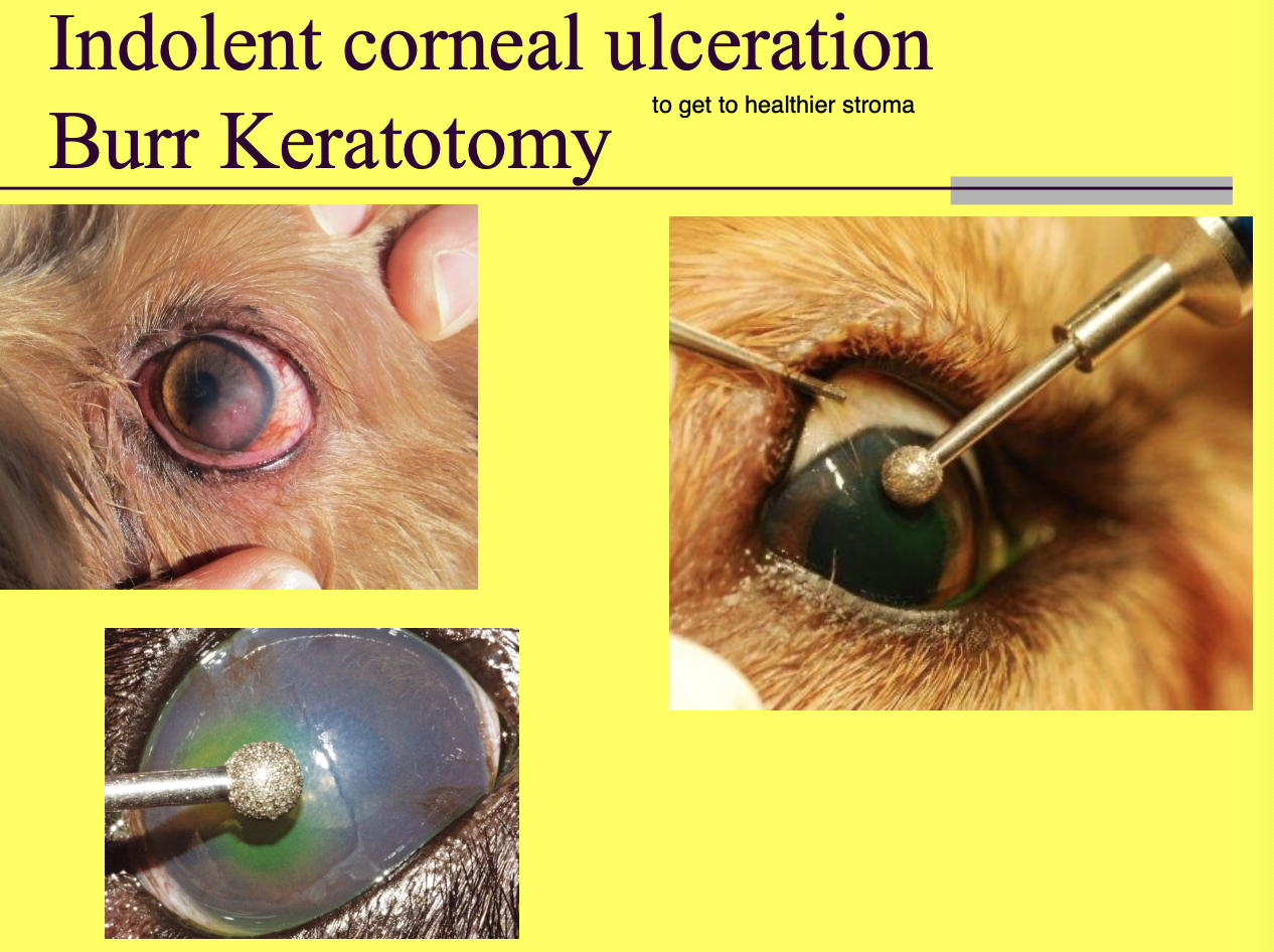
Indolent corneal ulceration
superficial corneal ulceration w/ loose epithelial edges (epithelium does not want to stick back to stroma)

Indolent corneal ulceration risk
Boxer.
middle aged.
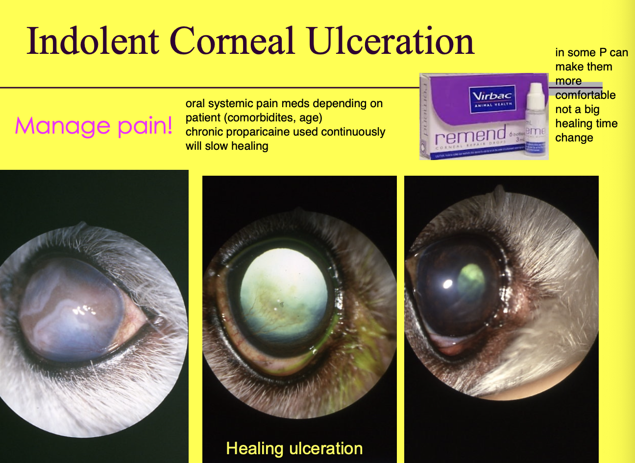
Indolent corneal ulcer Tx (3)
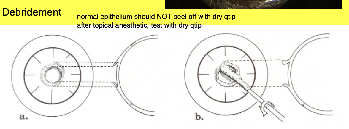
Debridement w/ removal of loose epithelial edges.
Manage pain - systemic v. atropine on surface v.
Punctate and grid keratotomy or Burr keratotomy.
How do you know if epithelial edge is loose
normal epithelium will not peel off w/ a Q-tip.

Considerations for Punctate and grid keratotomy or Burr keratotomy (2)
do not do if the ulcer is infected.
goal is to expose underlying healthy stroma to allow for healing.
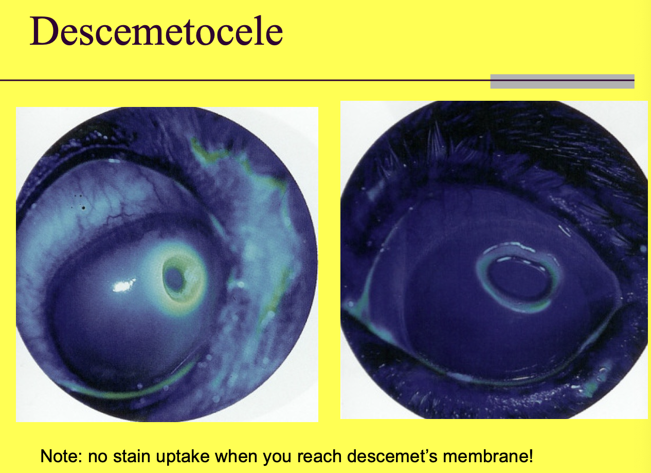
Descemetocele def
Ulcer that is down to descemet's membrane (central clearing is not a good sign).
High risk for rupture!
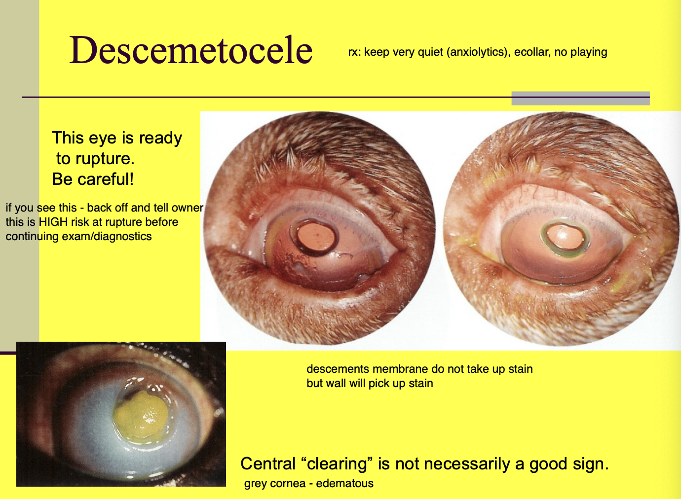
Descemetocele staining
Will stain the surrounding stroma but not Descemet's membrane.
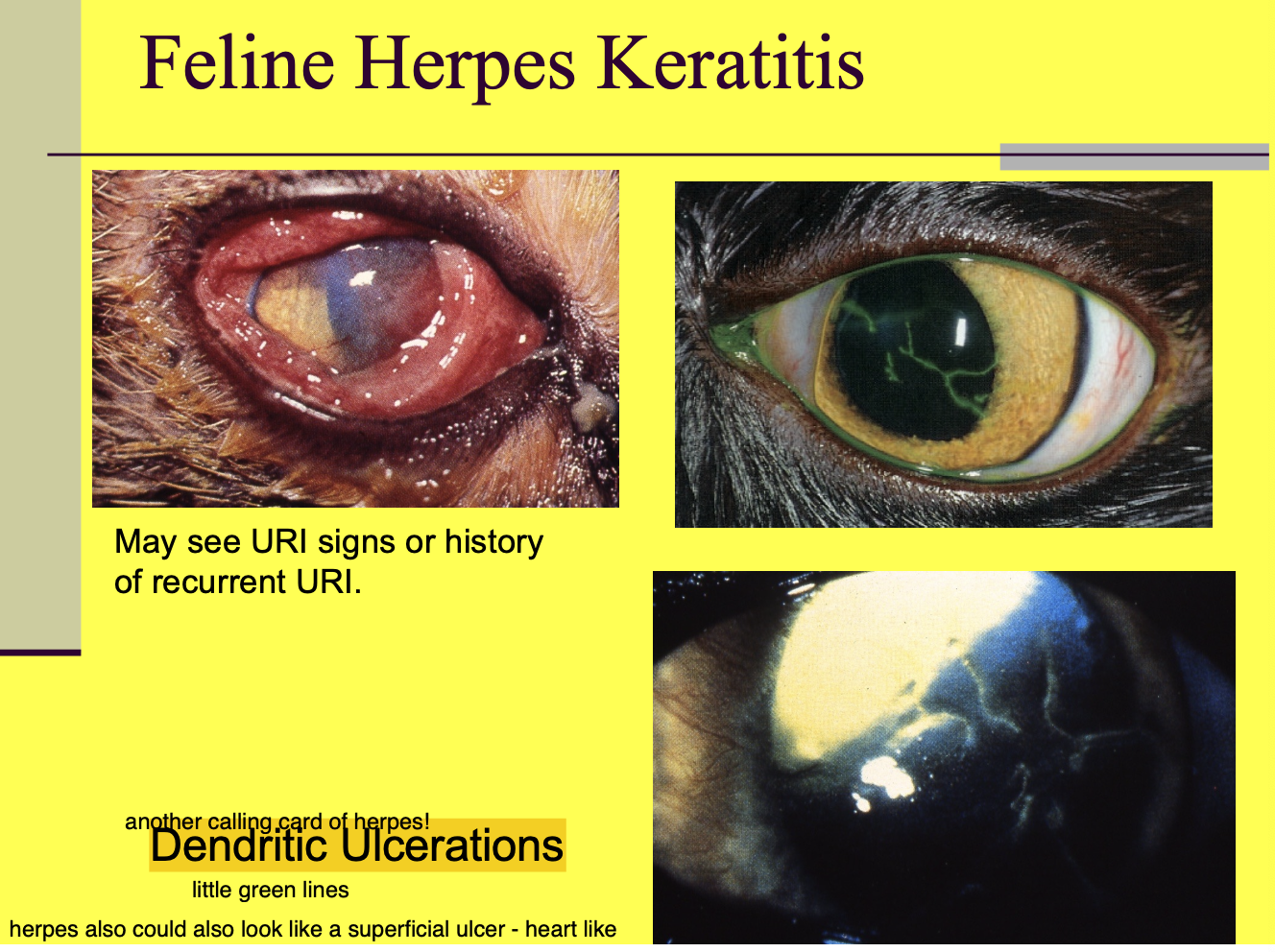
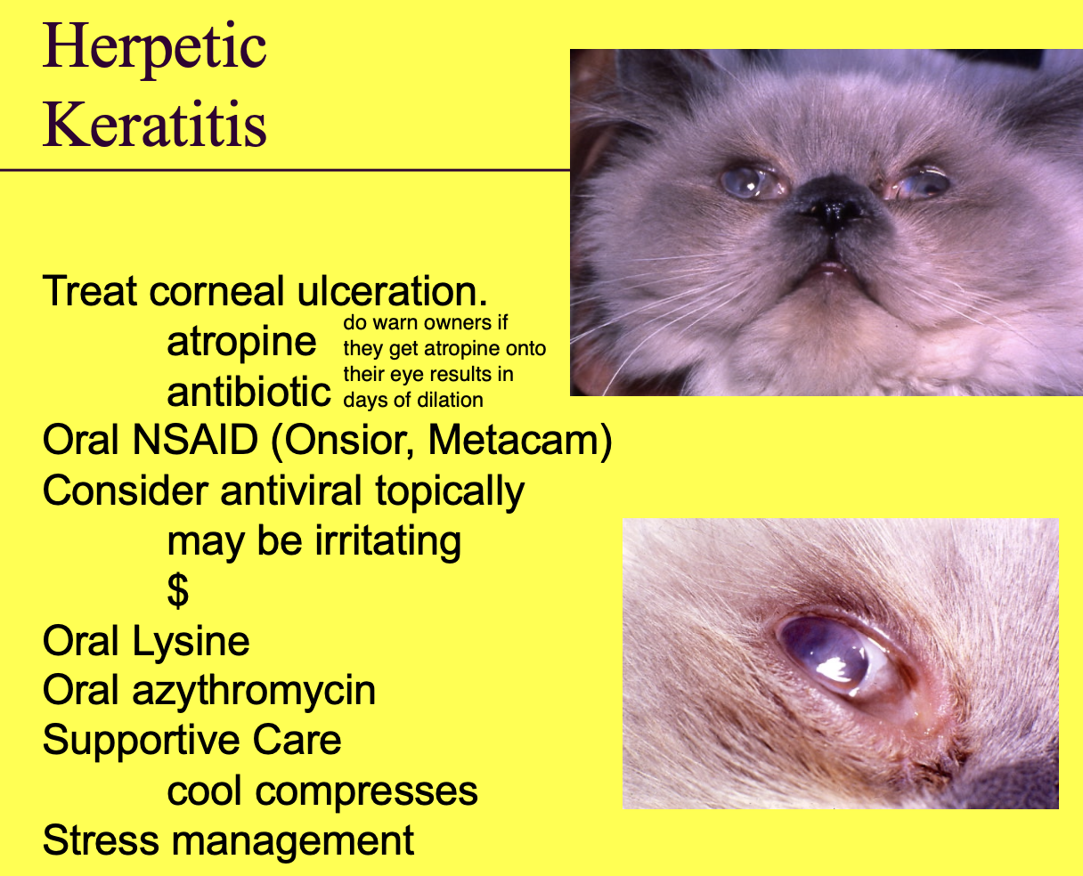
Feline herpes keratitis
Dendritic ulceration - ulcer that follow along the n. tract. (can also see large geographical ulcers)
Feline herpes keratitis tx (9)
Tx corneal ulceration.
Atropine.
ABX.
PO NSAID (Onsior, Metacam)
Consider antiviral topically - may be irritating, $$.
PO lysine.
PO azithromycin.
Supportive care w/ cold compress.
Stress management.
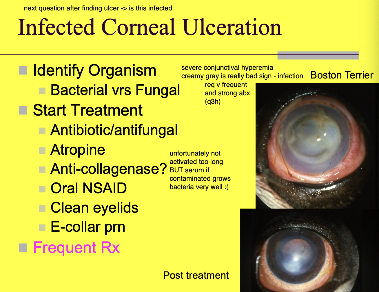
Infected corneal ulcers have a
cloudy, white (hypopyon) or green appearance to the ulcer/eye

Infected corneal ulceration can be
bacterial or fungal

Tx for Infected corneal ulcer (6)
Strong ABX/antifungals.
Atropine.
Anticollagenase?
PO NSAIDs.
Clean eyelids.
E-collar PRN.

Tx for infected corneal ulcers
needs to be frequent administration of ABX
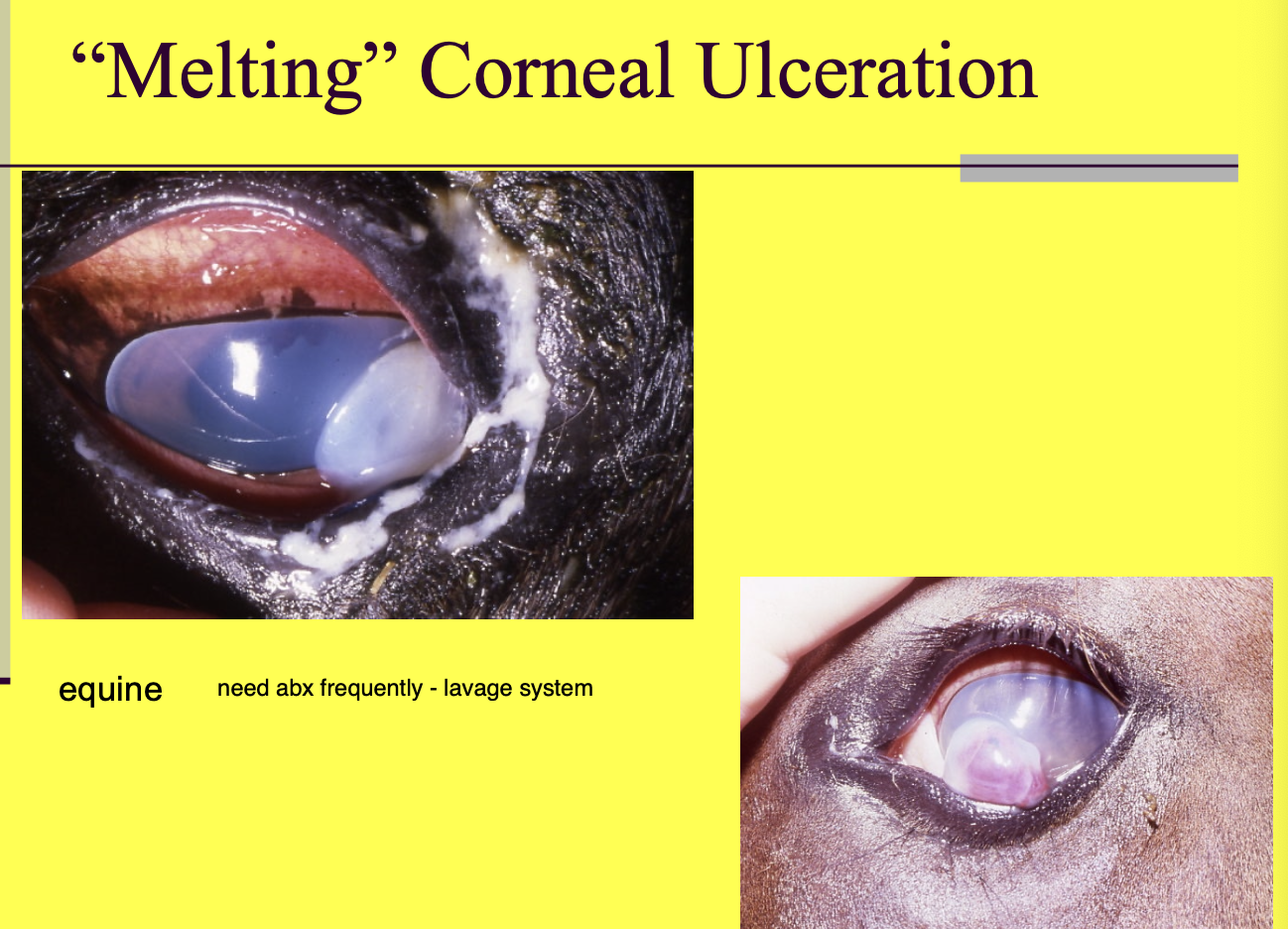
Melting corneal ulcers require
hospitalization, very frequent tx, monitoring.
Fungal keratitis may present w/
fungal plaques
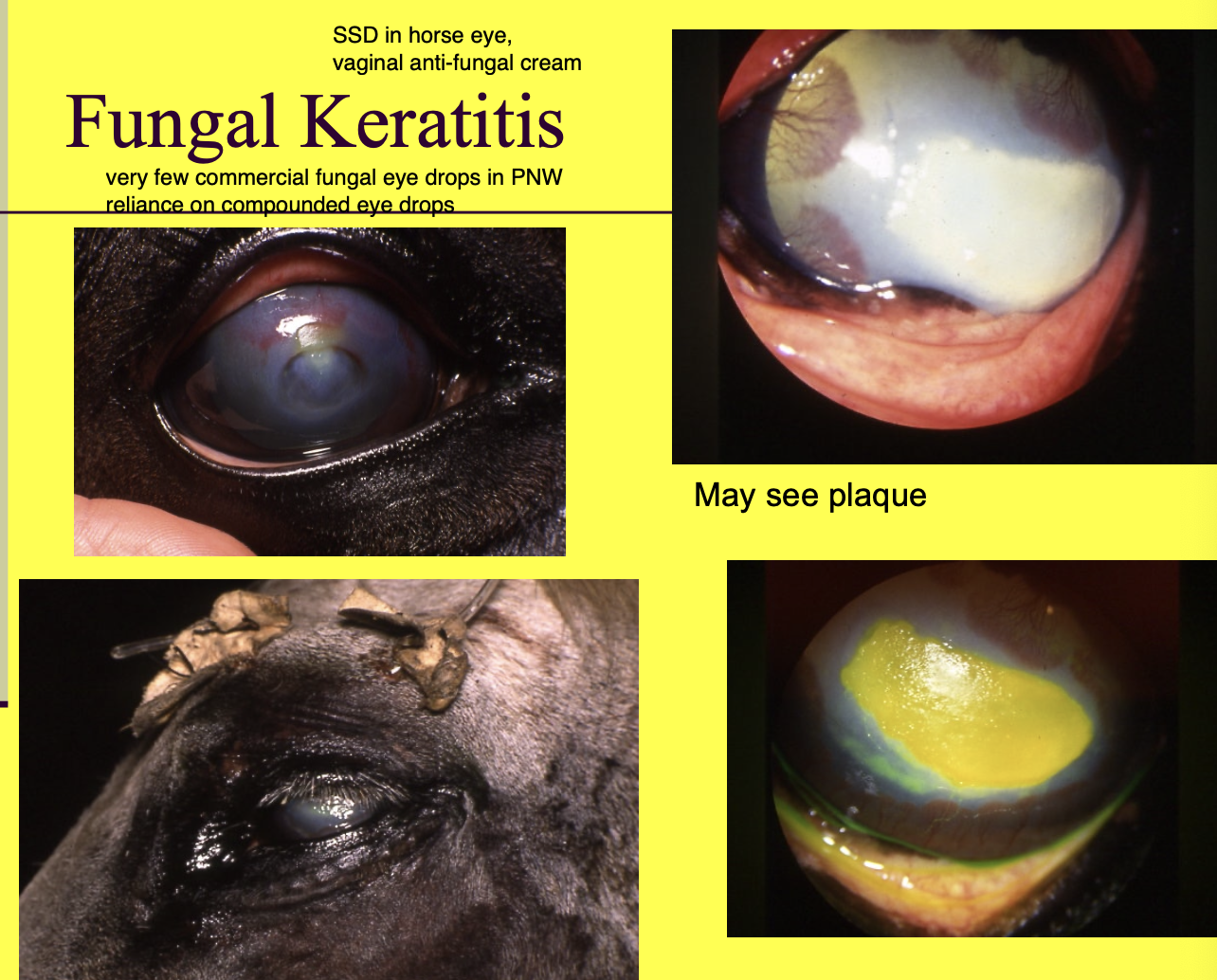
Fungal keratitis Dx (3)
Corneal scrape.
Culture.
Biopsy.
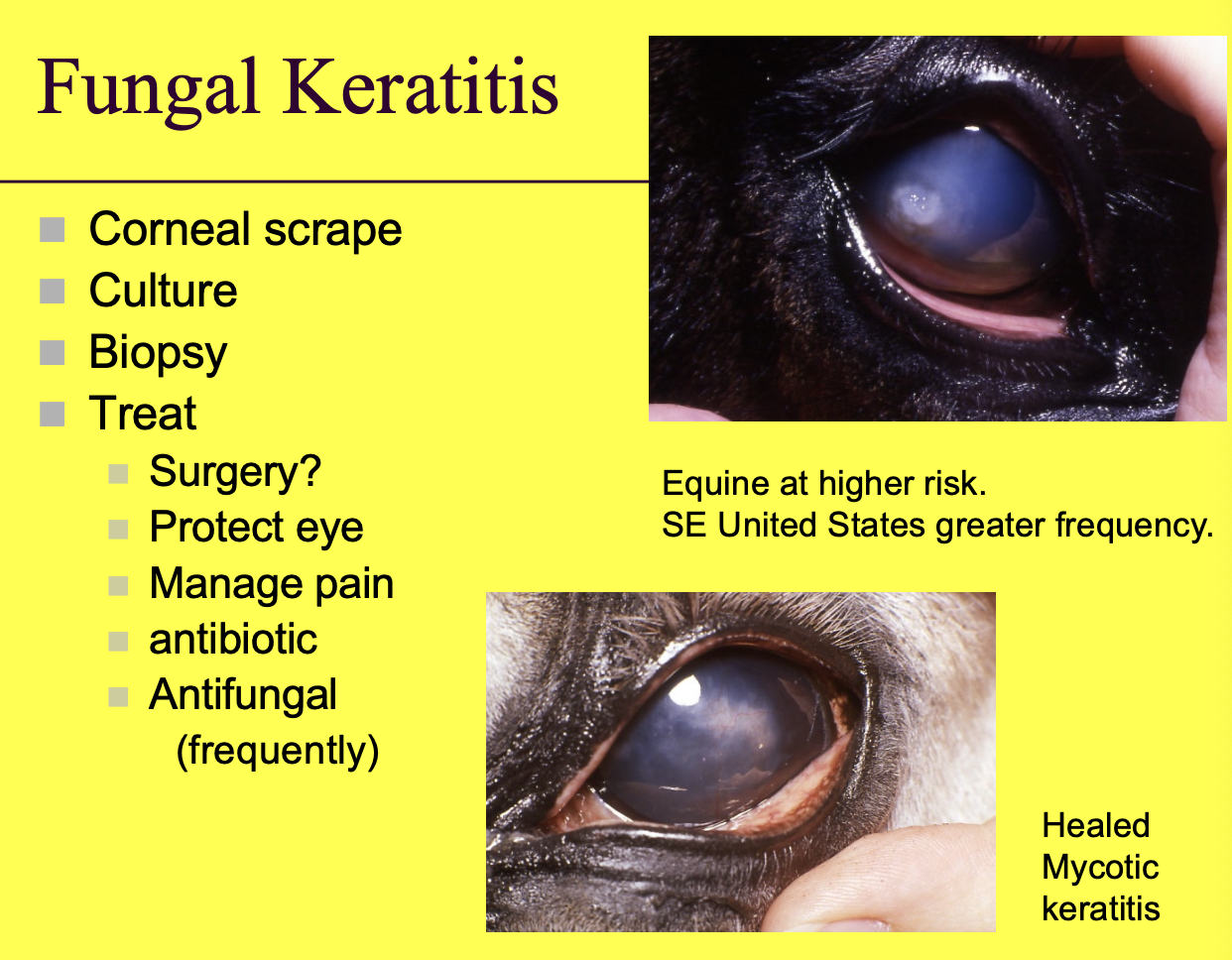
Tx for fungal keratitis (5)
Sx - removal of large plaques.
Protect eye.
Manage pain.
ABX.
Antifungal - frequent.

Corneal stromal abscess def
Cornea inoculated w/ organism. Abscess develops within the stromal sealed under an intact layer of epithelium.
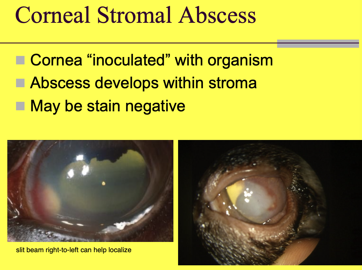
Corneal stromal abscess stain
may be negative.

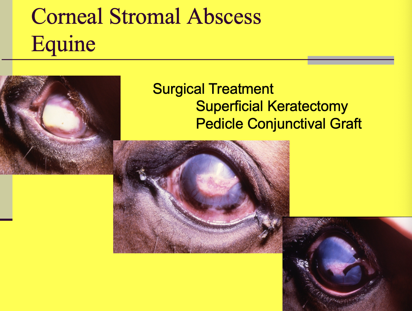
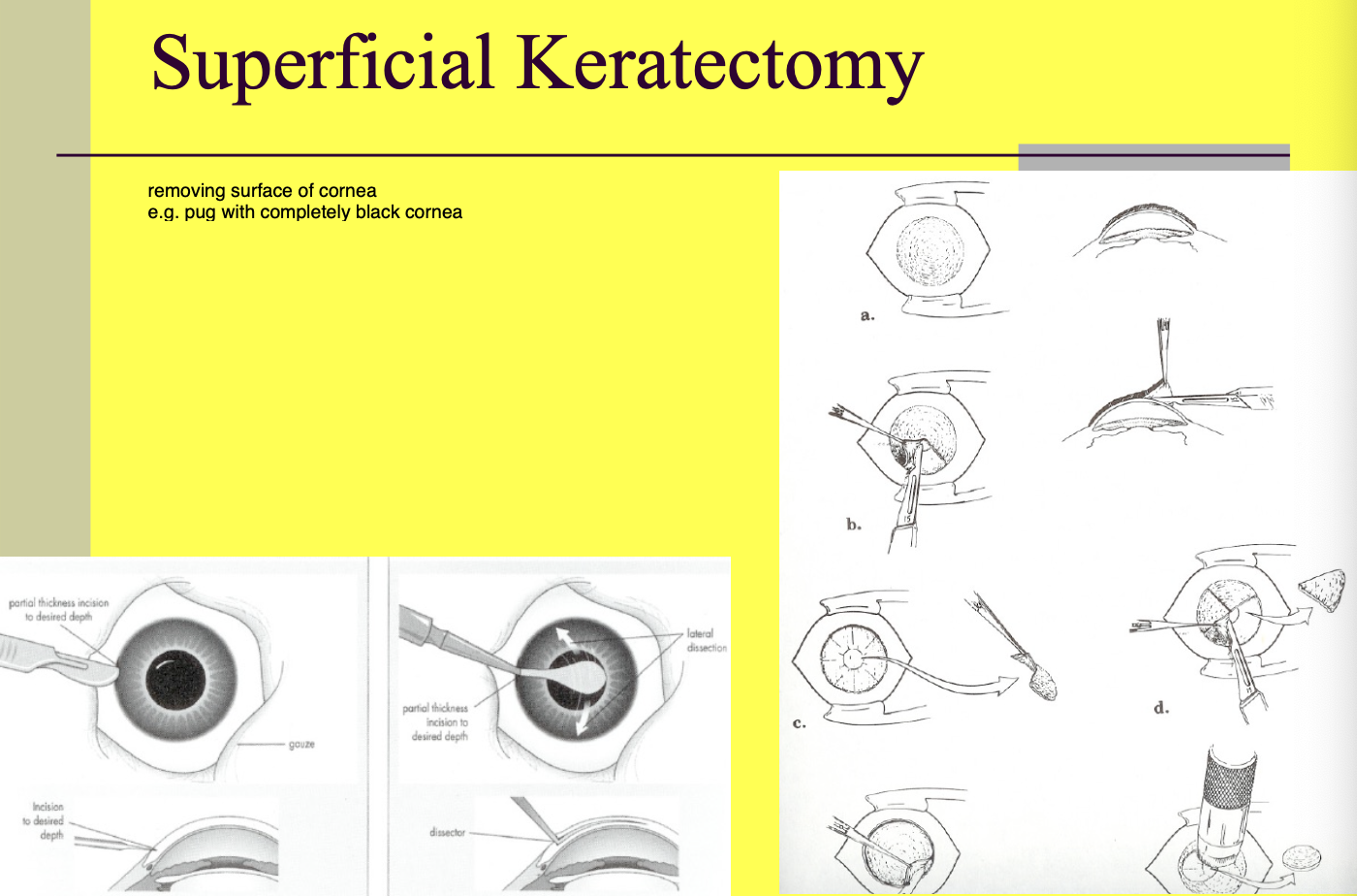
Corneal stromal abscess sx tx (2)
Superficial keratectomy.
Pedicle conjunctival graft.
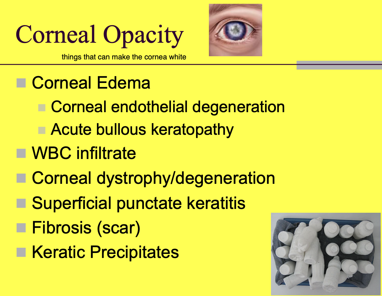
Corneal opacity can be caused by (6)
corneal edema - blue (corneal endothelial degeneration, acute bollous keratopathy)
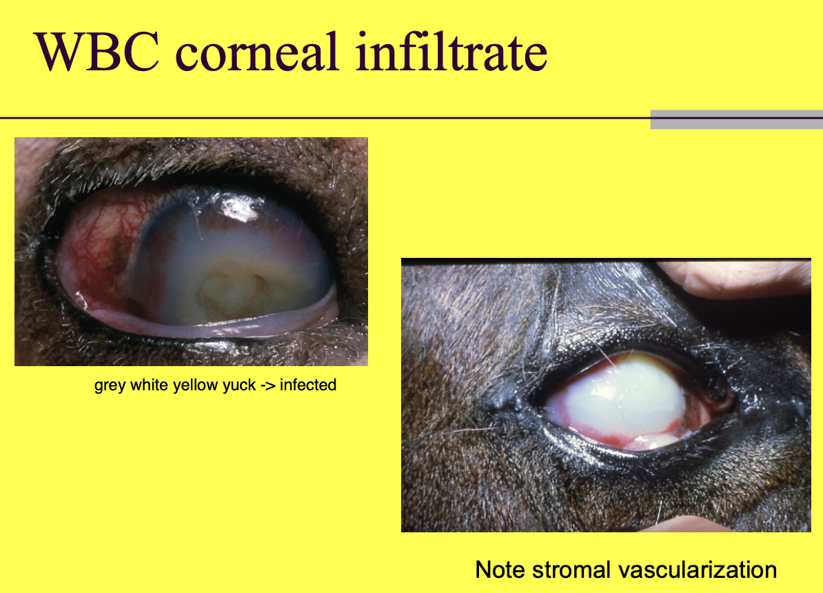
WBC infiltrates - white.
Corneal dystrophy/degeneration.
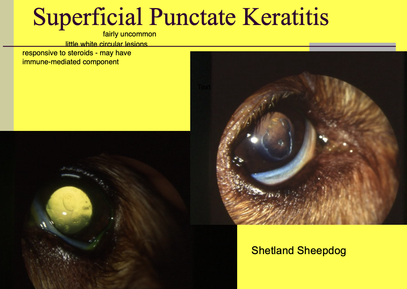
Superficial punctate keratitis.
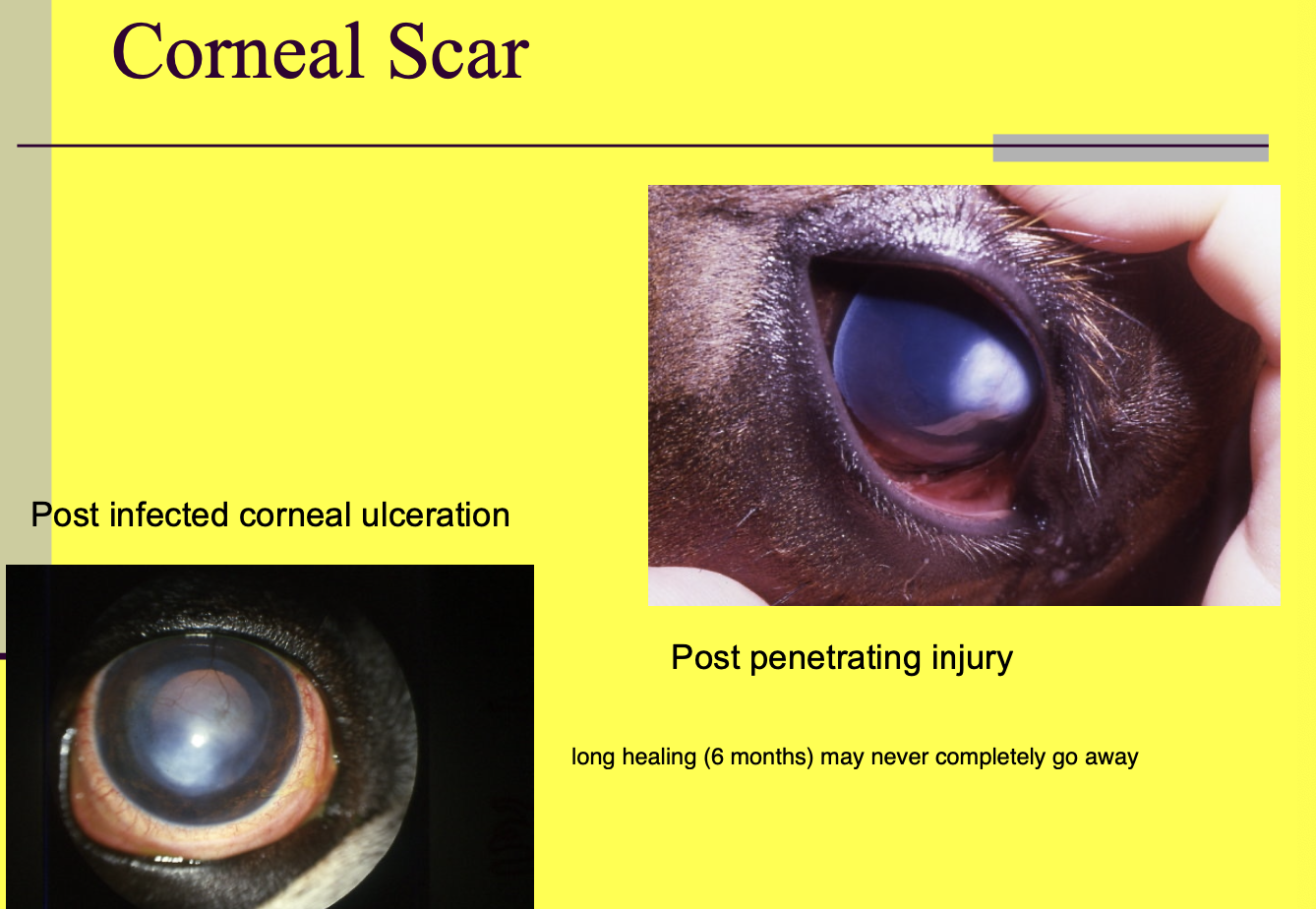
Fibrosis (scar).
Keratic precipitate.
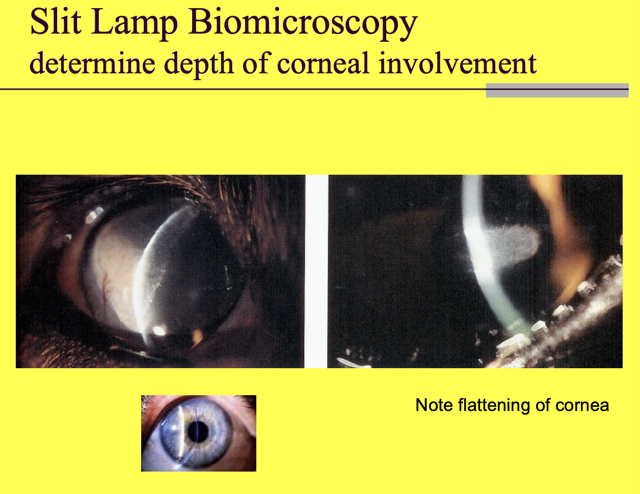
Which tool helps determine depth of corneal involvement
slit lamp biomicroscopy.
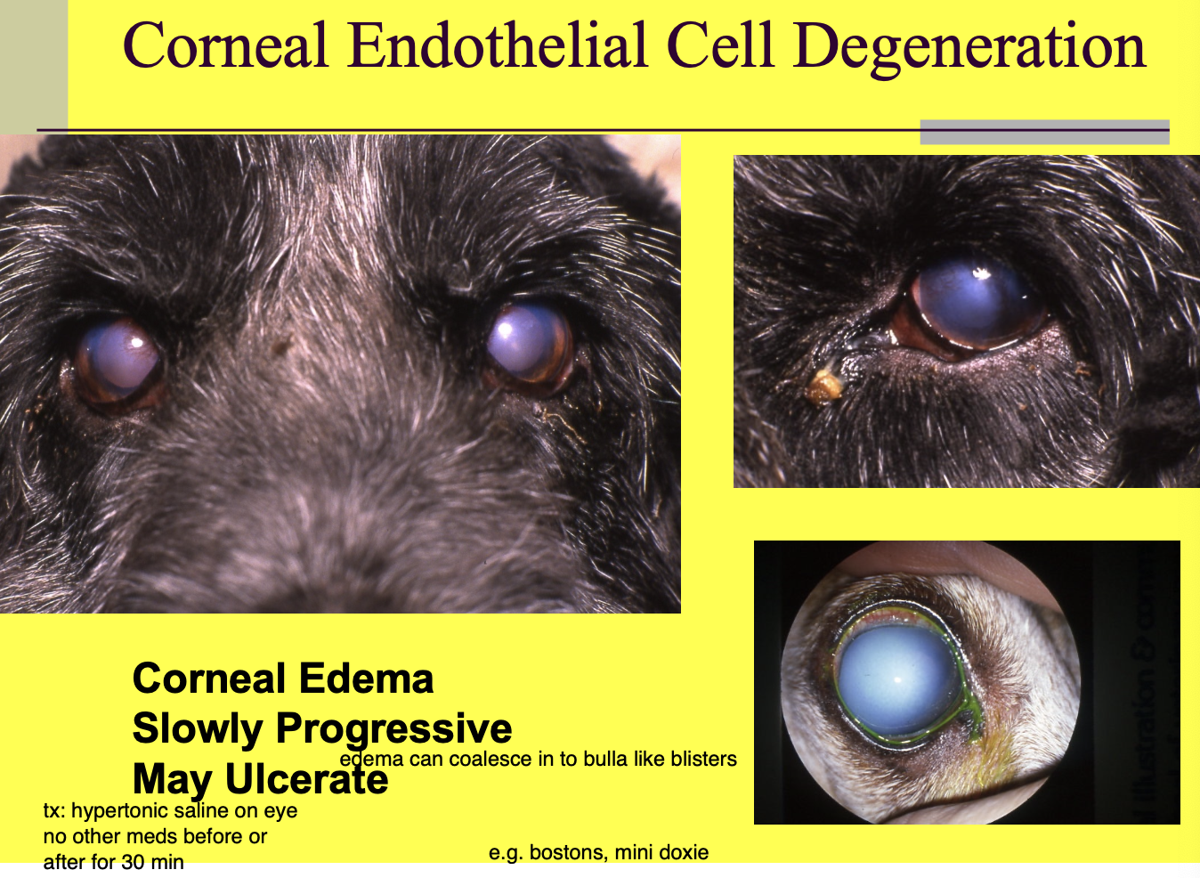
Corneal endothelial cell degeneration at risk breeds (2)
miniature doxies.
Boston terriers.

Corneal endothelial cell degeneration presentation (3)
corneal edema.
slowly progressive.
may ulcerate - use hypertonic saline to reduce risk of bulla formation (do not apply meds for 30m after).
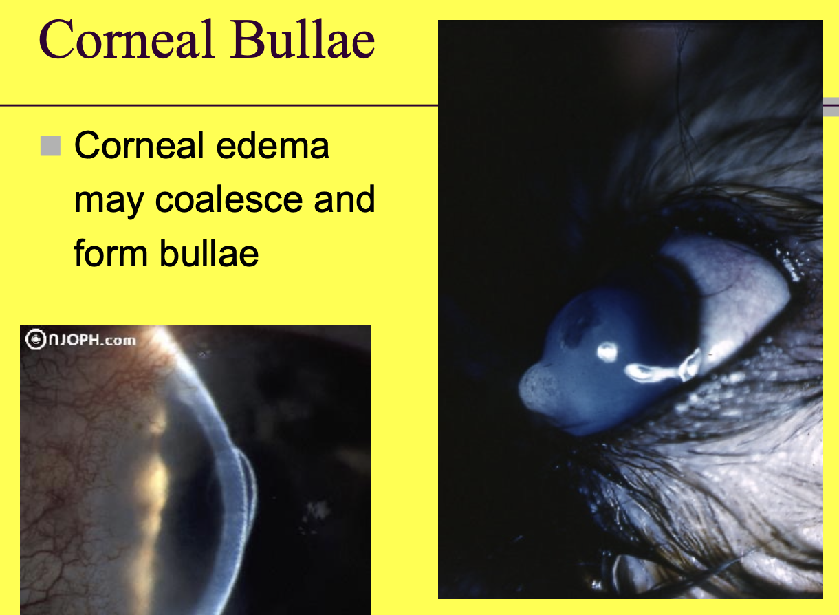
Corneal bullae
when corneal edema coalesces, it can form bullae
Acute bullous keratopathy (2)
Feline dz w/ unknown etiology.
A bullous on the cats cornea that occurs acutely - can impede ability to close eyelid.
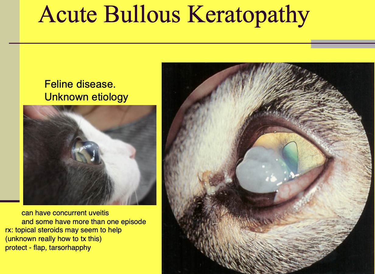
Acute bullous keratopathy possible tx (2)
topical steroids.
protection of the tissue - tarsorrhaphy, flap, or a lot of lube.

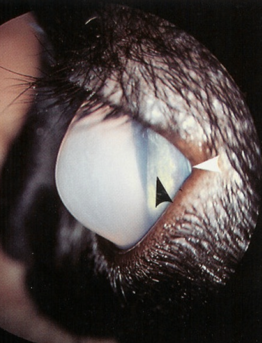
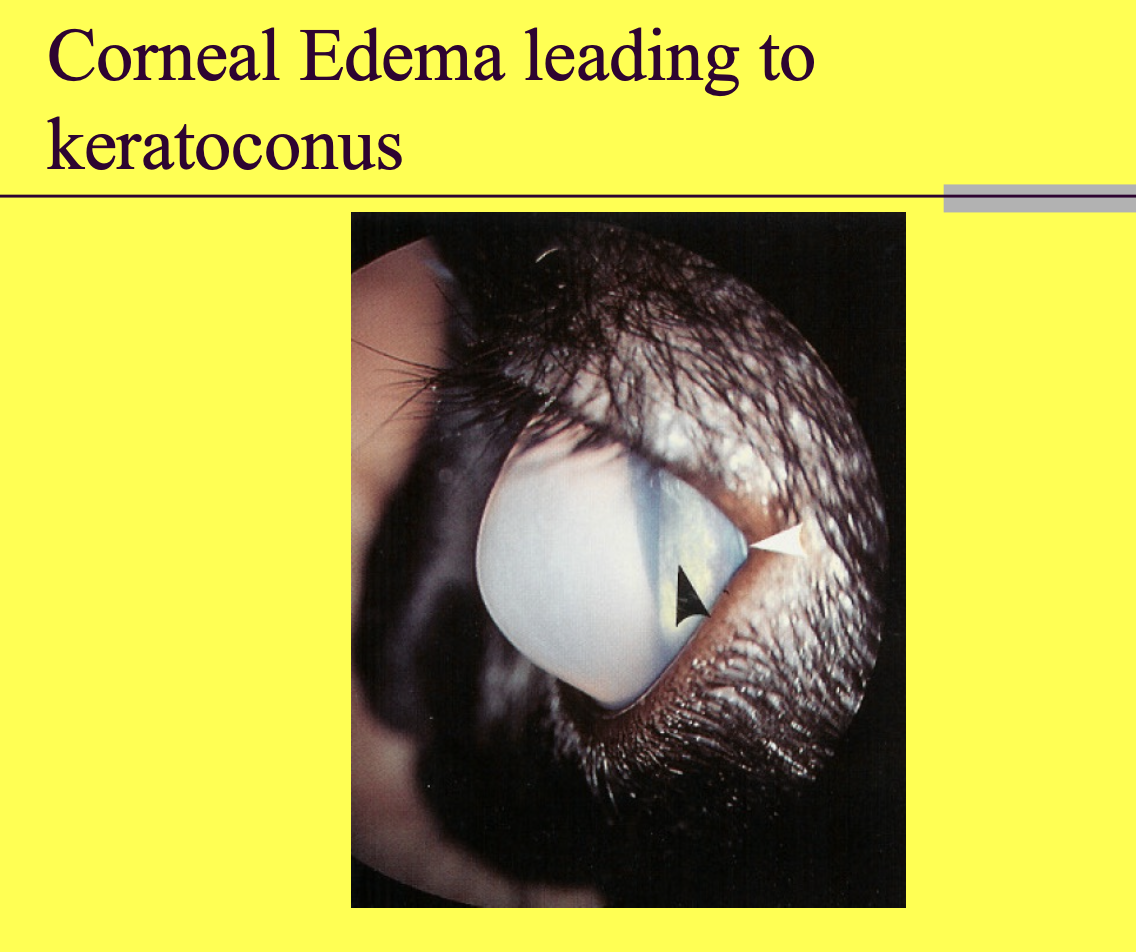
Corneal edema can lead to
keratoconus
WBC corneal infiltrates may the cornea
appear white (can range from grey to white)
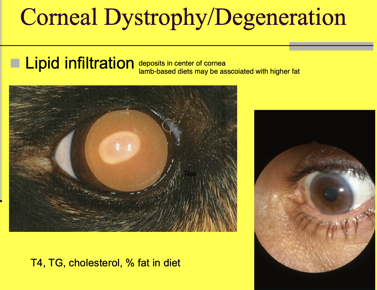
Corneal dystrophy/degeneration can be caused by (3)
lipid infiltration
calcium/mineral deposits - senile
drug deposits
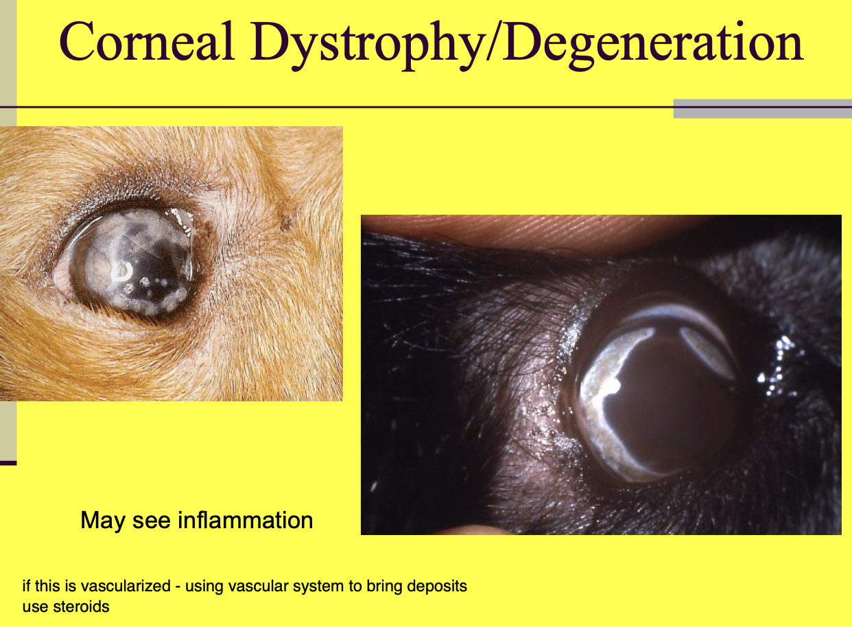
Corneal dystrophy/Degeneration from lipid infiltration
genetic predisposition.
deposits in center of cornea.

Lipid infiltriation causing corneal dystrophy/Degeneration may have dietary relation to
high fat diets such as lamb

What levels should be check w/ lipid infiltrates causing corneal dystrophy/Degeneration (4)
T4.
TG.
chol.
% fat in diet.
Senile corneal degeneration
tend to see calcium mineral deposits - flaky snow, spicular appearance.
usually not painful until ulceration.
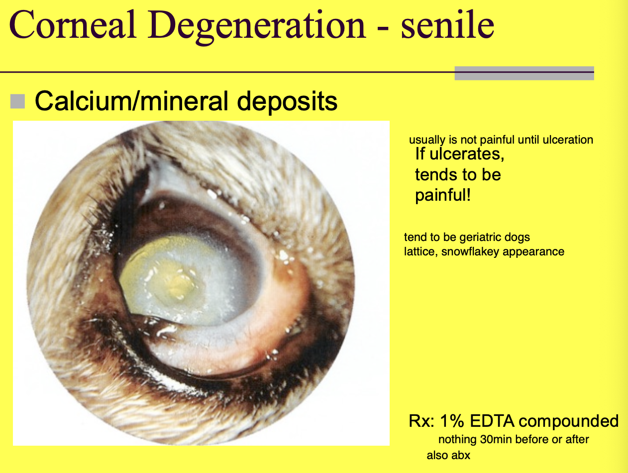
Tx for senile corneal degeneration
1% EDTA compounded

Superficial puncutate keratitis apperance
little white circular lesions
fairly uncommon

Superficial punctate keratitis Tx
responds well to steroids (suspected immune-mediated component)
Fibrosis appearance in the cornea
White - may remodel over the course of 6m.
Keratic precipitates is material that
leaks out of the uveal tract usually due to inflam.
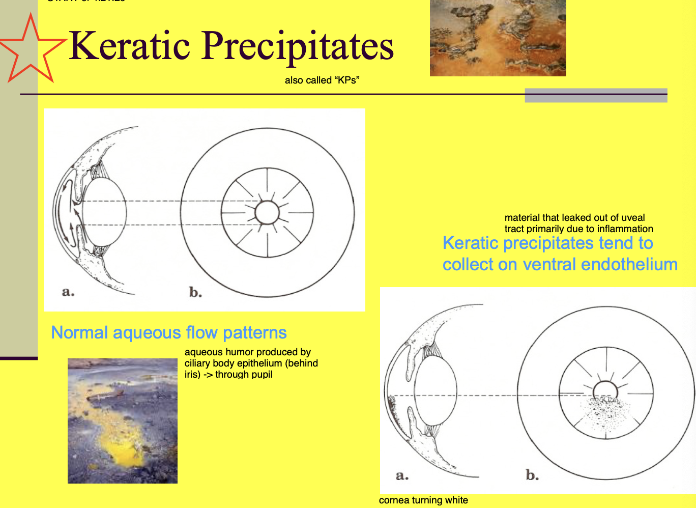
Keratic precipitates tend to collect
on ventral endothelium of the cornea

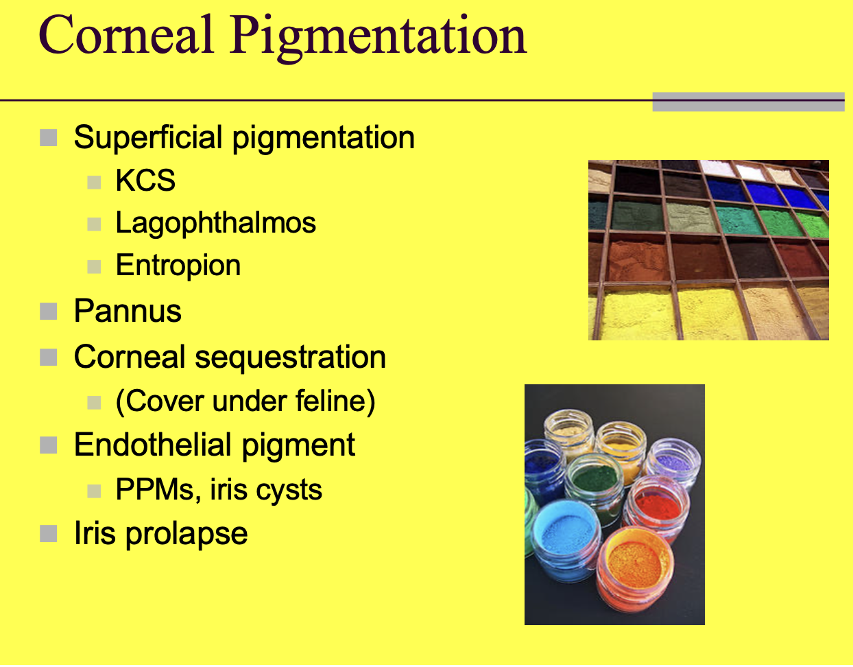
Corneal pigmentation Ddx (5)
Superficial pigmentation.
Pannus.
Corneal sequestration.
Endothelial pigment (PPMS, iris cysts).
Iris prolapse.

Superficial pigmentation of the cornea Ddx (3)
KCS.
Lagophthalmos.
Entropion.
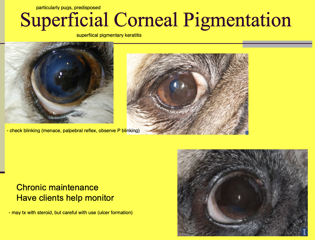
Superficial corneal pigmentation can become a problem b/c
it can affect the degree to which the dogs blink

Superficial Corneal pigmentation monitoring
have clients do at home, req chronic maintenance P blinking well
Chronic superficial keratitis is also called
pannus

Chronic superficial keratitis is when there is
pigment and vessels infiltrate below epithelium
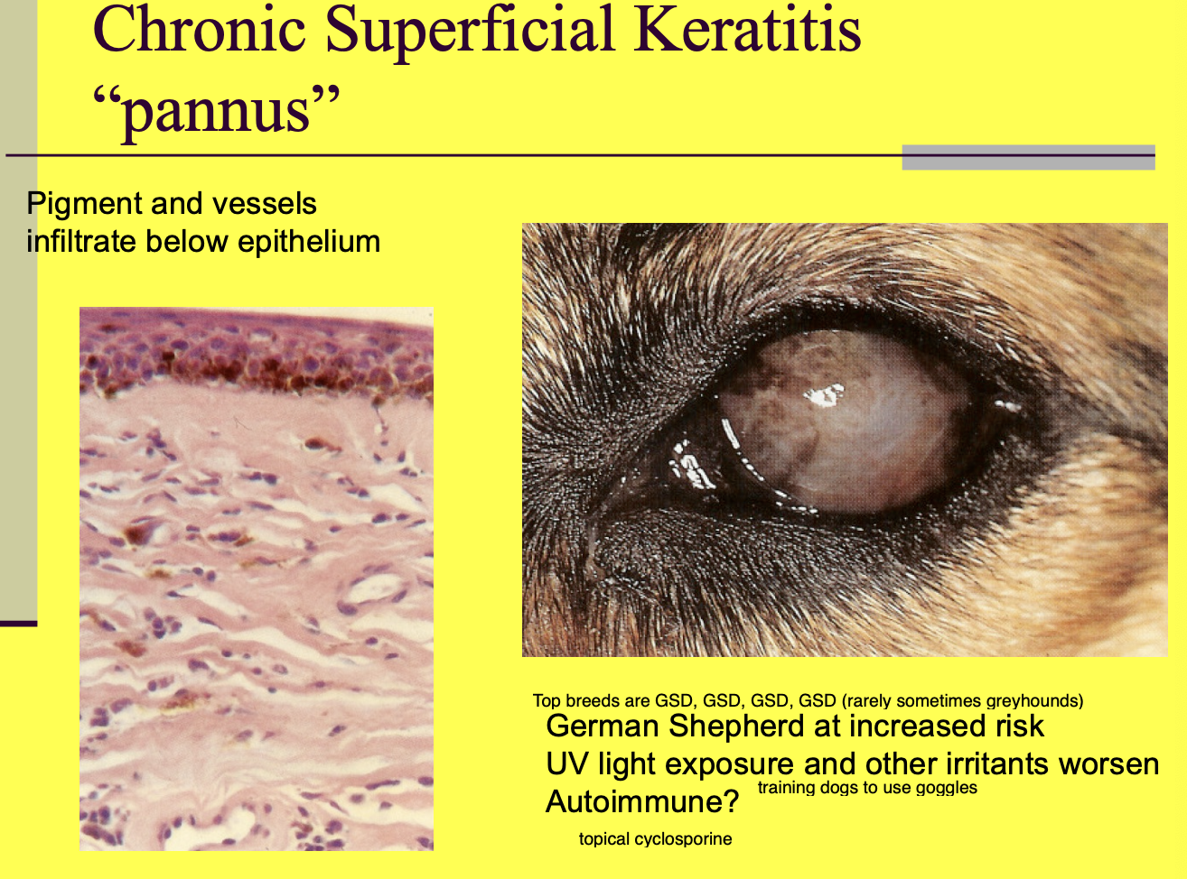
High risk breed for Chronic superficial keratitis
GSD

What worsens chronic superficial keratitis
UV light exposure and other irritants.

Chronic superficial keratitis usually starts
at temporal (limbus) cornea
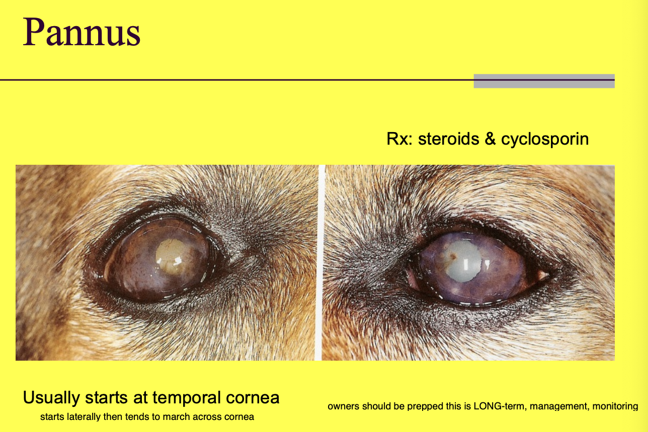
chronic superficial keratitis tx (3)
Steroids (autoimmune component).
Cyclosporin.
Will be long term tx w/ need for monitoring.

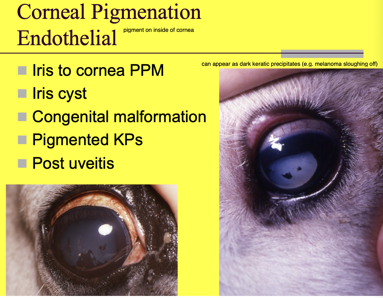
Corneal Pigmentation endothelial (inside of the cornea) Ddx (5)
Iris to cornea persistent pupillary membranes (PPM) (attach iris to inside of cornea).
Iris cysts (migrates from iris ands explodes on back of cornea).
Congenital malformation.
Pigmented KP (keratoprecipitates).
Post uveitis.
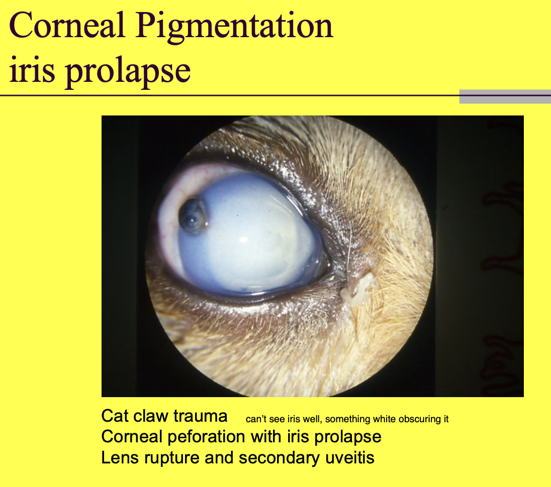
Corneal pigmentation from iris prolapse etiologies
cat claw trauma
corneal perforation with iris prolapse
lens rupture and secondary uveitis
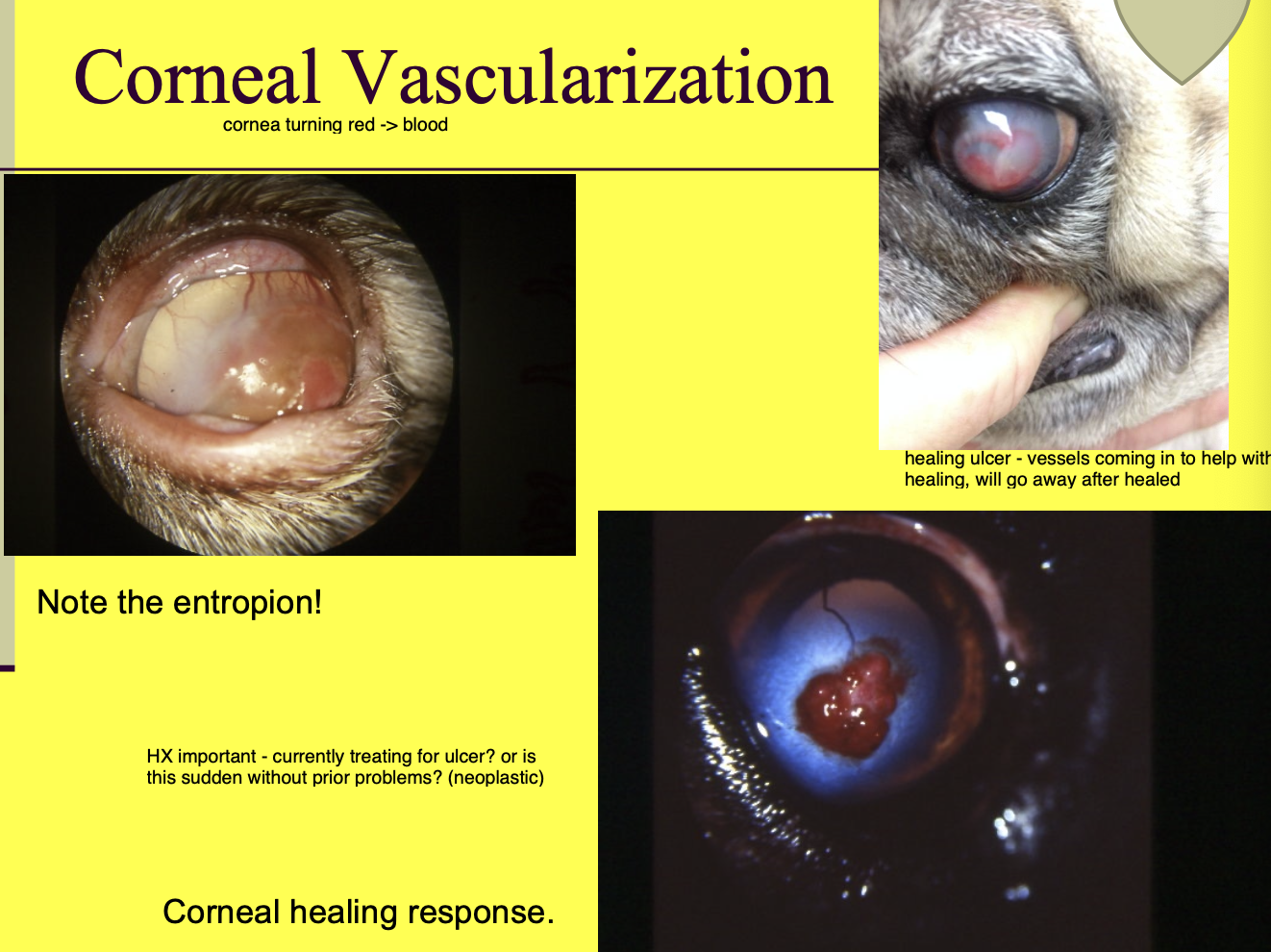
Corneal vascularization etiologies
healing ulcer
neoplastic
Feline corneal Dzs - primary dz that occur in cats (2)
corneal sequestration.
Eosinophilic keratitis.
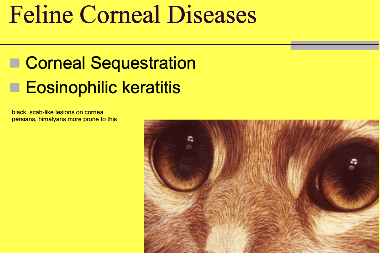
Corneal sequestration appearance
black scab like lesions on surface of corneal.

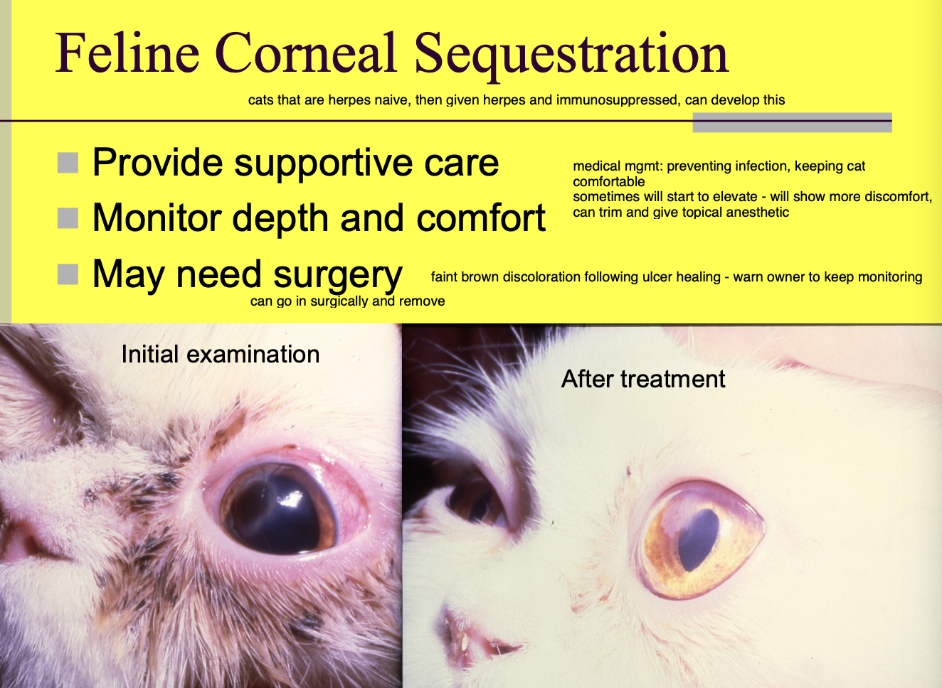
Corneal sequestration generally occur when
there is an abnormality of the eye (ex. can be previous injury or FHV but not every cat w/ FHV gets this)

Corneal Sequestration - response seen in the eye
sequestrum is viewed as foreign, so edema and neovascularization may be seen

Feline Corneal Sequestration Tx (3)
Provide supportive care.
Monitor depth and comfort.
May need Sx.
Eosinophilic Keratitis is seen in
felines - white eosinophilic infiltrates on the cornea (can look like plaques or spotting of white)
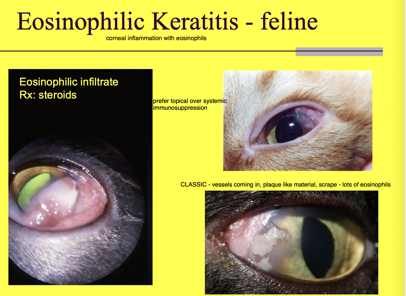
Tx for Eosinophilic Keratitis
Steroids.

Corneal Masses Ddx (4)
SCC.
Papilloma.
Inclusion cysts.
Fibroma.

Inclusion cysts
Epithelium of the cornea pokes into the stroma. Cell turnover results in buildup since the old epithelium can't fall into the tear film.
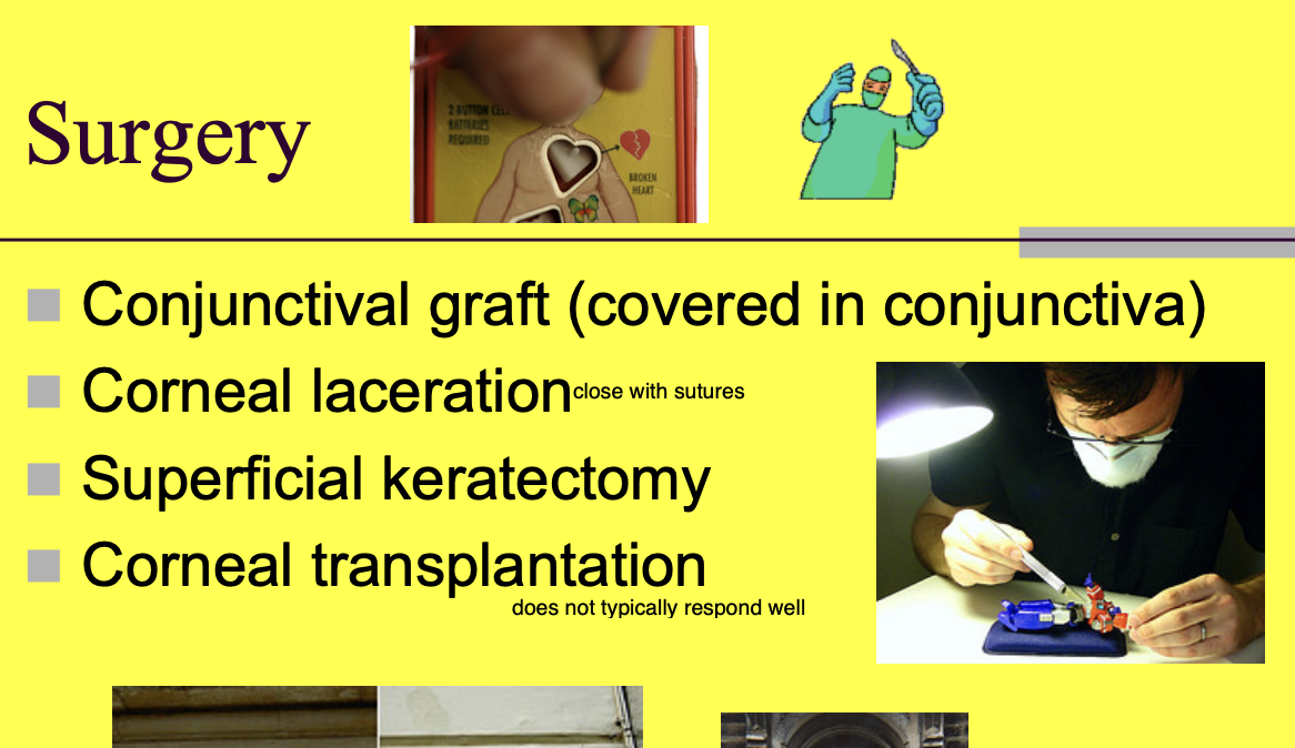
Sx of the cornea includes (4)
Conjunctival graft.
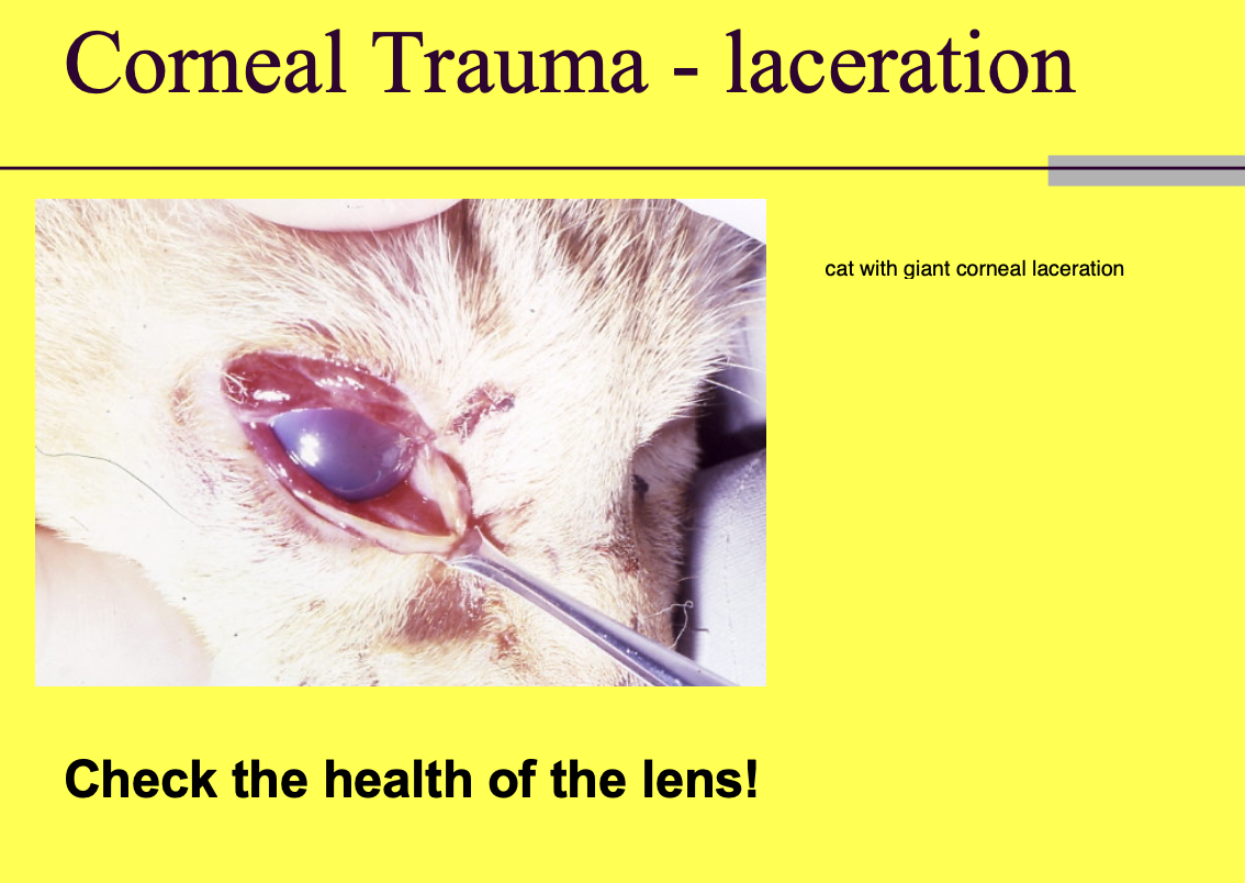
Corneal laceration.
Superficial keratectomy.
Corneal transplantation.
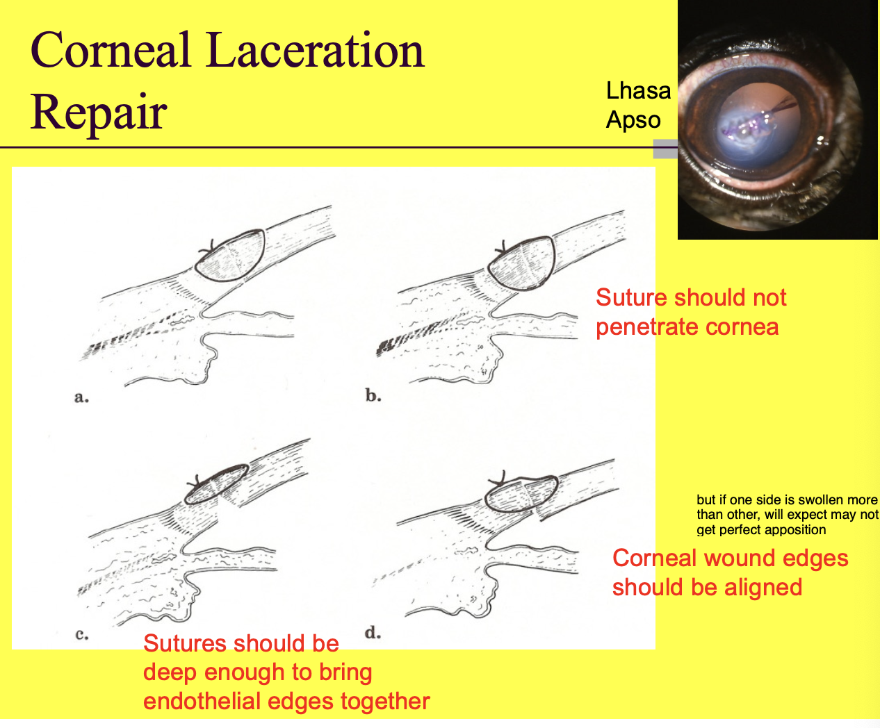
Corneal lac repair principles (3)
Sutures should not penetrate cornea (don't want to penetrate into anterior chamber).
Corneal wound edges should align.
Sutures should be deep enough to bring endothelial edges together.
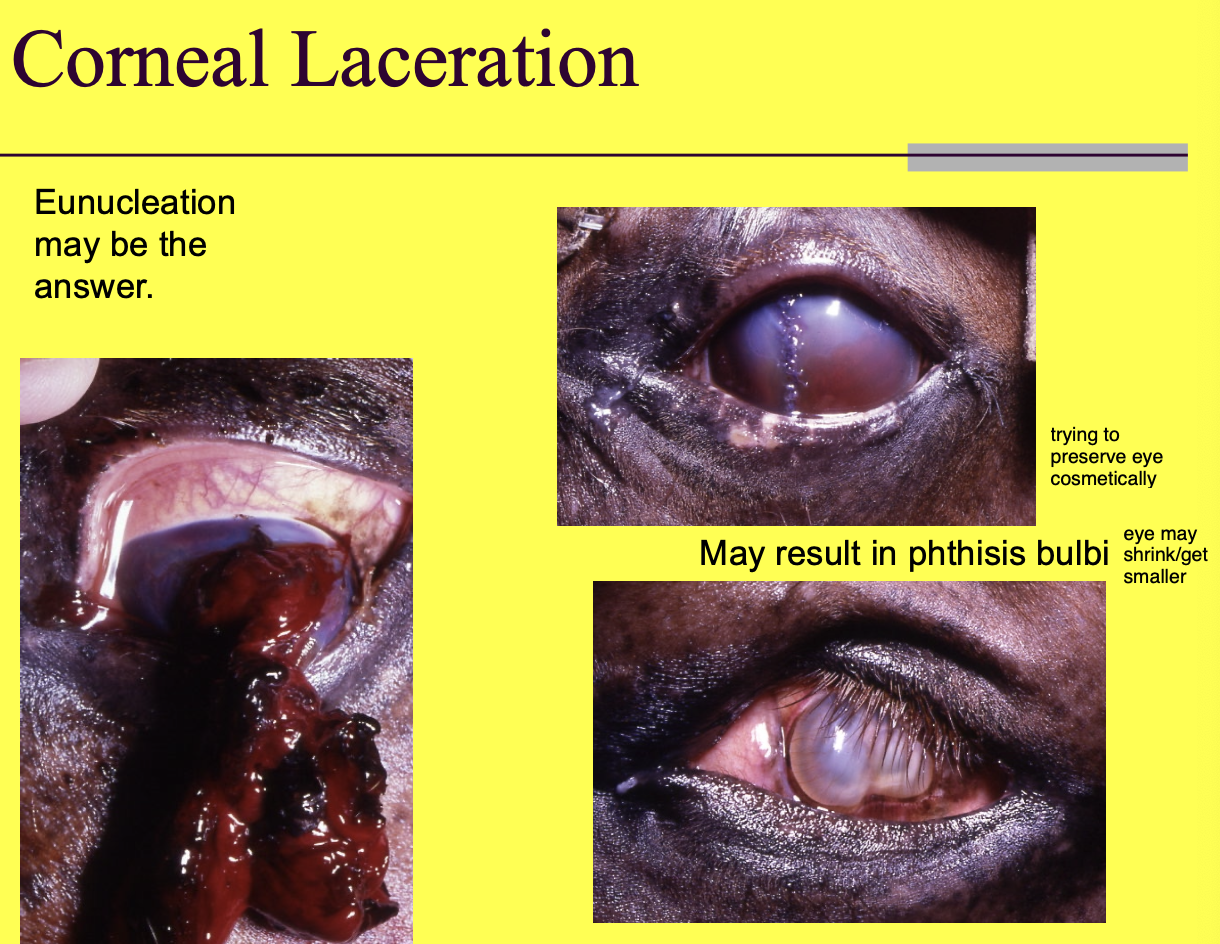
Corneal laceration can result in
phthisis bulbi.
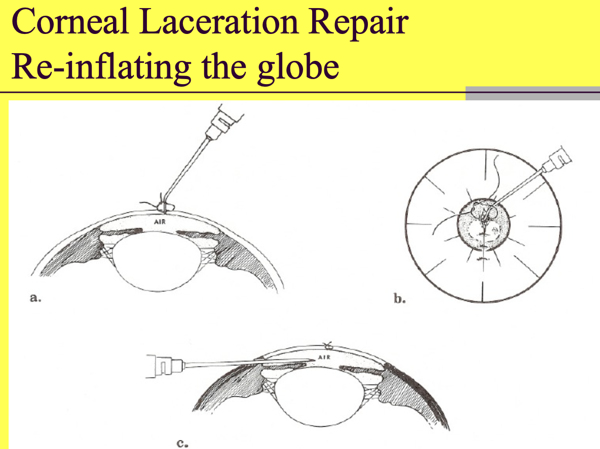
Re-inflating the eye can be done by
placing air in the anterior chamber - can be through incision w/ canula or in another location (difficult in a flat eye).
Superficial keratectomy is
removal of the superficial cornea surface
Corneal trauma - what decreases prognosis
involvement of the lens.
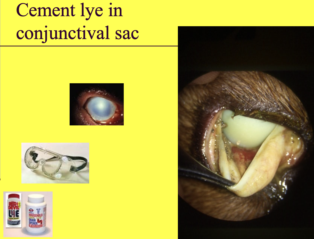
Cement lye when it gets into your eye
causes severe adhesions
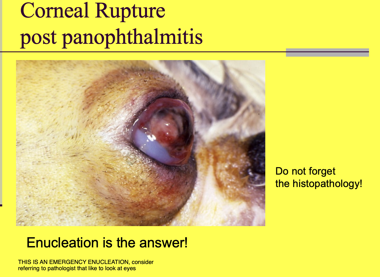
Corneal rupture post panophthalmitis tx
Enucleation - is highly infected and painful.
Remember to send in for histopath (look for underlying cause).
Protection of the cornea is done when a dog can't blink appropriately and might be done via (2)
Third eyelid flap.
Temporary tarsorrhapy.
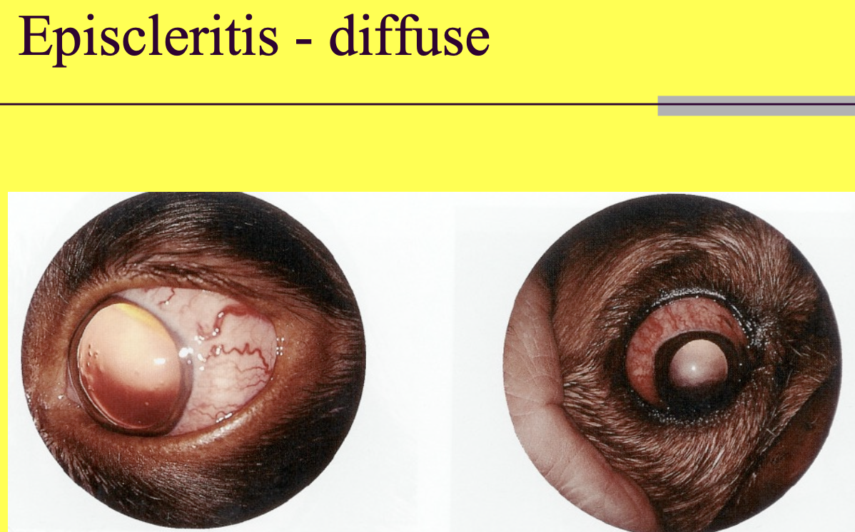
Episcleritis is often a
immune mediated process that can be chronic
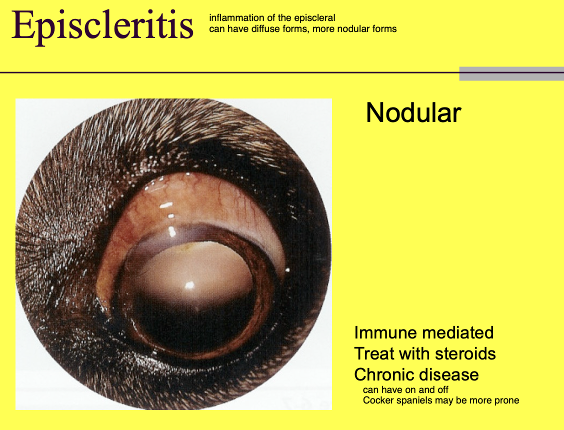
Different forms of episcleritis
necrotizing
proliferative (nodular) - e.g. NGE