phy3111 auditiory
1/28
There's no tags or description
Looks like no tags are added yet.
Name | Mastery | Learn | Test | Matching | Spaced | Call with Kai |
|---|
No analytics yet
Send a link to your students to track their progress
29 Terms
Describe three physical properties of sound waves and their perceptual correlates.
1. Frequency: frequency of a pure tone is the physical dimension related to the perceptual dimension of pitch, expressed in hertz (Hz)
2. Amplitude: Difference between the max and min sound pressure in a sound wave. The physical dimension of sound that is related to the perceptual dimension of loudness, measured in Decibels (dB)
3. Waveforms: A graphic representation of the shape of a wave that indicates its characteristics.
Waveforms perceptual correlates can be harmonic, which is a component frequency of a complex waveform that is an integer multiple of the fundamental frequency
Waveforms perceptual correlates can also be Timbre, which is the difference in sound quality between two sounds with the same pitch and loudness.
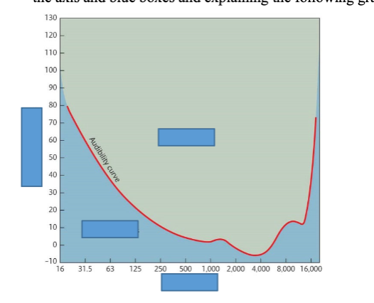
Describe the range of sound frequencies that a normal young human can perceive. Is perceiving sound frequency related to its intensity? Describe your response by labelling the axis and blue boxes and explaining the following graph.
Frequency range: Approximately 20 Hz to 20,000 Hz (20 kHz) for a normal young adult. The range decreases with age (presbycusis), with high frequencies lost first.
Frequency and intensity relationship: Yes, the threshold for detecting a sound varies with frequency. The audibility curve (absolute threshold curve) shows the minimum amplitude (dB SPL) needed to just hear a tone at each frequency.

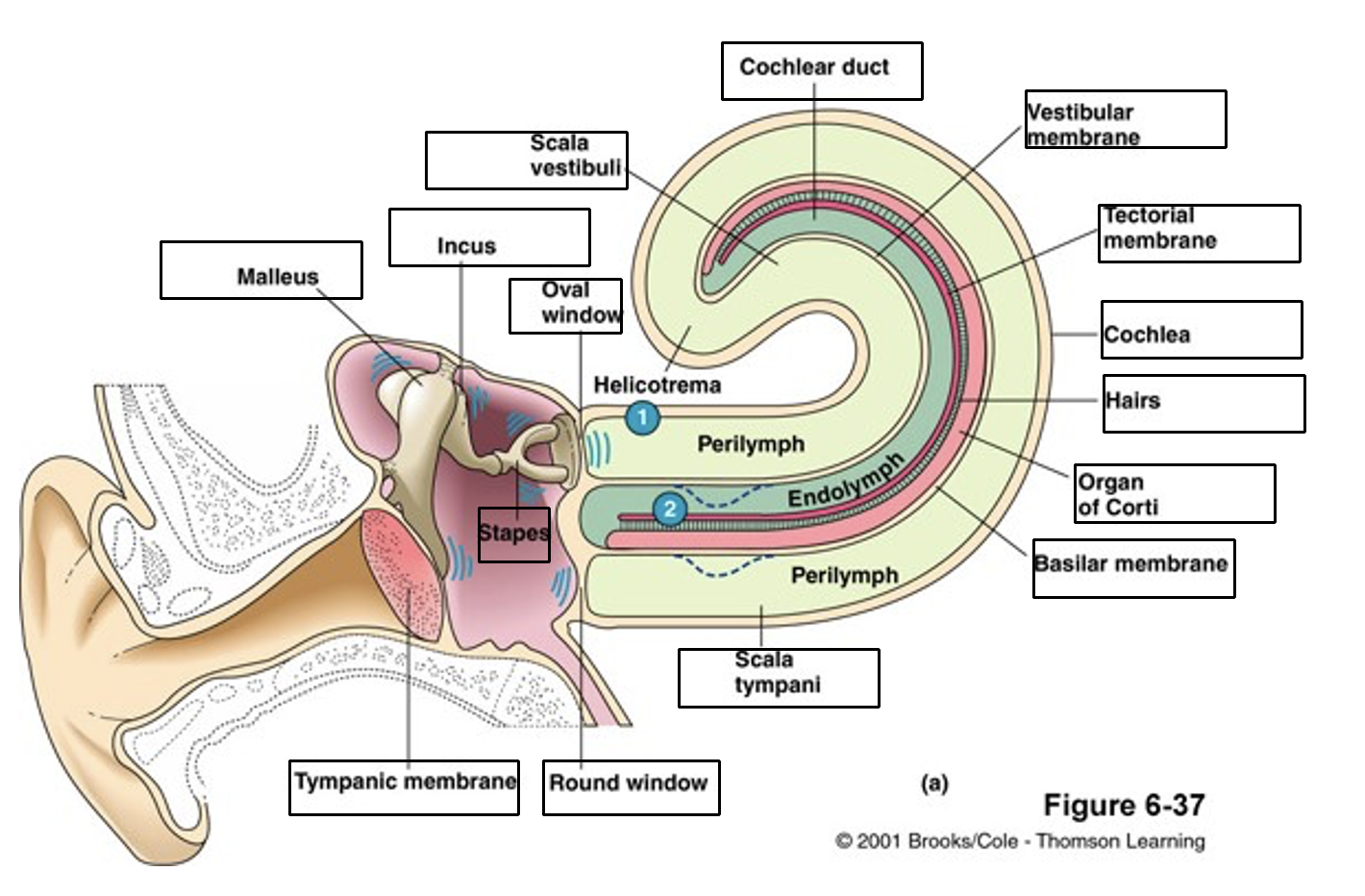
What is the function of ‘PINNA’ in hearing sounds?
Pinna is the outmost portion of the ear
Sound collection and funnelling: The pinna gathers sound waves from the environment and funnels them into the auditory canal, directing them toward the tympanic membrane.
What is the function of ‘TYMPANIC MEMBRANE’ in hearing sounds?
The tympanic membrane (eardrum) is a thin, elastic diaphragm at the inner end of the auditory canal. It vibrates in response to the fluctuating air pressure of incoming sound waves, converting them into mechanical vibration.
It forms an airtight seal between the outer ear and middle ear, ensuring that sound energy is efficiently transferred to the ossicular chain (beginning with the malleus, which is attached to the tympanic membrane).
Describe ‘impedance matching’ and how middle ear structures contribute to that.
The tympanic membrane and ossicles overcome the impedance mismatch between air and cochlear fluid, preventing most sound energy from being reflected.
Impedance matching is one of the important functions of the middle ear
The middle ear transfers the incoming vibration from the comparatively large, low impedance tympanic membrane, to the much smaller, high impedance oval window.
This converts low pressure, high displacement vibrations into high pressure, low displacement vibrations suitable for driving cochlear fluids
What is the main function of middle ear Ossicles in hearing sounds?
The three ossicles (malleus, incus, stapes) form a mechanical linkage chain that transmits vibrations from the tympanic membrane to the oval window of the cochlea.
Their main function is impedance matching amplifying the pressure of sound vibrations so that sound energy can efficiently transfer from low-impedance air to the high-impedance fluid of the cochlea.
The sequence is: tympanic membrane → malleus (hammer) → incus (anvil) → stapes (stirrup) → oval window → cochlear fluid.
Describe the “Acoustic reflex”
The acoustic reflex begins with sound presented to the ear.
Transduction of sound occurs in the cochlea, resulting in action potential that is transmitted along the auditory nerve to the cochlear nucleus in the brain station.
Interneurons in the ventral cochlear nucleus project directly or indirectly to middle ear muscle motor neurons located within the brain steam. Motor neurons provide efferent innervation to the middle ear muscles.
Describe the role of middle ear muscles.
There are two middle ear muscles, the stapedius and the tensor tympani
The stapedius contracts in response to intense low frequency acoustic stimuli, exerting forces perpendicular to the stapes and increasing middle ear impedance and attenuating the intensity of sound energy that reaches the cochlea (inner ear).
The tensor tympani contracts in response to self-generated sound (speaking, chewing, swallowing)
Describe the role of “Eustachian tube” in hearing sounds.
The Eustachian tube connects the middle ear cavity to the nasopharynx (back of the throat). It is normally closed but briefly opens during swallowing or yawning.
Its primary role is to equalize air pressure between the middle ear and the external environment. Proper vibration of the tympanic membrane requires the air pressure on both sides to be approximately equal.
When ambient pressure changes rapidly (e.g. aircraft ascent/descent, elevator), the pressure differential causes the tympanic membrane to bulge inward or outward (muffled hearing, ear pain). Opening the tube (swallowing, yawning) allows pressure equalisation.
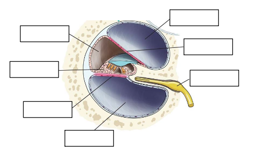
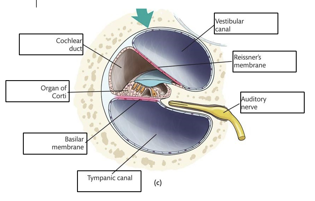
Please label the arrows in the following image (a cross section of Cochlea).
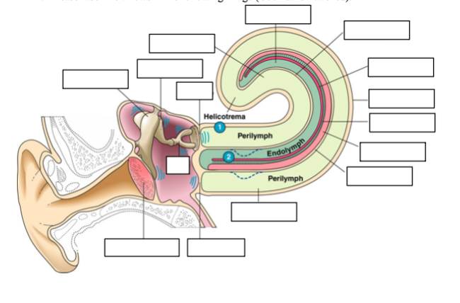
Please label the arrows in the following image (Cochlea is flattened).
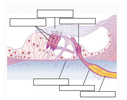
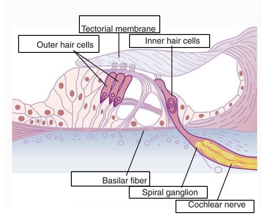
Please label the arrows in the following image (Organ of Corti).
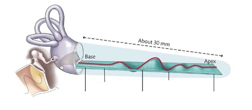
Referring to the following picture, please describe how the frequency of sounds are detected and encoded within Cochlea.
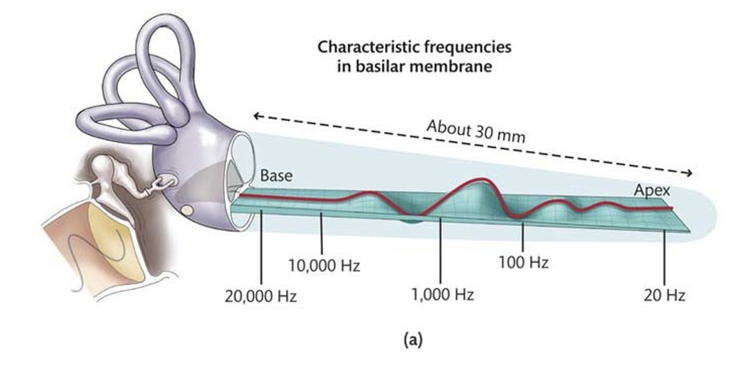
What is the meaning of “characteristic frequency” of the basilar membrane?
The characteristic frequency (CF) of a location on the basilar membrane is the specific sound frequency to which that location responds most readily
The base of the basilar membrane responds best to high frequency waves
The apex responds best to low-frequency waves
The characteristic frequency at each location determines the displacement of the membrane in response to a given frequency.
Describe how hair cells on the Corti organ transduce sound into neural signals.
The basilar and tectorial membranes have different anchor points. So, when the basilar membrane moves, the tectorial membrane moves in opposition, creating a shearing force that causes the stereocilia of the outer hair cells to bend and the stereocilia of the inner hairs cells bend as they sweep through the endolymph due to the movement of the basilar membrane.
The bending of the stereocilia of hair cells increases the tension on the tip links that connects them.
This tip link opens up calcium and potassium channels, allowing calcium and potassium ions to enter the hair cells.
Action potentials are generated and sent along to type 1 auditory nerve fibers that are connected to the inner hair cells and this causes transduction of sound into neural signals
Describe and compare two characteristics of the inner and outer hair cells.
Inner hair cells:
Connected to type I auditory nerve fibers
Responsible for transduction of sound into neural signals
Outer hair cells:
Connected to type II auditory nerve fibers
Serve to amplify and sharpen the responses of the inner hair cells
What is the meaning of “Place code” in transducing sound into neural signals?
The frequency that is represented by the displacement of different locations of the basilar membrane.
Varying degrees of displacement result in correspondingly different rates of action potentials being sent along the type 1 auditory nerve fibers at those locations.
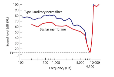
Describe the following diagram which shows the activation of basilar membrane and an auditory afferent nerve fibre.
Describe two mechanisms for the “Determination of Loudness”.
As sound becomes louder, the amplitude of vibration of the basilar membrane and hair cells also increase, and excitation of nerve endings is exaggerated
As the amplitude of vibration increases, this causes more hair cells on the neighbouring regions of the resonating portion to be stimulated. Therefore, many nerve fibers change their firing rate, also known as spatial summation.
additional:Outer hair cells become activated at higher vibration level and they may also contribute to perception of loud sounds
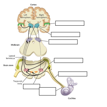
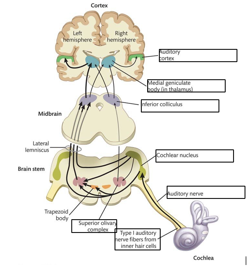
Please label the boxes in the following image:
What is the meaning of tonotopic organization of auditory cortex?
The auditory cortex is made up of
the primary auditory cortex (A1)
the rostral core
the rostro-temporal core
These regions have a tonotopic organisation, meaning that specific regions will respond to specific characteristic frequencies, ranging from low to high frequencies
Describe two mechanisms for perceiving the left–right location of sound.
Interaural time difference (ITD)
Refers to the difference in time of sound arrival in both ears.
The direction-dependent differences in path lengths that sound must travel to reach each ear from the source will generate different times of arrival of the sound at the two ears.
Interaural intensity / level difference (IID /ILD):
Due to shadowing of sound wave by the head, sounds coming in from a source located closer to one side of the head will have a higher intensity at the ear nearest the sound source
Describe the anatomical location of the primary auditory cortex in the human brain?
The primary auditory cortex (A1, Brodmann areas 41 and 42) is located on the superior surface of the temporal lobe, specifically on Heschl's gyrus (transverse temporal gyrus).
It lies tucked within the lateral sulcus (Sylvian fissure), which separates the temporal lobe from the frontal and parietal lobes; so it is largely hidden from lateral view unless the temporal lobe is pulled down.
Surrounding A1 is the auditory belt cortex (secondary auditory cortex), which in turn is surrounded by the parabelt.
All three regions have tonotopic organisation. The 'what' pathway (identifying sounds) runs anteriorly toward the temporal pole and prefrontal cortex; the 'where' pathway (localising sounds) runs posteriorly toward the parietal cortex.
Describe a mechanism for perceiving the up–down location of sound.
The human PINNA
Provides information used to judge elevation
A PINNA-induced modification in a sound's frequency spectrum provides information about the elevation of the sound source.
Describe a mechanism for perceiving the distance of sound.
Doppler effect:
The frequency of a sound emitted by a moving sound source is higher in front of the sound source behind it.
The frequency rapidly decreases as the sound source passes the listener
Additional: Echolocation:
Sound localisation based on emitting sounds and then processing the echoes to determine the nature and location of the object that produced the echoes
Describe how semi-circular canals contribute to the control of balance/posture.
Rotation of the head causes the endolymph within one or more of the semi-circular canals to move
This movement causes displacement of the cupula, causing the stereocilia of the hair cells to bend, signalling the position to the vestibular nerve and then trasmitting through the vestibulocochlear nerve and into the brainstem and cerebellum. Therefore, affecting balance/posture
Describe how Utricle & Saccule contribute to the control of balance/posture.
Tilting of the head causes the otolithic membrane to slide over the macula in the direction of gravity.
This then bends some of the stereocilia, causing depolarisation of the hair cells.
The difference of inertia between the hair cells stereocilia and the otolithic membrane creates a shearing force and causes the stereocilia to bend in the direction of that linear acceleration
The exact position of the head is interpreted by the brain based on the pattern of hair-cell depolarisation
Name three brain areas that receive information from the vestibular system.
Vestibular nuclei (brainstem): the primary relay station; coordinates vestibulospinal and vestibulo-ocular reflexes.
Cerebellum (flocculonodular lobe / vestibulocerebellum): uses vestibular input for balance, posture, and coordination of eye movements.
Thalamus (and via thalamus to the vestibular cortex in the parietal lobe / insular cortex): contributes to conscious perception of head position and balance, and spatial orientation.
How do we perceive linear acceleration with closed eyes (e. g. when a car starts moving and then stops).
Linear acceleration is detected by the utricle (horizontal acceleration) and saccule (vertical acceleration).
When a car accelerates forward, inertia causes the heavy otolithic membrane to lag behind the hair cells-it slides backward (posteriorly) relative to the macula. This creates a shearing force on the stereocilia, depolarising hair cells whose stereocilia bend in the direction of acceleration, and hyperpolarising those bending in the opposite direction.
The resulting pattern of increased and decreased firing in vestibular nerve afferents signals the direction and magnitude of linear acceleration to the brainstem and cerebellum.
When the car decelerates (brakes), the otolithic membrane slides forward, bending stereocilia in the opposite direction and producing the opposite pattern of nerve firing — signalling deceleration.
The brain integrates the otolith signal with neck proprioceptors and visual information (when eyes are open) to maintain awareness of body position and to trigger appropriate postural adjustments via vestibulospinal pathways.