Micro Lab Midterm
1/68
There's no tags or description
Looks like no tags are added yet.
Name | Mastery | Learn | Test | Matching | Spaced | Call with Kai |
|---|
No analytics yet
Send a link to your students to track their progress
69 Terms
Two Tube Transfer
smack that thing
pick up and place tubes in hand
sterilize loop
uncap tubes
sterilize tubes- 3 or 4 s
transfer culture
recap tubes
sterilize loop
place everything back
cationic dyes
positive ion that exhibits color
methylene blue or crystal violet
anionic dyes
negative ion that exhibits color, doesnt penetrate cell, no heat fixation
acid fuchsin, congo red, or nigrosin
how long should heat fixation be for
10 seconds
resolving power of our microscopes vs average
around 0.2 µm vs 1.0
resolution equation
d = λ / 2NA
NA is numerical aperture or light gathering capacity of the lens
example of vibrio
V. cholerae
example of spirochete
Spirillum volutans
example of club shaped
Corynebacterium diptheriae
what type of stain is a gram stain
differential
history of gram stain
developed by danish Hans Christian Gram in mid 1880s
tried to differentiate bacteria causing pneumonia from lungs of deceased patients
considered stain a failure
pour plate
culture diluted in molten agar, poured into empty sterile petri dish and solidified
primary and secondary streak
primary: isolation of mixed colonies from each other on first plate
secondary: isolation of a single colony from first plate
general purpose media
maintain culture
nutrient agar (NA), tryptic soy agar (TSA), brain heart infusion agar- enriched (BHI)
selective media
inhibitors prevent growth of certain organisms
phenyl ethyl agar (PEA), eosin methylene blue (EMB), MacConkey agar (Mac)
differential media
growth of many microbes differentiated w/ indicators like dyes (fermentation causes pH change and color diff)
blood agar (BA), eosin methylene blue (EMB), MacConkey agar (Mac)
combination
selective and differential
Mac and EMB
complex media
undefined chemical composition, nutrients are extracts or digests from naturals
BHI and TSA
PEA
phenyl ethyl agar
selective only, inhibitor: phenylethyl alcohol, prevents gram neg cells
BA
blood agar
differential only, indicator: rbc, ex. TSA w/ 5% sheep’s blood, detects hemolysis, Beta- Best, Alpha- Average, Gamma- Garbage
cannot detect gram reaction
Mac
MacConkey agar
combination, indicator: neutral red for lactose and non-lactose fermenters, inhibitor: crystal violet and bile salts, block gram pos, used to show gram neg enterics (Enterobacteriaceae)
EMB
eosin methylene blue
combination, inhibitor and indicator: eosin and methylene blue, lactose and non-lactose fermenters, screens for coliforms (fecal, also UTIs)
2,3-butanediol in EMB
pale pink lavender color
used by Enterobacter aerogenes
mixed acid in EMB
produce very dark, black, green metallic sheen
motility agar
detect mobile flagella, soft agar, dye is optional (tetrazolium chloride) and turbidity or cloudiness is used, single line slab- motile spread away from line
antimicrobial agents 3 kinds
chemicals that kill or inhibit growth of microbes
disinfectants: strong, inanimate surfaces, not as effective as sterilization, may not kill spores
antiseptics: safely applied to living tissue, prevent infection
antibiotics: systemic circulation, most are synthetic analogs of naturals
disk diffusion asssay
tests effectiveness of antimicrobial compounds
E. coli and S. aureus
sterilize foreceps, dip in alcohol and ignite over burner, always downward facing, pick up disks and dip in betadine, hydrogen peroxide, or lysol bleach, transfer to plates
kirby bauer test
highly standardized testing antibiotic sensitivity
lawn inoculation, stamped with antibiotics and incubated on MH
zone of inhibition measured in mm and compared to table
mueller hinton (MH) agar or broth
MH agar or broth
mueller hinton
used in kirby bauer test, simple nutrient composition, neutral pH (7.2-4), 4mm depth for lateral antibiotic diffusion
too low of bacteria inoculated kirby bauer
overestimation of sensitivity to the antibiotics
too high of bacteria inoculted kirby bauer
swamp antimicrobial or deplete nutrients
agar plates that test for exoenzymes
lipase
milk agar
starch agar
tube media that tests for metabolic processes
litmus milk
sugar fermentation broth
Kligler’s Iron agar
nitrate broth
gelatin
urea broth
phenylalanine
SIM
IMViC
litmus milk
contains: lactose, casein and other peptones, litmus pH indicator
test for: fermentation lactose, metabolism proteins, degradation casein, litmus reduction
results in: acid production (pink), alkaline production, (blue) curd, redox (white is reduced), proteolysis (clearing)
in litmus milk, what does deamination of peptones produce
a blue, alkaline production usually at the top of the tube due to oxygen dependence
will litmus milk function as a pH indicator if it is in a reduced state
no, only when oxidized, so it will be white if reduced
what results in proteolysis for litmus milk test
caseinase enzyme to degrade casein
Phenyl Red (PR) Sugar fermentation broth
contains one sugar: glucose, lactose, or mannitol
indicators: phenyl red and a durham tube
fermentation: phenol red turns yellow, gas bubble in tube
can’t ferment: phenol turns pink (cerise), peptone degradation releases ammonia
Kligler’s Iron Agar (KIA)
tests for: lactose, glucose fermentation, and sulfur reduction
ingredients: 1% lactose, 0.1% glucose, 1% peptone (cysteine- ammonia sulfur)
indicators: phenol red, iron from ferric ammonium citrate (combines w/ H2S to form black precipitate)
lactose fermenters: 1% lactose + 0.1% glucose > 1% peptone, whole tube yellow
glucose only: 0.1% glucose < 1% peptones, need O to breakdown so red on top, bottom is yellow
reversion: oxidation of weaker less stable acids at top of tube, Enterobacter aerogenes
sulfur reduction: H2S combines w/ iron, black precipitate at bottom of tube
lipase plate
differential, tests for enzyme lipase
lipase: hydrolyzes fats into glycerol and fatty acids
indicator: spirit blue
hydrolysis produces dark blue zone around growth w/ no oily surface
milk agar plate
differential, tests for enzyme caseinase
caseinase: hydrolyzes casein (milk protein) into amino acid products
clearing is a positive result
starch agar
differential, tests for enzyme amylase
amylase: breaks down starch into simple sugars
indicator: iodine added after colony growth
iodine and starch make a darker purple ring, and a clearing
biochemical tests
catalase
oxidase
fermentation v respiration similarities
both see conversion of molecules into ATP, both see glycolysis (glucose into pyruvate)
respiration
final e- acceptor O: aerobic
final e- acceptor Nitrate, Sulfate, etc: anaerobic
glycolysis → kreb’s cycle → ETC
ETC
transfer e- to molecules that have a more positive reduction potential, indirectly testing for presence of ETC
flavoprotein → FeS cluster → ubiquinone → cytochrome C → cytochrome C oxidase → final e- acceptor
catalase test
aerobic metabolism produces ROS: superoxide radical, H2O2
aerobic and facultative aerobic break down ROS w/ SOD and catalase
2 H2O2 → 2 H2O + O2
add 3% H2O2 to edge of colony, kills organism, bubbles means catalase is present
cytochrome C oxidase test
cytochrome C oxidase: removes e- from cytochrome C (oxidizes) and transfers to O (reduces)
chromogenic reducing agent: dimethyl-p-phenylenediamine, aromatci amine
^^ when oxidized by cytochrome C oxidase, changes color to purple blue and passes e- back to cytochrome C
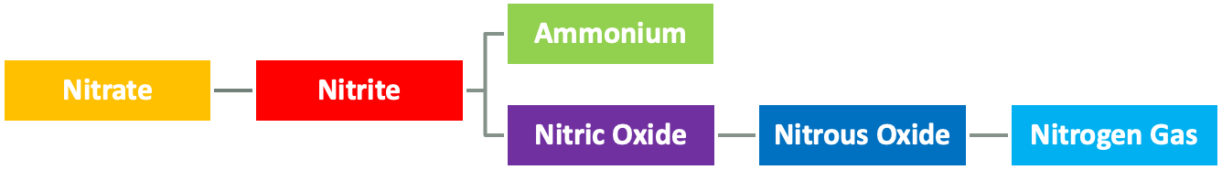
nitrate respiration test
nitrate (NO3) used as final inorganic e- acceptor
needs enzyme nitrate reductase to convert nitrate to nitrite
or another to reduced nitrate to N2 (g) (denitrification)
or nitrate to ammonium for incorporation (assimilatory nitrate reduction)
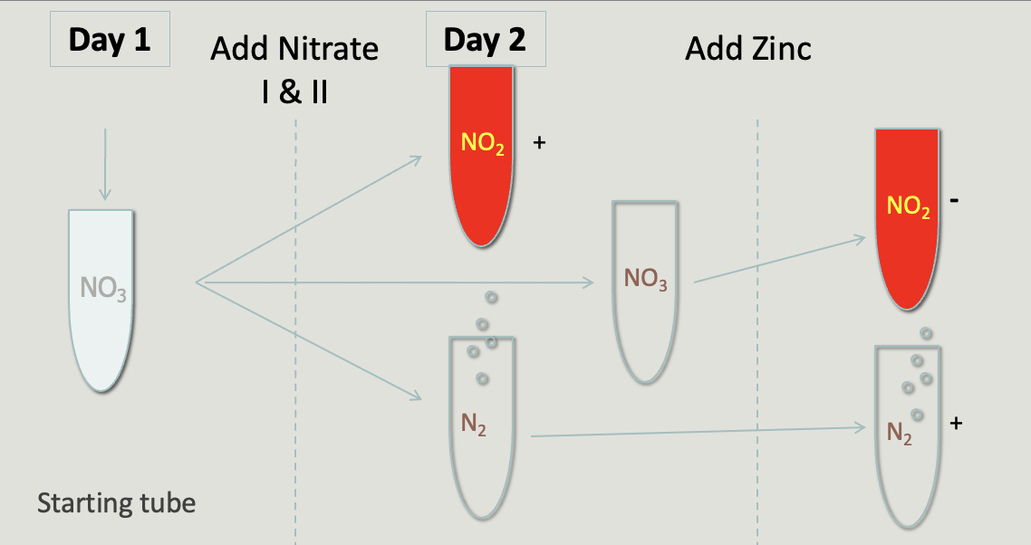
nitrate broth
detects nitrate reductase (nitrate to nitrite, NO3 → NO2)
reagents: nitrate 1 (sulfanilic acid) and nitrate 2 (dimethyl-alpha-naphthylamine)
^^ reacts with nitrite to form brick red color
zinc powder can be used as a catalyst for rxn if color doesnt change to confirm that microbe is negative for nitrate reductase
^^ if tube remains turbid clear it has been reduced to N2 (g)
urea broth
detects urease: degrades urea → 2 ammonia (NH3) and CO2
pH increases to more alkaline, changes color to cerise (hot pink)
indicator: phenol red
pH > 8.1 (cerise); neutral (red); decreases (yellow)
gelatin
detects presence of gelatinase: catalyzes hydrolysis gelatin → aa
viewed after chilling in ice bath
liquid is positive result
phenylalanine slant
detects production of enzyme phenylalanine deaminase
breaks phenylalanine → phenylpyruvic acid (PPA) + ammonia (NH3)
reagent: 5-10 drops 10% ferric chloride (FeCl3)
positive reaction changes yellow to green (pp in pool turns green)
SIM
3 results in single tube
S: Sulfide; bacteria produces H2S and it reacts w/ Fe → FeS; black precipitate
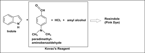
I: Indole; breakdown of tryptophan by tryptophanase produces this; 3-5 drops Kovac’s reagent; positive produces pink color
M: Motility; determined by turbidity
fermentation
final e- acceptor is organic molecule
does not produce as much energy as respiration
e- onto pyruvate → general end products organic acids and gases
mixed acid fermentation
2,3-butanediol fermentation
fermentation general term
breakdown of polysaccharides into monomers that can be fermented
lactose → glucose and galactose → pyruvate
MR-VP broth
two separate tests, but same exact medium (tested after incubation)
MR: methyl red tests for mixed acid fermenters; 3-4 drops; detects low acid (< 4.4); positive stays red, negative turns yellow
VP: vogues-proskauer; tests for 2,3-butanediol; barritt’s reagents (VP I and VP II) react w/ acetoin intermediate → acetoin; reacts w/ guanidine to produce red color
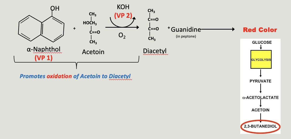
mixed acid v 2,3-butanediol
2,3-butanediol produces less acid and products are more easily converted to neutrality
(glucose → → acetoin → 2,3-butanediol)
baritt’s reagents
used in VP broth
VP I: alpha naphthol, intensifies red color
VP II: KOH, alkaline condition, favors acetoin oxidation → diacetal + O2, red color
tryptone broth
detects tryptophanase: hydrolyze tryptophan → indole, pyruvate, + ammonia (NH3)
3-5 drops Kovac’s reagent (p-DMABA and HCl dissolved amyl or butyl alcohol); positive is pink top (cerise)
simmons citrate slant
detects utilization of citrate as sole C source for growth, initially green
indicator: bromothymol blue
blue rxn almost always accompanies growth, but is not primary indicator, growth is
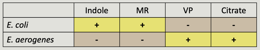
IMViC
set of four tests to differentiate E. coli from Enterobacter aerogenes
indole (SIM or tryptone)
methyl red
voges proskauer
citrate
dilution factor
A / (A+B)
A: volume transferred
B: volume being added to
plate dilution formulas
amount plated (mL) / 1 mL
dilutions for bacterial population counts
add 0.1 mL stock into 9.9 mL (1:100)
dilute 0.1 mL again into 9.9 mL (1:10,000)
do it once again (1:100,000)
0.1 mL (1:10,000,000) and 1 mL (1:100,000) of final tube onto plates
0.1 mL (1:100,000) and 1 mL (1:10,000) of 2nd tube onto plates
standard plate count
30 and 300 colonies in CFU/mL
which organism had negative results for phenol red sugar broth tests, which had all positives
negative: Pseudomonas aeruginosa
positive: E. coli, and gas production
which organism was able to degrade casein w/ proteolytic enzymes
Bacillus cereus, because there was a clearing
organism that caused a reversion in KIA slant
Enterobacter aerogenes, uses 2,3-butanediol fermentation