Bio 14 Exam 2
1/47
There's no tags or description
Looks like no tags are added yet.
Name | Mastery | Learn | Test | Matching | Spaced | Call with Kai |
|---|
No analytics yet
Send a link to your students to track their progress
48 Terms
homeostasis
A self-regulating process for maintaing internal stability while adjusting to changing external conditions
Direction of Homeostasis
1.) Sensors: Detect the issue to homeostasis
2.) Integrators: process info from multiple sensors
3.) Effectors: do something about the stimulus
Negative Feedback
A thing acts as its own signal to counteract something else, stabilizes systems
positive feedback
a thing acts as its own signal to do something even more, self-reinforcing. Generally not important to homeostasis
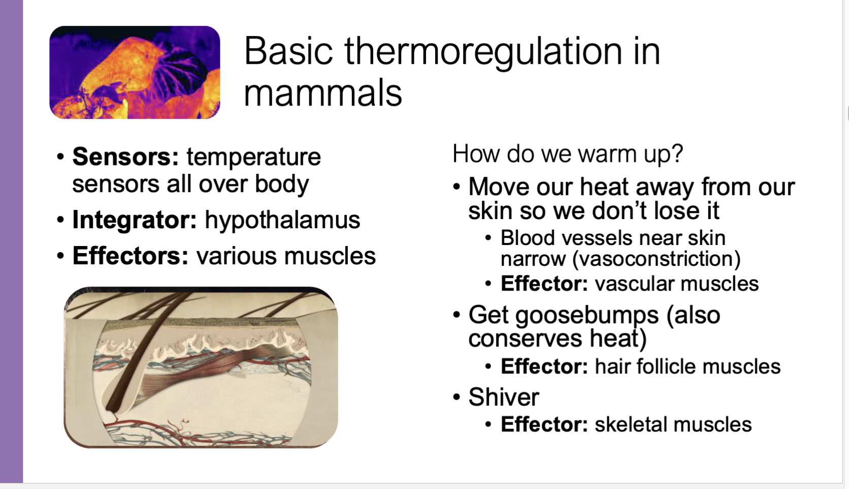
Examples of Homeostasis for thermoregulation of mammals
Isotonic
Solutions have equal solute concentration; no net movement of water (cell stays the same).
hypertonic
A solution with higher solute concentration than another; water moves out of the cell (cell shrinks).
Hypotonic
A solution with lower solute concentration; water moves into the cell (cell swells, may burst).
counter-gradient flow
when two fluids move in opposite directions, which helps maintain a constant gradient so transfer (like heat or oxygen) is more efficient. Uses energy to push molecules against the concentration gradient.
Osmosis
the passive movement of water across a selectively permeable membrane from an area of low solute concentration (high water) to high solute concentration (low water) until equilibrium is reached.
selective permability
Membrane proteins control what can and can’t cross, allows for solute gradients to form and allows for osmosis
Passive transport
No energy, facilitates flow down a gradient. Gradient flow mechanism
Active transport
Costs energy, counter-gradient flow mechanism
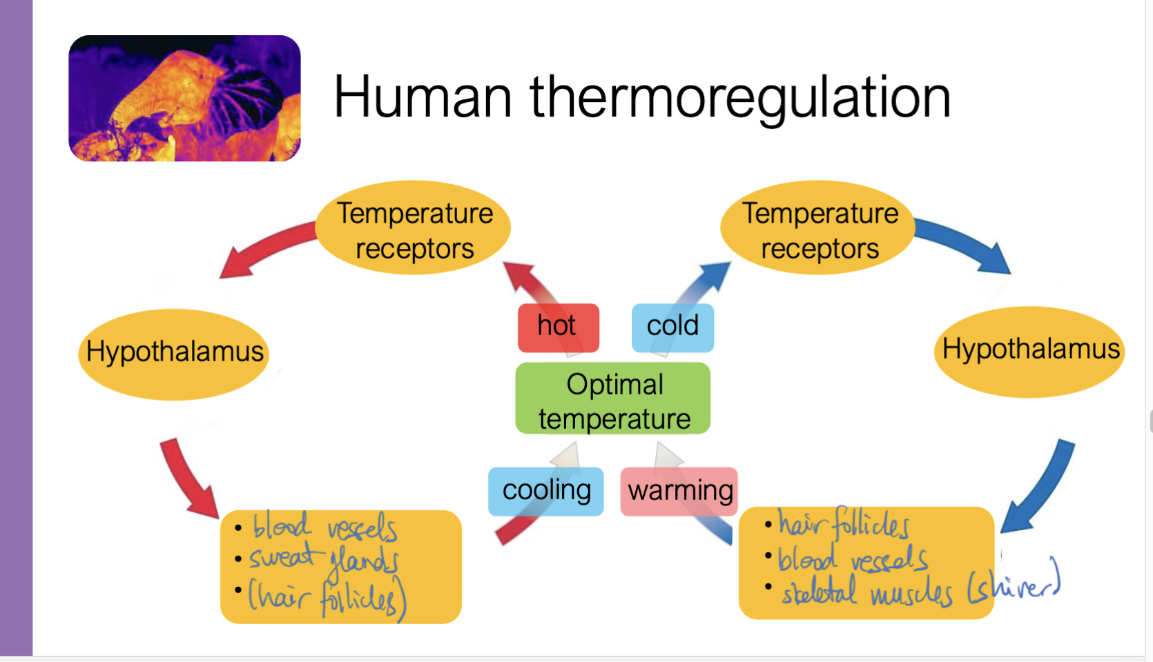
Human Thermregulation diagram
endotherms
internal heat generation regulated ot maintain temperature
ectotherms
can’t switch on internal heat, regulate temperature in other ways
homeotherms
keeps temperature very consistent
poikilotherms
tolerates some changes in temperature
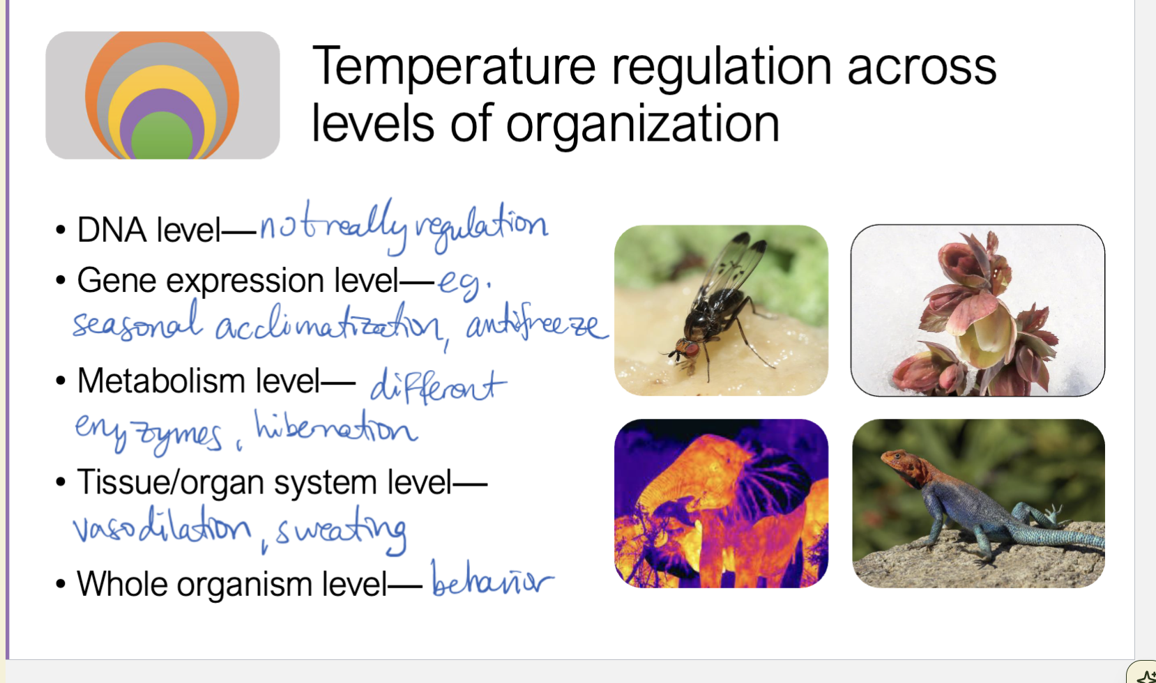
Tempterature regulation across levels of organization
Internal communication
Chemical and electricity based communication within the body with chemical being slower and electric being faster
Chemical signals
Longer lasting, more general over the entire body, slower
Electric signals
Fast, short-lived, targeted to specific regions of the body
Hormone
A signal molecule that triggers a particular response in distant target cells, a type of chemical signal
Endocrine signals
Hormones carried by blood, a type of chemical signal
Blood glucose regulation
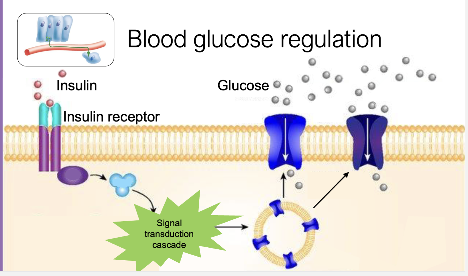
Blood Glucose Regulation (cellular level)

Key points about Blood Glucose Regulation
1.) insulin binds to a receptor on cell which amplifies the signal, positive feedback
2.) glucose transport proteins move towards cell membrane
3.) glucose enters the cell through glucose transport proteins via facilitated diffusion
Big Picture: neuron anatomy
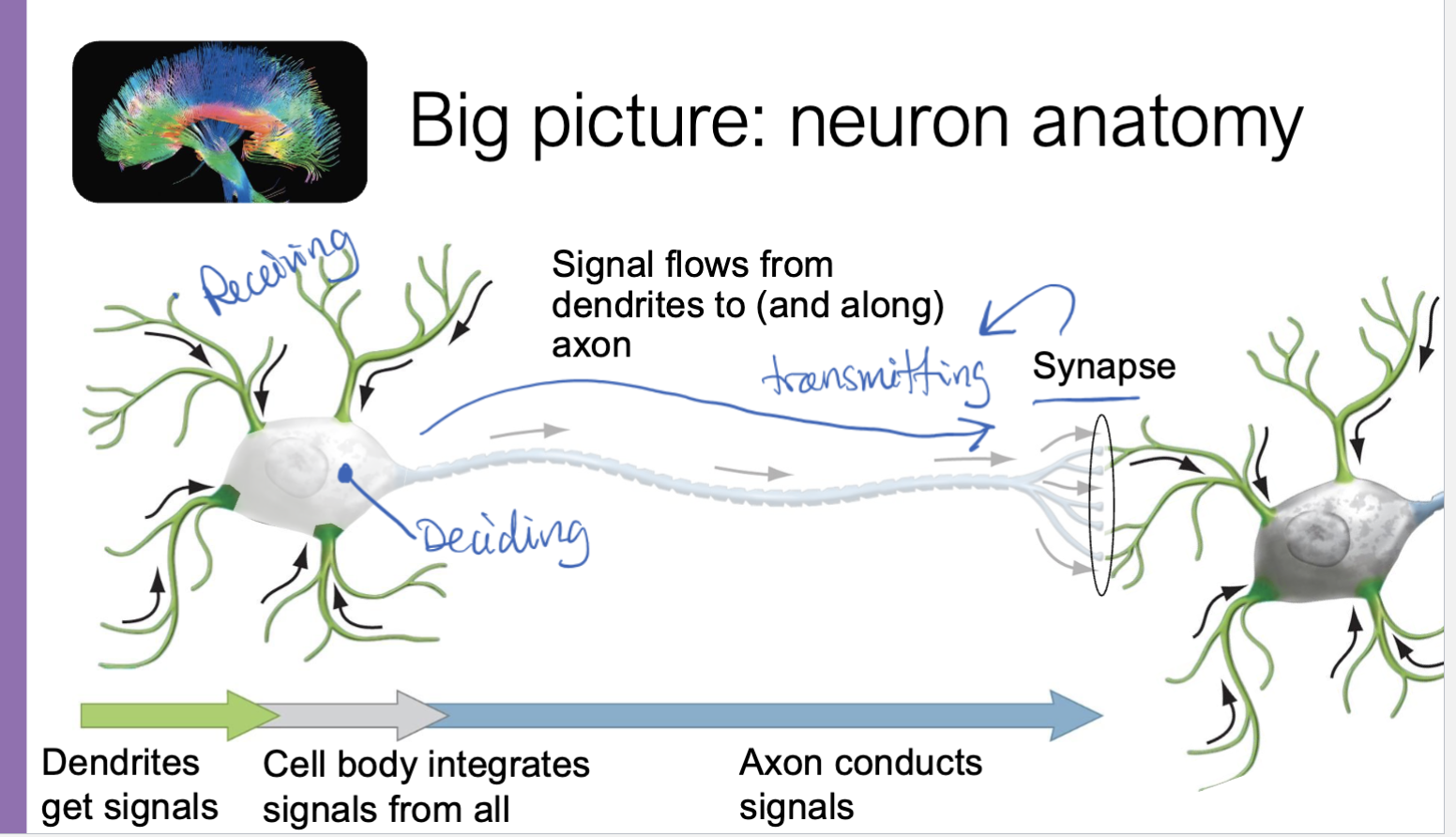
Dendrite
short, branch-like extensions of a neuron that receive signals from other cells and carry them toward the cell body.
Axon
a long fiber of a neuron that sends signals away from the cell body to other neurons, muscles, or glands.
Cell body (soma)
the central part of a neuron that contains the nucleus and organelles; it processes incoming signals and maintains the cell’s basic functions.
Signal
Electricity or voltage difference. Signals are either on or off (binary).
Signal strength
Comes from frequency (action potentials)
action potential
a rapid, temporary change in a neuron’s membrane potential (electrical charge) that travels down the axon as a nerve signal. It occurs when the neuron reaches a threshold, causing Na⁺ to rush in (depolarization) followed by K⁺ moving out (repolarization). Occurs because of gated channels
Signal consequence
Depends on which cell receives signal and which cell sent it
Electric potential
Separation of charge, potential to do work.
Cell membrane potential
charge difference across the membrane
Resting potential
typical membrane potential the cell maintains
Chemical gradient
Cells have a chemical gradient of potassium and sodium ions. Lots of potassium ions on the inside and sodium ions on the outside. This creates a resting potential and a charge difference.
Communication Mode: Chemical
Signal: Hormones
Sender: Organs and Tissues
Receiver: Any cell w/ right hormone receptor
Medium/Route: Blood
Communication Mode; Electrical
Signal: Action potentials
Sender: Neurons and other “excitatory cells”
Receiver: Specific, connected cells primed to receive signal
Medium/Route: Axons + synapses
Voltage gated channels
protein channels in the cell membrane that open or close in response to changes in membrane potential (voltage).
When the membrane reaches threshold, these channels open
Voltage-gated Na⁺ channels open first → Na⁺ rushes in → depolarization
Then voltage-gated K⁺ channels open → K⁺ leaves repolarization
Feedback in Neurons
Positive: transmitting down the axon
Negative: for repolarlization
presynaptic neuron
the neuron that sends the signal. It releases neurotransmitters from its axon terminal into the synapse.
Postsynaptic neuron
the neuron that receives the signal. It has receptors on its dendrites or cell body that bind those neurotransmitters and respond.
Action potential and release of neurotransmitters
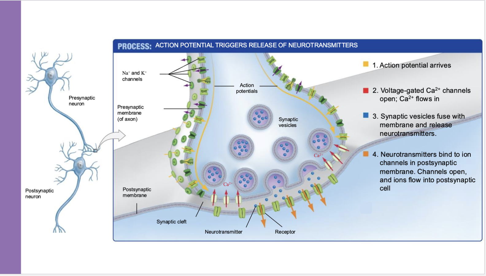
Neurotransmitters
chemical messengers released by neurons that carry signals across the synapse to another neuron, muscle, or gland.
How they work (quickly):
Released from the presynaptic neuron
Cross the synapse
Bind to receptors on the postsynaptic cell
Either excite (increase chance of firing) or inhibit (decrease chance)
Examples:
Dopamine → reward, motivation
Serotonin → mood, sleep
Acetylcholine → muscle movement
GABA → inhibitory (calming)