Extremities Nonvascular & Superficial Soft Tissue Structures
1/10
There's no tags or description
Looks like no tags are added yet.
Name | Mastery | Learn | Test | Matching | Spaced | Call with Kai |
|---|
No analytics yet
Send a link to your students to track their progress
11 Terms
muskuloskeletal structures
equipment: linear array transducer
must place transducer parallel to tendon to avoid anisotropy (artifact)
image symptomatic and asymptomatic areas for comparison
evaluate in its entirety in SAG and TRV planes
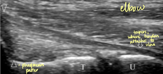
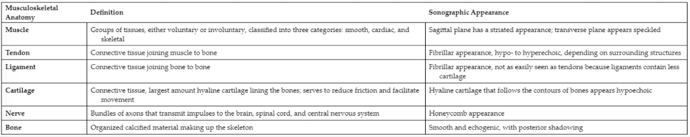
MSK structure: tendons
bundles of connective tissue that attach muscle to bone
proximal attachment is called the origin, and distal attachment is the insertion
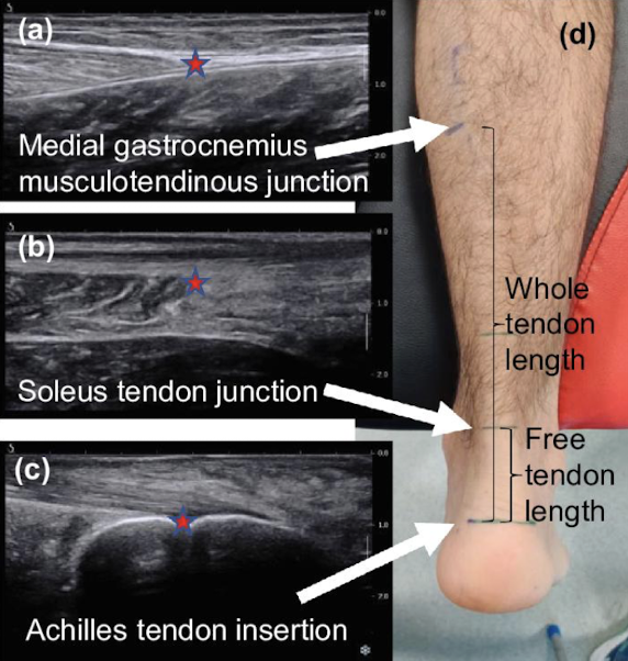
portion of the attachment to the muscle is called the musculotendinous junction
point of attachment to the bone is the osteotendinous junction
SONO: hypo-hyperechoic fibillary appearance; surrounded by thin, echogenic peritoneum
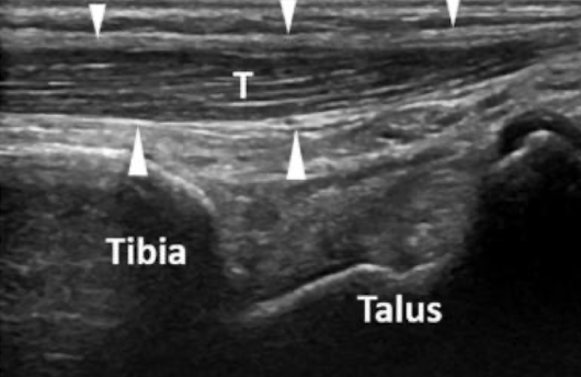
MSK structure: achilles tendon
MC injured ankle tendon
connects the calf muscle to the calcaneus (posterior surface of the feel)
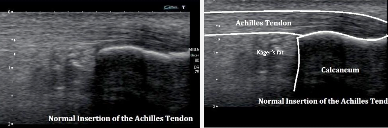
how to image achilles tendon
start high, then go downwards towards heel
look for breaks, thickening, fluid, etc. on the way down
MSK structure: ligaments
connective tissue that attach bone to bone
types: collagenous, not flexible, or elastic
elastic ligaments can stretch and recoil as the bone moves
SONO: echogenic fibrillar appearance; not easily seen because they contain less cartilage
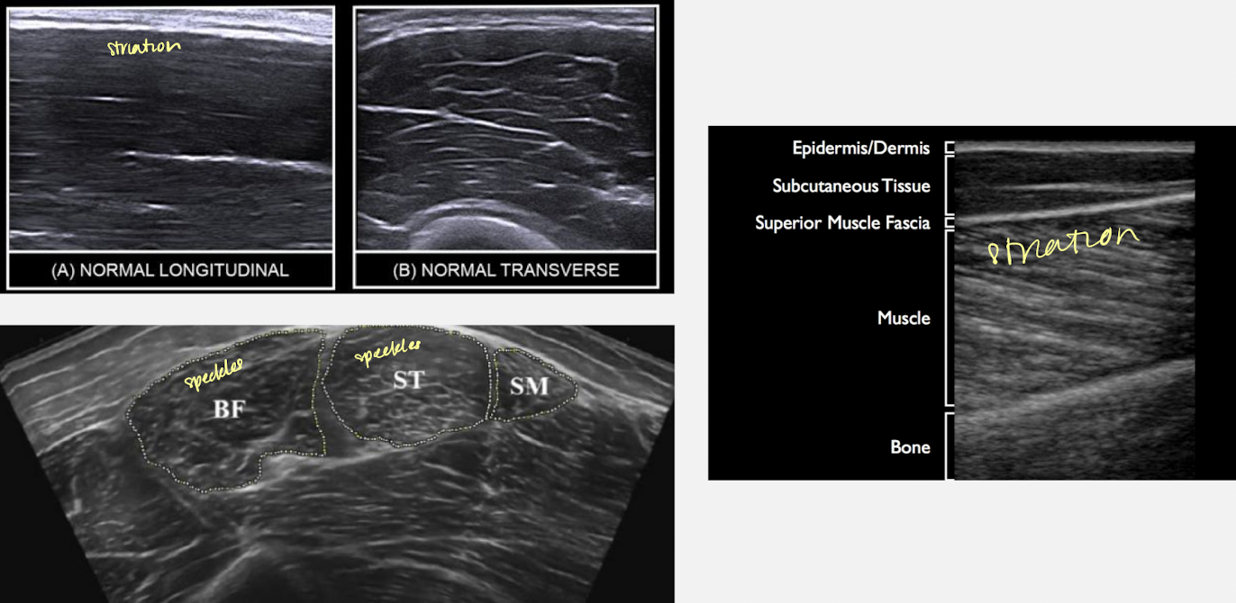
MSK structure: muscles
muscles are voluntary or involuntary (controlled by Autonomic Nervous System)
3 types: skeletal; smooth; cardiac
skeletal: controlled through the nervous system; attached to bones
cardiac: smooth muscle involving the heart; involuntary
smooth: attached to internal organs and muscles surrounding the blood vessels
SONO:
hypoechoic with linear, echogenic strands/striations when imaged SAG
speckled “starry night” appearance when imaged TRV
tendonitis
inflammation of the tendon
S/S: pain, swelling
SONO:
thickened and hypoechoic tendon (diffuse) or enlarged hypoechoic area within tendon (local)
possible hyperechoic flow within the tendon
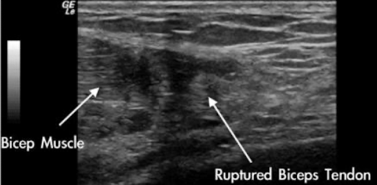
tendon rupture
aka “tear”
S/S: significant pain; edema; fluid accumulation
SONO:
partial tear=focal hypoechoic area within tendon
complete tear=anechoic or heterogeneous area within the tendon, indicative of hematoma
complete ruptures also show shadowing in area of the separated tendon, with fat, a hematoma, or granulomatous material filling the gap created by the tear
**MRI is gold standard
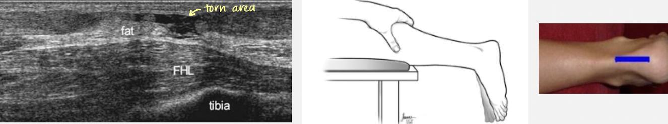
achilles rupture
S/S: posterior ankle pain
transducer is placed misline above the heel; light touch; ample amount of gel; dorsiflexion (foot lifted towards the chin)
thompson to determine complete or partial tear (can’t flex if theres a complete tear)
pt. prone, squeeze calf → foot should plantar flex when a complete tear is not present