3: Exchange with the Environment
1/62
There's no tags or description
Looks like no tags are added yet.
Name | Mastery | Learn | Test | Matching | Spaced | Call with Kai |
|---|
No analytics yet
Send a link to your students to track their progress
63 Terms
how does surface area:volume affect exchange rate in organisms
the larger an organism gets, the lower its SA:V is, but the greater its metabolic rate(energy expedned by the organism in a time period/day), so multicellular organism have evolved more complex mass transport and exchange systems
unicellular vs multicellular organisms exchange
unicellular: large SA:V, short difffusion distance
+can exchange materials directly with the environment bc all of the cell has surface exposed to the outside
-loses heat energy and water quickly, cannot survive extreme heat/cold
multicellular: small SA:V, large diffusion distance
+loses less energy as heat so can survive in the cold more easily
-some cells have no surfaces exposed to the outside so need internal mass transport systems, in hot environments they need adaptations to cool down
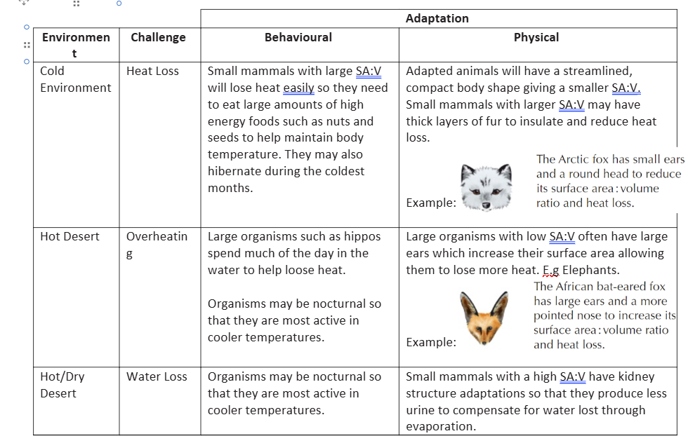
behavioural and physical adaptations for hot deserts and cold environments
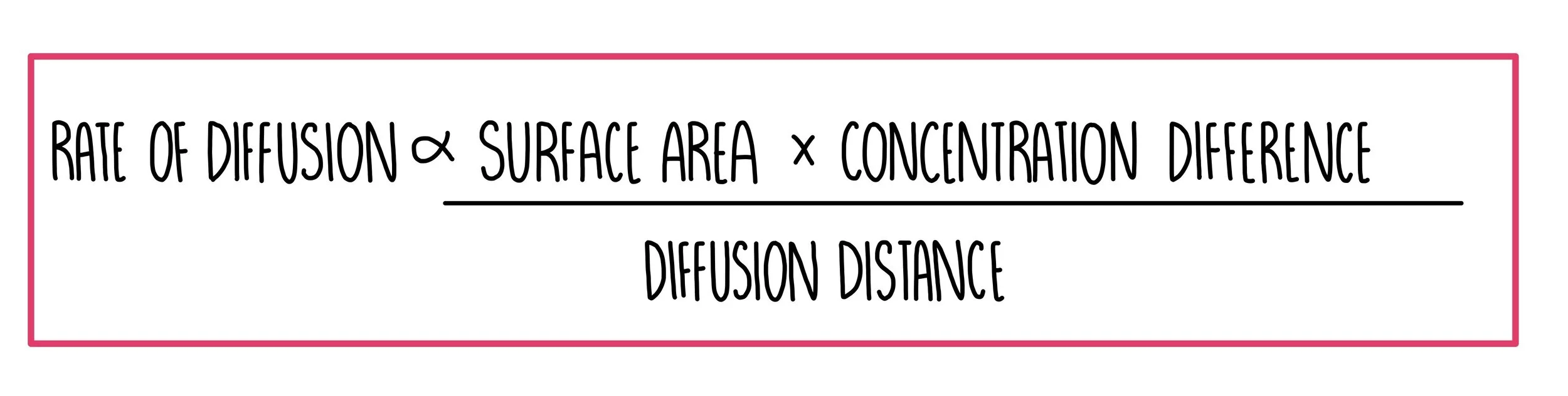
what is Fick’s law
diffusion is proportional to
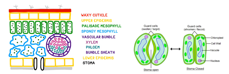
cross section of leaf and functions of each tissue
waxy cuticle- transparent layer on the top of the leaf
upper epidermis-transports water and minerals
palisade mesophyll- layers of palisade cells for photosynthesis, air spaces between them for gas exchange to occur
spongy mesophyll-tiny pores on the underside of the leaf for gases to diffuse in/out, water vapour is also lost from here
veins/vascular bundle: contains xylem-for water transport phloem- transports dissolved sugars in the plant
lower epidermis- to prevent water loss
stomata and guard cells-allow water and gases in/out of the cell. When plants have enough water guard cells are turgid which keeps the pores open, but when plants are dehydrated the guard cells become flaccid, causing the hole to close
xerophytic adaptations
xerophyte-plant with extra adaptations to prevent too much water loss when their stomata are open, mostly by reducing transpiration
small leaf surface area: reduced surface area for evapouration and fewer stomato
sunken stomata: maintain humid air around the stomata to reduce the water potential gradient
stomatal hairs(trichores): maintains humid air around the stomato to reduce the water potential gradient and reduce evapouration
rolled leaves: reduces the effects of wind to reduce the water potential gradient and reduce evapouration
extensive root systems: maximises water uptake, helps to increase chances of contact with water, often shallow but wide area to absorb rainfall. Some (Succulents) have swollen stems to store collected water
reduced number of stomata: reduce the amount of places water can evaporate from
thicker waxy cuticle: waterproof leaves and stems to reduce evapouration
investigating stomatal density
1.peel a thin layers of epidermis
2. mount and on a slide and add a drop of water and examine under a microscope
3. examining under the microscope, the number of stomata in an area of leaf tissue can be calculated as stomatal density per mm2.
use multiple fields of view and calculate a mean
pi*radius of microscope lens2 = area
mean number of stomata/area
How to find the surface area of a leaf
place the leave ln 1cm² sqaured graph paper
Trace around the leave in pencil
Count the number of squares, partial squares count as 0.5
Number of squares x2 bc there are 2 sides of the leave
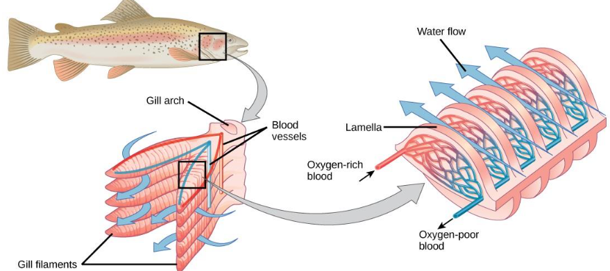
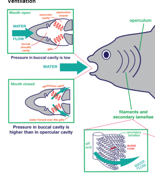
gill structure
filaments- attached to the gill arch and feather out to create a large surface area
lamellae- flattened disc like membranes at right angles to the gill filaments, which increase the surface area of the gills. They contain capillaries. Gas exchange happens at the lamellae
adaptations for gas exchange in fish
thin epithelium/walls of lamella- shorten diffuusion distance of gases to blood
large number of filaments and lamella- increase surface area for gas exchange
large number of capillaries around lamellae-circulation constantly removes oxygenated blood to maintain the steep concentration gradient
ventilation by operculum- ensure constant fresh water flow over gills to replace lost oxygen and maintain steep concentration gradient
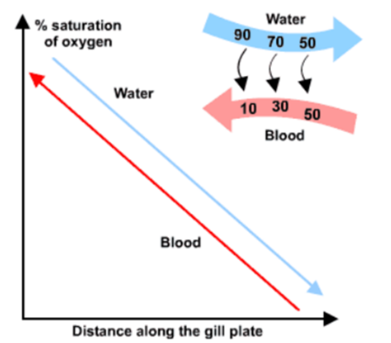
countercurrent flow
water flows over the lamellae in the opposite direction to blood flow in the lamellae capillaries
the water always has a higher oxygen concentration than the blood, so diffusion can happen across the full length of the lamellae as the conc gradient is maintained
the blood absorbs more oxygen as it moves along, but since the there still a concentration gradient more oxygen can flow into the blood
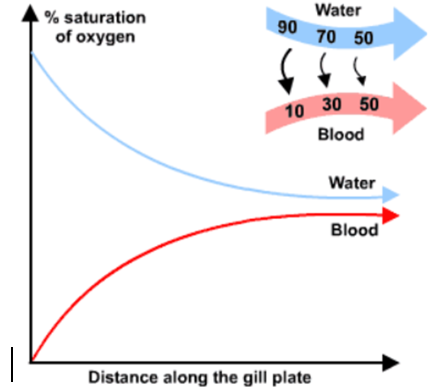
concurrent flow
water flows over the lamella in the same direction the blood flows through the lamellae
at first there is a very large concentration gradient as water has a much higher oxygen concentration, so diffusion occurs
as they flow along the lamellae the concentration gradient is decreased until equillibrium is reached and no more oxygen diffuses into the blood
less oxygen is absorbed into the blood overall becuase diffusion only happens in the first part of the lamellae
ventilation
the internal gills are protected by an operculum and need to be actively ventilated, by the fish taking in water
the mouth opens so the operculum shuts
water enters the buccal cavity(space in fish’s mouth) due to decreased pressure and increased volume
mouth closes and the operculum opens
this means that there is increased pressure and decreased volume, which forces water out over the gills
deoxygenated water leaves through the opperculum
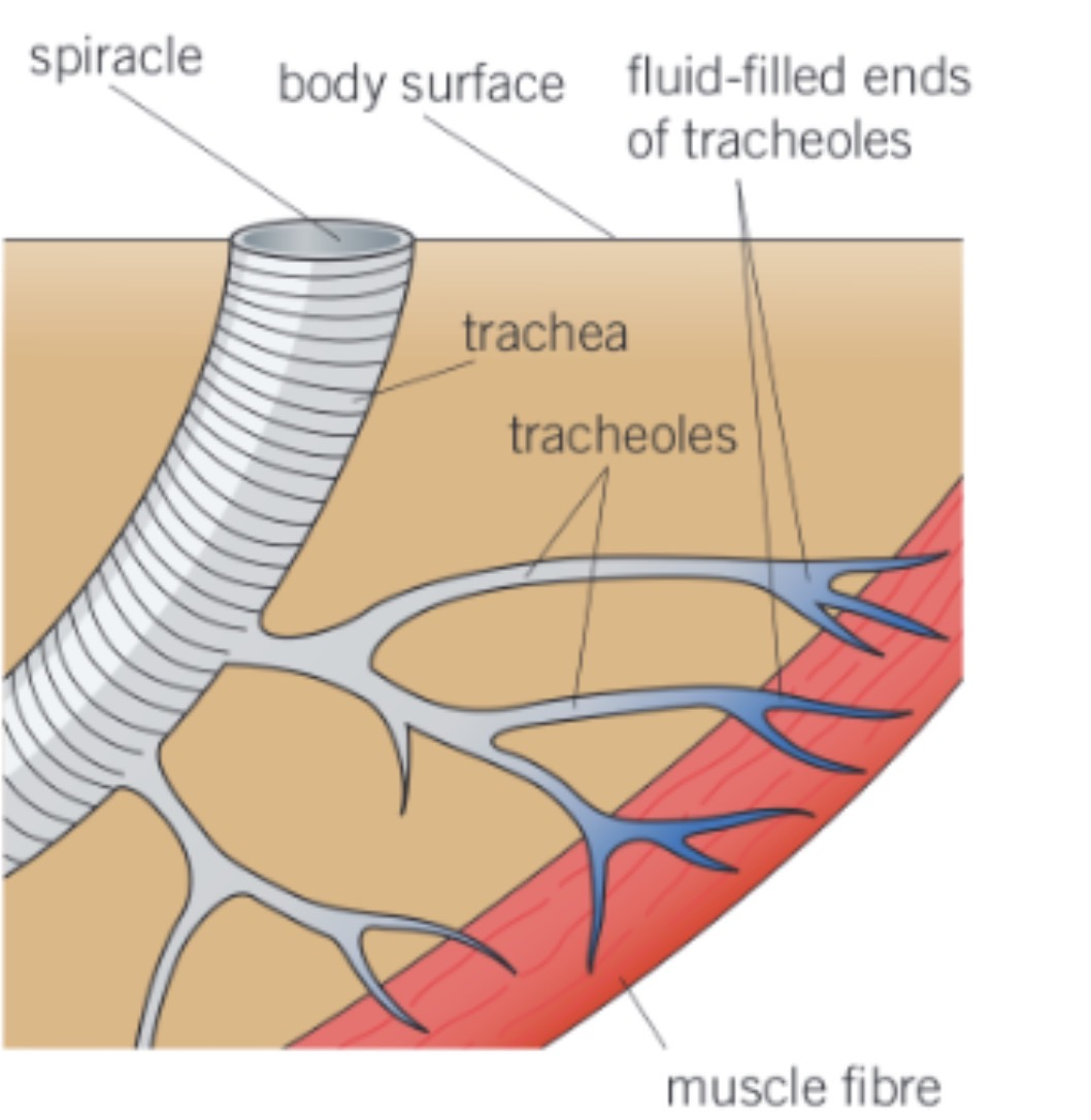
what is the tracheal system in insects, and advantages of it
Tracheal system:
The Trachea is an internal network of tubes in insects for gas exchange
They are supported by strengthened rings and chitin to prevent them collapsing
The trachea divide into small tubes with dead ends called tracheoles(not enforced w chitin so gas exchange can freely happen across lining) which extend throughout all thr body tissue in the insect
Advantagea:
Large surface area
Short diffusion pathway from a tracheole to any body cell
Oxygen from environmental air is brought directly to respiring tissue
3 ways respiratory gases move in/out of the tracheal system
Along a diffusion gradient:
respiring cells use up oxygen
The O2 concentration at ends of the tracheoles is low, creating a diffusion gradient
Oxygen diffuses in
Carbon is produced by cells so the diffusion gradient is in the opposite direction
CO2 diffuses back into the atmosphere from tracheoles and trachea
Mass transport:
Speeds up respiration by mass movement of air in/out
Abdominal muscles squeeze the tracheal system, creating a pressure gradient to pump air
More oxygen/less CO2 so conc gradient is maintained for diffusion
Ends of tracheoles are filled with water:
Muscle cells around the tracheoles respire anaerobically which produces lactate
Lactate is soluble so lowers the water potential to allow water to move in to the cells by osmosis
Water in the ends of the tracheoles decreases in volume, so more air can be drawn in
Gases diffuse faster through air than through water
How do gases leave the trachea
Spiracles are tiny pores that allow gases to leave the trachea. They open or close by a valve. When open they allow evaporation, but when closed they prevent water loss
How is water loss prevented in insects
Thick exoskeleton made of nitrogen-containing polysaccharide chitin covered by a waterproof cuticle
Spiracles
Small surface area to volume ratio to minimise the area in which water can be lost
why do mammals have lungs
mammals have evolved specialised surfaces-lungs- for efficient gas exchange between the air and their blood bc they need to have a large volume of o2 absorbed and co2 removed bc:
they are large organisms with a large volume of living cells
they maintain a high body temperature and have a high metabolic and respiratory rate
lungs are in the body bc air is not dense enough to support and protect these delicate structures and the body as a whole would lose a lot of water and dry out.
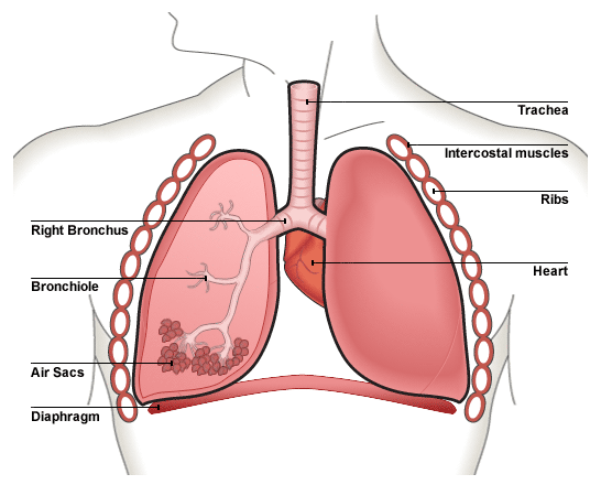
structure of the lungs
lungs:
each lung is surrounded by a membrane and the space between them(pleural caitiy) is filled with pleuraln fluid to lubricate the lungs and help them adhere to the walls of the thoracic cavity by water cohesion, so the lungs can expand with the chest during inhalation
trachea:
flexible airway supported by rings or cartilage.
Cartilage prevents trachea collapsing when the air pressure falls when u breath in.
Trachea walls are made of muscle, lined with celiated epithelium and goblet cells
bronchi:
similar structure to trachea
also produce mucus to trap dirt and have cilia to move mucus towards the throught
as the bronchi get smaller there is less cartilage
Bronchioles
walls of muscle lined with epithelial cells. Muscles allow them to to constrict so they can control the flow of air in and out of the alveoli
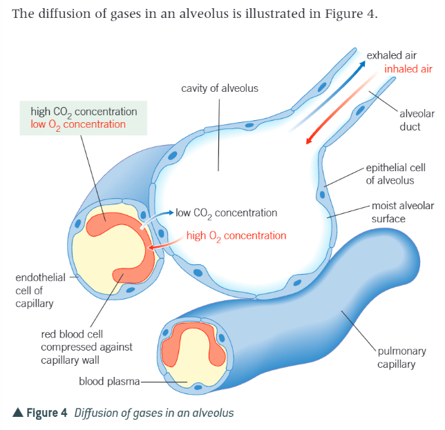
alveoli:
air sacs lined with epithelium with collagen and elastic fibres between them to allow the alveoli to stretch as they fill with air and spring back during breathing to expel carbon dioxide rich air.
the alveolar membrane is the gas-exchange surface
specialised tissues and cells in the lungs to help them function
cartilage: in trachea and bronchus, provides strength to trachea and bronchus, holds airway open to prevent collapse when the air pressure falls
surfactant: coats surface of lungs, phosphlipid layer that maintaisn moisture but reduces surface tension to stop alveoli collapsing when air pressure falls
smooth muscle: lining trachea to bronchioles, can contract to constrict the airways
goblet cells: lining trachea to bronchioles, secrete mucus to trap dust and bateria that are breathed into the lungs
ciliated epithelial cells: lining trachea to bronchioles, beat regularly to move mucus up the aireays towards the mouth to be removed, helps keep the airways clean and prevent infections, contain lots of mitochondria to provide energy required to move cilia
elastin(protein): lining of all airways and alveoli, allows lung tissue to stretch when breathing in and filling up the lungs, recoil when breathing out to help force air out of the lungs, allows alveoli to return to original shape after exahiling
squamous epithelium: lining alveoli, given a short diffusion distance pathway for oxygen and carbon dioxide in the alveoli
alveoli adaptations
alveolar epithilium and capillary endothelium are only 1 cell thick/very thin)→ shortens diffusion distance of gases from alveoli to blood as it only has to diffuse through 2 cells
large number of alveoli→increases surface area for gas exchange
capillaries that surround the alveoli are very narrow→red blood cells are slowed down to squeeze through one at a time, increasing the time fro diffusion
large number of capillaries around the alveoli→circulation constantly removes oxygenated blood to maintain steep concentration gradient
constant ventilation of air in and out of lungs→ensures concentration of oxygen in alveoli is higher and concentration of carbon dioxide is lower than blood and therefore maintains steep concentration gradient
muscles involved in ventilation
ventilation=breathing
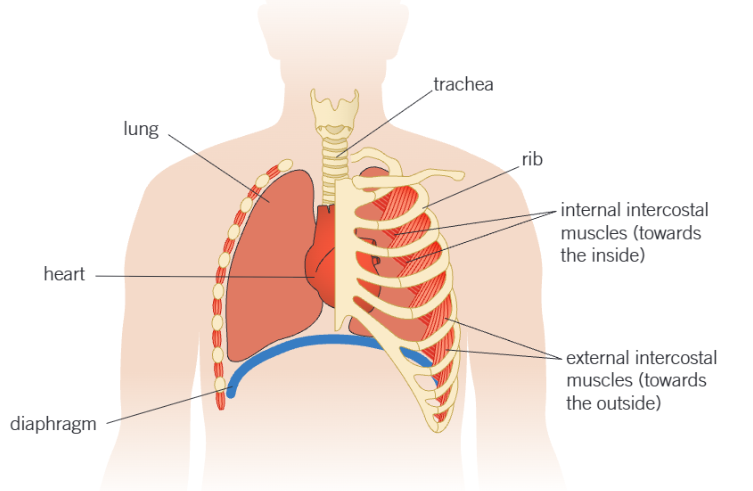
diaphragm- sheet of muscles separating the thorax from the abdomen
intercostal muscles- lie between the ribs
internal intercostal muscles- contraction leads to expiration(breathing out)
external intercostal muscles-contraction leads to inspiration(breathing in)
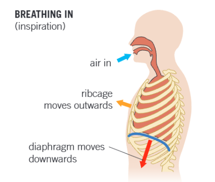
inspiration
breathing inn is an active process that uses energy
the external intercostal msucles contract, while the internal intercostal muscles relax
the ribs are pulled upwards and outwards increasing the volume of the thorax
the diaphragm muscles contract causing it to flatten,further increasing the volume of the thorax
the increased volume of the thorax results in reduced pressure of the lungs
atmospheric pressure is now greater than pulmonary pressure so air is forced into the lungs
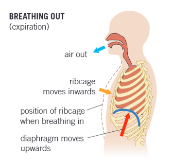
expiration
breathing out is passive
the internal intercostal muscle contract whilst the external intercostal muscles relax
the ribs move downwards and inwards, decreasing the volume of the thorax
the diaphragm muscles relax and so it is pushed up again by the contents of the abdomen that were compressed during inspiration, further decreasing the volume of the thorax
the decreased pressure of the thorax increases the pressure in the lungs
the pulmonary pressure is greater than that of the atmosphere, so air is forced out of thelungs
during normal breathing the recoil of the elastic tissue in the lungs is the main cause of air being forced out, but under strenuous conditions such as exercise the various muscles play a major part
graph to see lung function: defien ventilation rate, tidal voulume, total lung capacity, pulminary ventalation, FEV, FVC
ventilation rate-how many breaths per minute,
tidal volume-volume of air that enters or leaves the lungs at normal resting breath
there is a certain volume of air to makesure they never fully deflate-residual volume
total lung capacity=vital+residual
pulminary ventilation-total vol of air thart moves in and out of lungs in one minute= tidal volume*ventilation rate(breaths per minute)
You can measure lung function using a spirometer and measure ventilation rate and tidal volume from a speriometer trace
the health and function of a person’s lungs can be measured by looking at their:
-forced expiratory volume(maximum volume of air that can be breathed out in 1 second)FEV1 air breathed out in the first second
-forced vital capacity(max vol of air possible to forcefully breath out of the lungs
restrictive vs obstructive lung diseases
restrictive e.g fibrosis, make it difficult to fully breath in(affects elastic tissue), severley reduce FVC as breathing in is difficult but FEV1 is less affected bc breathing out is normal
obstructive e.g asthma make it difficult to breath out as airways are blocked. FVC and FEV1 are both much lower than normal
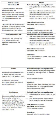
causes and effects of tuberculosis, pulmonary fibrosis, asthma and emphysema
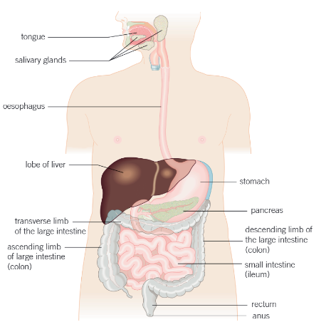
parts of the digestive system
oesophagus- carries food from the mouth to the stomach
liver-produces bile
gallbladder- stores bile
pancreas-
stomach- muscular sac with an inner layer that produces enzymes. Stores and digests foods, especially proteins. Has glands that produce protein-digesting enzymes
ileum(small intestine)- long muscular tube where food is further digested by enzymes. Inner walls are folded into villi to increase surface area. Microvilli- projections on the epithelial cell of each villus to increase SA for absorption into bloodstream. This increases SA, decreases diffusion distance and maintains the conc gradient
large intestine- absorbs water, most of the water that is absorbed is from gland secretions
rectum- final section of the instestines, where feces are stored before being removed via the anus in egestion
physical and chemical digestion
physical digestion: breaking large foods into smaller pieces using teeth and muscles in the stomach to churn foods, this increases its surface aerea for chemical digestion
chemical digestion: hydrolyses large insoluble molecules into smaller soluble ones using enzymes. The general names for these enzymes are carbohydrases(carb→monosaccarides), lipases(lipids→glycerol and fatty acids), and proteases(protein→amino acids)
digestion of carbohydrates
starch→maltose:amylase in pancreas and saliva
glycogen:kinase
maltose→2x glucose:maltase in epithilial cells in ileum
lactose→glucose + galactose: lactase
sucrose→glucose + fructose: sucrose
begins in mouth, then duodenum(first part of small intestine), then ileum
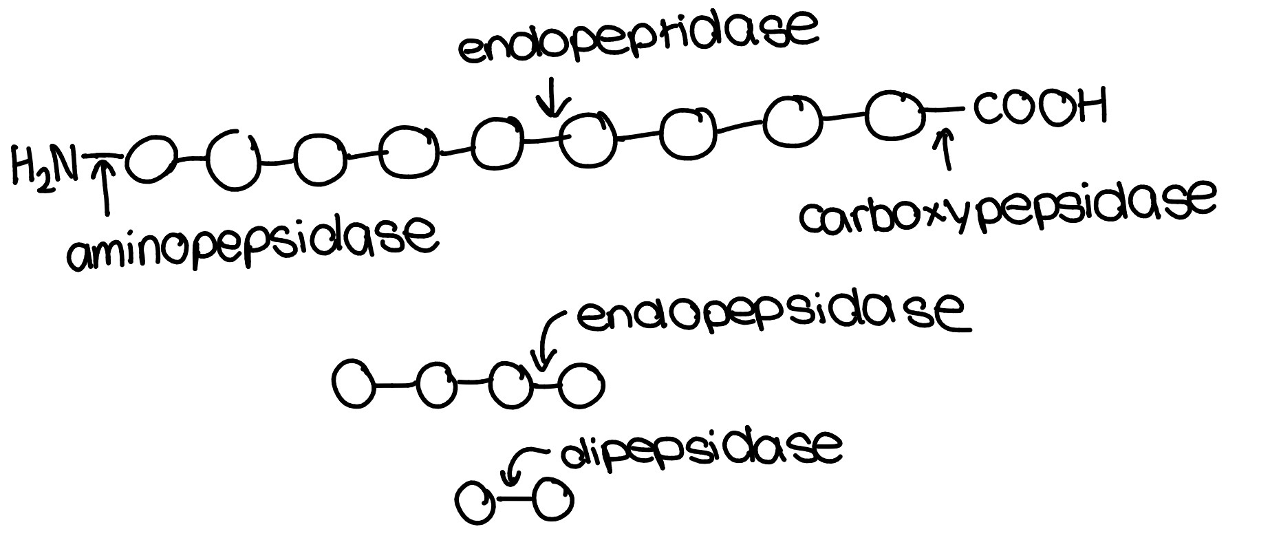
digestion of proteins
endopepsidase- hydrolyses peptide bonds between amino acids in the central region of the protein e.g trypsin made and secreted in pancreas, pepsin in stomach ph2
exopepsidase- hydrolyses peptide bonds on the terminal amino acids of the protein e.g carboxypeptidase removes amino acids from the carboxylic acid end, aminopepsidase removes amino acids from the amino group end
dipepsidase- hydrolyse the peptide bond in a dipeptide
starts in stomach, then duodenum, then ileum
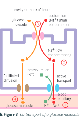
absorption of monosaccharides and amino acids
through co-transport
sodium ions actively transported out of epithelial cells by Na+-K+ pump into blood using a protein carrier on the cell-surface membrane of an epithelial cell
this maintains a higher conc of Na+ in the lumen of the intestine than in the epithelial cells
Na+ diffuse into the epithelial cell down the conc gradient using a different protein carrier. As the Na+ ions diffuse, they carry either amnio acid molecules or glucose molecules w them
the glucose/amino acids pass into the blood plasma by facilitated diffusion using another type of protein carrier
both sodium ions and glucose move into blood but Na+ ions move down their conc gradient and the amino acids/glucose moves against their conc gradient. This movement against the conc gradient is powered by the Na+ conc gradient, rather than ATP directly unlike in step 1, so is indirect form of active transport
digestion of lipids
lipase produced in pancreas hydrolyses ester bonds in triglycerides to form monoglycerides or glycerol and fatty acids
bile salts produced in liver- emulsify lipids to form droplets called micelles to increase their SA for faster hydrolysis by lipase
micelles are water soluble vesicles that deliver fatty acids, glycerol and monoglycerides to the epithelial cell of ileum for absorption
absorption of lipids
micelles carrying fatty acids/monoglhycerides to epithilal cells
monoglycerides and fatty acids can diffuse into ileum through plasma membrane bc they are non-polar and lipid soluble
once they are in, they reform triglycerides in golgi apparatus so they can be used or
fatty globule combined w protein inside golgi to form a chylomicron
chylomicron released inside golgi vesicle and moves towards other end of epithelial cell iand released inro blood through exocytosis
lacteal/lymph vessel transports chylomicrons/lipids
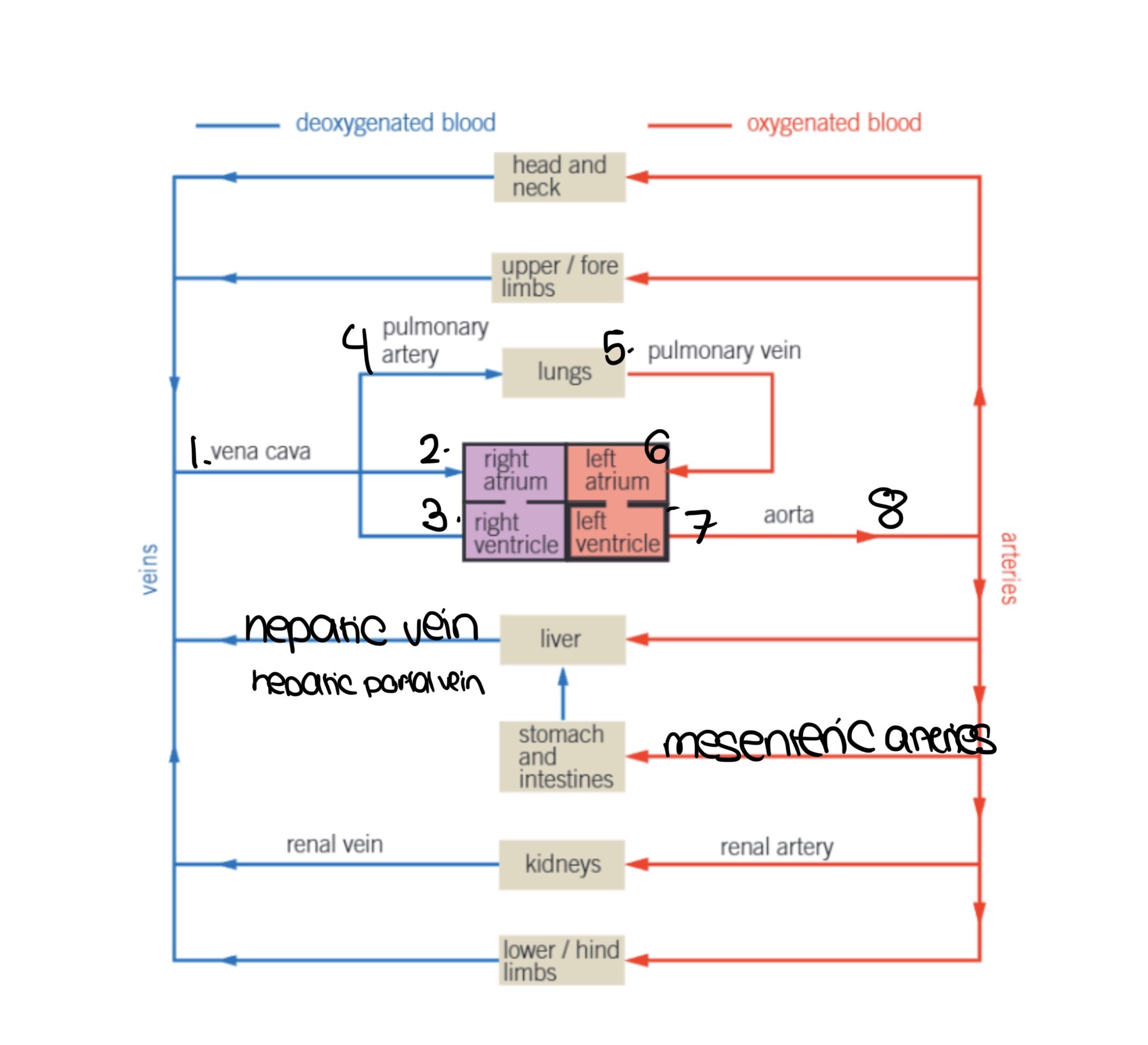
what is a closed circulatory system
what is a double circulatory system
closed circulatory system- where the blood is confined to blood vessels only so it has increased pressure and speed of blood flow
double circulatory system- blood is confined to vessels and passes twice through the heart for each complete circuit
why do multicellular organisms require a transport system
they have a low SA:V and their diffusion distance so they can’t rely on diffusion to supply their cells and tissues with everything they need or to remove waste such as CO2 and urea
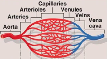
blood vessel system
arteries (away from the heart) branch into arterioles(smaller branches) that form a network throughout the body. Arterioles decrease pressure, so the same volume of blood is split between many arterioles so there is an increased cross sectional area
they can contract to restrict blood flow and relax to increase blood flow
this directs blood to different areas
arterioles branch to capillaries which form networks called capillary beds
once excahnge has occured, deoxygenated blood(except pulmonary vein) flows from capillaries to venules and then veins and then back to the heart
type of tissue in blood vessels
innermost part- endothelium: smooth to reduce friction
wall is made up of:
elastic layer: let artery stretch and recoil to maintain constant blood pressure
smooth muscle: can contract to constrict lumen(vasoconstriction) or relax to dilate lumen(vasodilation)
collagen: provides strength and helps maintain shape, prevents bursting
structure of the different blood vessels
arteries: small lumen, thick walls-strong and flexible, structured to handle high pressure from heart. Elastic arteries have more elastic fibres, and muscular arteries have more smooth muscle
arterioles: similar to arteries but larger lumen, thinner wall(less elastin and collagen, but lots of smooth muscle
capillaries: smallest blood vessels, very thin wall-only 1 layer endothelial cell so decreased diffusion distance, very nnarrow lumen-to bring the red blood cells close tissues , lowest blood vessels bc between arterioles and venules, very branched-maximum surface area for diffusion
venules: similar to veins but smaller,
veins: low blood pressure, large lumen, thin walls(thin elastic and smooth layer), collagen helps them hold their shape and prevent collapse, pocket valves to prevent back flow by opening when blood flows towards the heart and closing when blood flows away from the heart
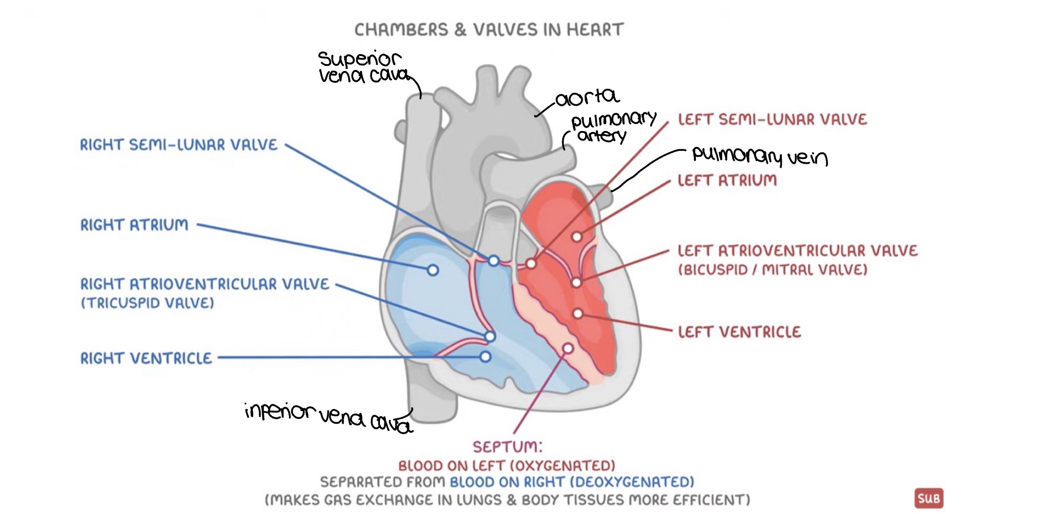
structure of heart
ventricles have thicker walls than atria because they pump blood a longer distance(out of heart to other organs whilst atria only pump into ventricles) so need more pressure
left ventricle has the thickest wall to pump blood all around body, but right ventricle only 2 lungs
septum separates oxygenated blood on left from deoxygenated blood on right for more efficient gas exchange
cardiac cycle
sequence of contraction(systole) and relaxation(diastole) that causes the heart muscles to pump blood
1.atrial systole:
atria contract increasing atrial pressure(bc same vol of blood but less space in cavity)
increase in pressure opens atrioventricular valve
bc ventricular pressure is lower than atrial pressure blood is pushed into ventricles
2. ventricular systole:
ventricles contract and atria relax, increasing ventricular pressure and decreasing atrial pressure
this pressure forces the atrioventricular valves to close to stopping backflow and semi lunar valves to open
blood is pushed through semi-lunar valves into arteries
3.diastole:
heart muscles in atria and ventricles relax so ventricular pressure decreeases
high pressure in arteries and low pressure in heart forces semi-lunar valves to close, prevent back flow
blood also passively flows from veins into atria to start the cycle again
how to calculate cardiac output
cardiac ouput=volume of blood pumped by 1 ventricle in 1 minute
heart rate-number of beats per minute. We can calculate this from a cardiac cycle graph bc 1 heart beat=time for one cardiac cycle in s. 60/time for one cardiac cycle in s=heart rate in bpm
stroke volume-volume of blood pumped out of ventricle with each beat
cardiac output=heart rate x stroke volume
blood flow path
deoxygenated blood enters vena cava
right atrium contracts to push blood into right ventricle
right ventricle contracts to pump blood to pulmonary artery which carries blood to lungs to become oxygenated
pulminary vein carries oxygenated blood to heart
left atrium contracts to pump blood to left ventricle
left ventricle contracts to pump blood to aorta
oxygenated blood flows from aorta to the rest of the body
define hydrostatic pressure and osmotic pressure
hydostatic pressure- outward force of a fluid at rest due to gravity. hydrostatic pressure=blood pressure
osmotic pressure- pressure that needs to be applied to prevent the inward flow of water across a semi permeable membrane(opposes hydrostatic pressure)
tissue formation
at the arteriole end there is a high blood pressure so therefore high hydrostatic pressure
hydrostatic pressure>osmotic pressure so more outward pressure force and water/O2/ions are forced out of the capillery- this forms tissue fluid. Tissue fluid can build up if there is too much for the lympatic system to quickly drain it away
plasma and red blood cells are too big to leave the venistration
at the venule end there is a low bp and hp bc water and small molecules left the capillery at the arteriole end
plasma proteins are water soluble so decrease the water potential in the venule end of the capillery so it is lower than in the fluid
so osmotic pressure>hydrostatic pressure and water moves into venule end carrying carbon dioxide and other waste e.g urea
any excess tissue fluid that is not reabsorbed is collected into the lympthatic system which returns it to the circulatory system
lympthatic system
some tissue fluid enters lymph vessels and becomes lymph
lymph has less nutrients and plasma proteins, but more fatty acids and more white blood cells(lymphocytes)
lymph moves through muscle contractions, and has valves to prevent back flow to move in one direction
lymph nodes filter out pathogens and dead cells
filtered lymph is returned to the blood stream to maintain the fluid balance
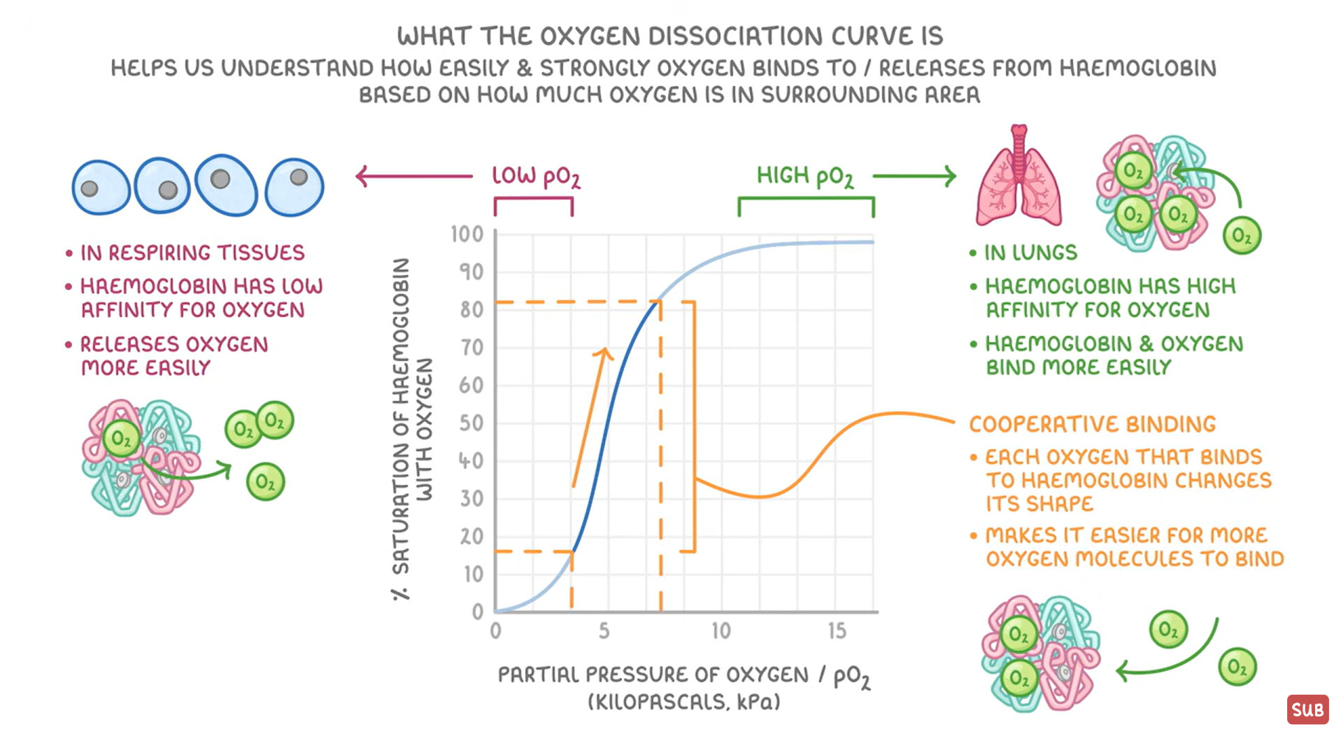
how does hemoglobin bind to oxygen, where is oxygen loaded and unloaded
hemoglobin is a protein in red blood cells that binds to oxygen. It has a quaternary structure of 4 polypeptide chains. each chain has a heam group with an iron ion that can bind to one oxygen, so one hemoglobin can carry up to 4O2 molecules at once
oxyhemoglobin-the name for when hemoglobin binds to oxygen through loading/association
oxygen is loaded in the lungs at high p.O2
oxygen is unloaded at respiring cells at low p.O2 and a high p.CO2
oxygen dissociation curve
hemoglobin’s affinity to O2(how easily it binds to it)- based on pO2, pCO2 and hemoglobin saturation
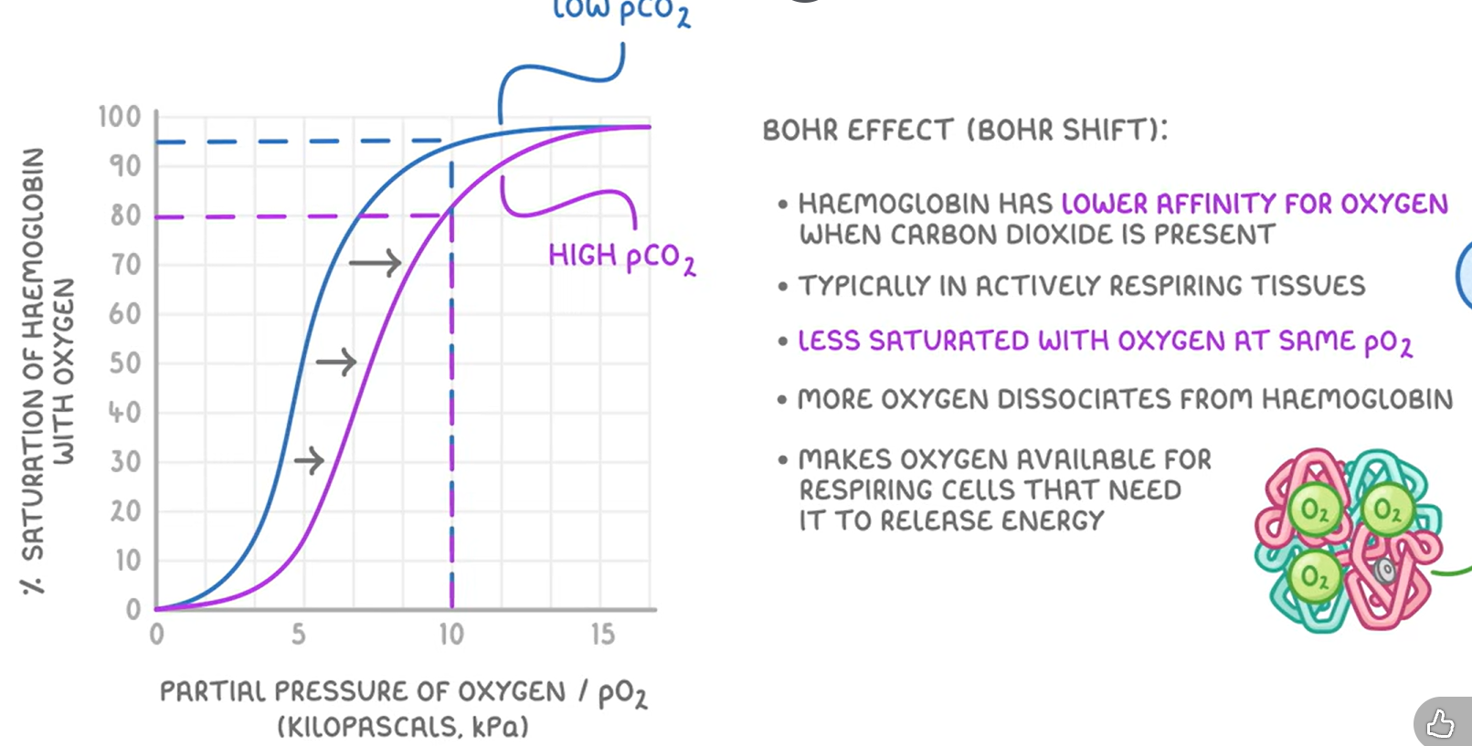
bohr shift
hemoglobin has a lower affinity for oxygen when CO2 is present, typically in respiring cells
it is less saturated at the same pO2
this means that oxygen is more available for respiring cells that need it to release energy to meet higher energy demands
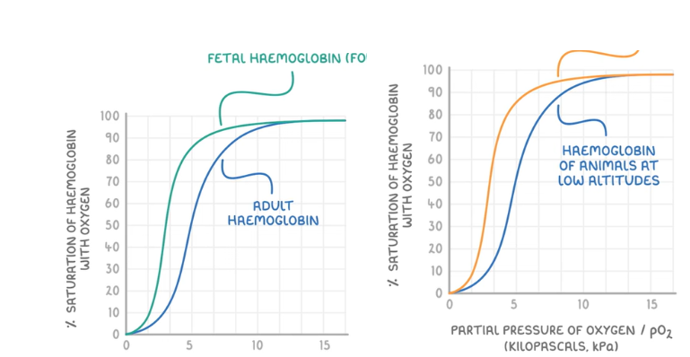
fetal hemoglobin and hemoglobin at high altitudes
fetal hemoglobin:
higher affinity for oxygen because oxygen dissociated from mothers hemoglobin and assoiciates with fetal hemoglobin instead
hemoglobin in animals at high altitudes:
hemoglobin has a higher affinity for oxygen and binds to oxygen more easily even at a lower pO2, as an adaptiation because oxygen has a lower pO2 at higher altitudes
heart diseases
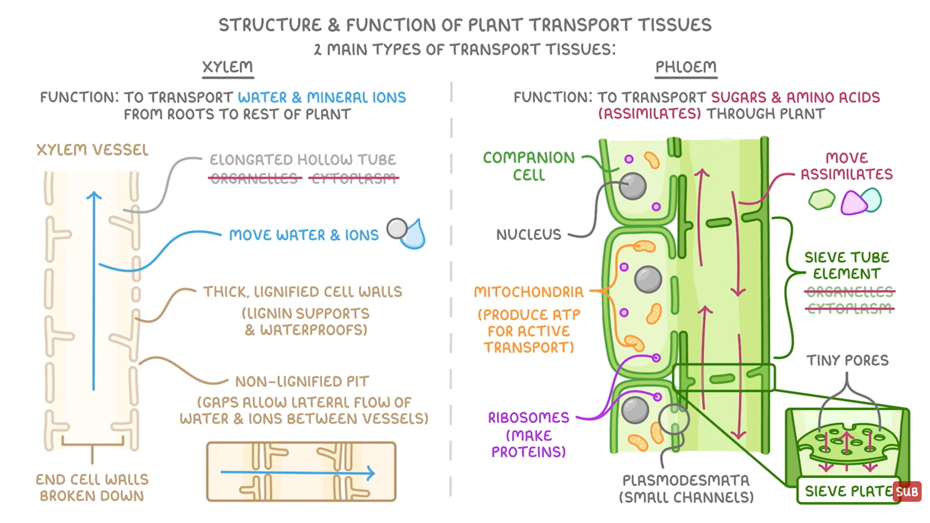
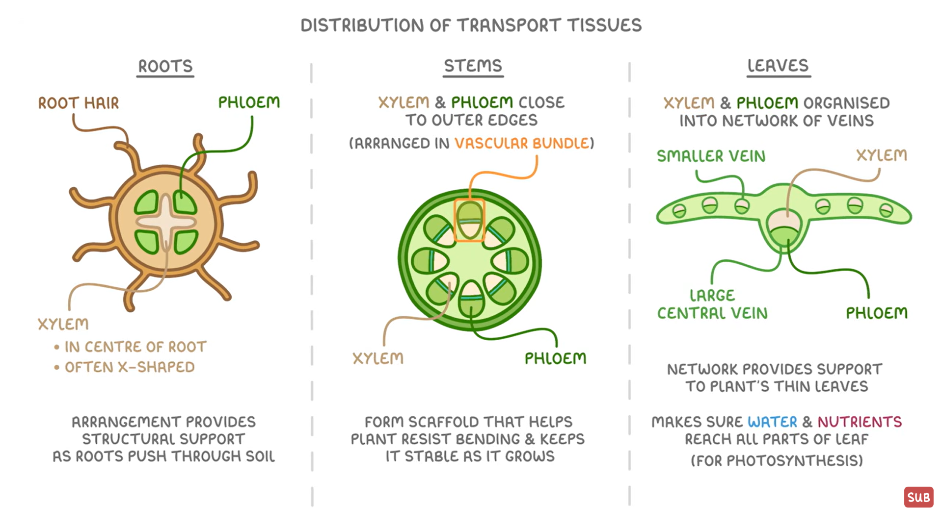
phloem and xylem structure
in vascular system to move substances quiclkly and efficiently
xylem: to move water and mineral ions from roots to rest of the plant
no organelles or cytoplasm→elongated hollow tube for water flow
no end cell walls→continuous column so water can continuously flow
cell walls strengthed with lignin(dead cells)→waterproofing and support so it stays upright and no water escapes plant, also so it doesn’t collapse from tension in transpiration
non lignified pit→aps to allow lateral flow of water and ions between adjacent vessels
phloem: to transport dissolved assimilates(sugar and amino acids) through plant
sieve tube cells joined end to end→form long columns to carry assimilates
no organelles or cytoplasm in sieve tubes→hollow for easier flow
sieve plates have tiny pores/gaps →let assimilates flow through
plasmodesmata→small channel to let substances pass betwseen companion celll and sieve tube
companion cell has lots of mitochondria to produce ATP for active transport, ribosomes make proteins that the phloem needs to function
distribution of transport tissue in plants
water transport in plants
1)water enters plant through its roots:
root hair cells have a long thin shape to increase SA for more water absorption
mineral ions enter by active transport bc higher conc in root, lower conc in soil, which lowers water potential in root so water can enter by osmosis
water moves from an area of high wp in soil to lower wp inside(bc of the minerals) root by osmosis
2) water moves towards xylem,
3)water moves up through xylem due to cohesion tension theory:
cohesion- hydrogen bonds form between water molecules creating a continuous column of water
adhesion- hydrogen bonds form between water molecules and cellulose in xylem walls, heloping water to move upwards against gravity
transpiration pull-water evaporates from leaves via transpiration, which creates tension that is transmitted through the colum of water in the xylem(like a suction straw)
xylem are strengthed with lignin so they don’t collapse under the tension
4)water exits the plant through transpiration
guard cells open stomata, when they are open water vapour can evapourate through the stomata
transpiration factors
increases rate of transpiration:
higher light intensity bc more photosynthesis so plant needs more CO2 so more stomato open
higher temperature bc it increases the rate of evapouration
higher wind speed bc fast winds remove water surrounding leave, maintaining a steep conc grad
decreases:
humidity bc there is a higher conc of water vapour outside the leaf so the conc grad isn’t as steep
evidence to support cohesion-tension theory
diameter of tree trunks decreases when transpiration rate increases bc there is more tension in xylem so the walls are pulled inwards, causing trunk to shrink in diameter
if a xylem vessel is broken and air enters it the treee can’t draw up water bc the continuous column of water is broken so water molecules no longer stick together by cohesion
when a xylem vessel is broken, air is drawn in and water doesn’t leak out bc it is under tension
using a potometer to measure the rate of water uptake
cut the leafy shoot under water, but don’t get any on the leaves
fill the potometer with water, making sure there are no air bubbles
fit the leafy shoot to the potometer under water, using a rubber tube
remove the potometer from under the water and seal all joints w jelly
introduce an air bubble to the cappilery tube
distance moved by air bubble/time = speed of water uptake, repeat and calculate a mean
mass flow hypothesis
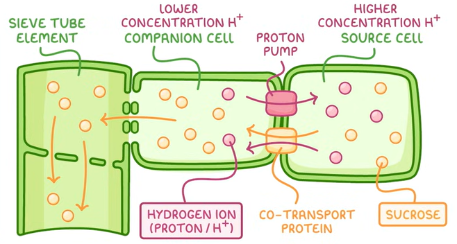
in the source, sucrose is actively transported into the phloem by companion cells
high concentration of glucose and sucrose in phloem decreasing the water potential
xylem has a higher wp so water moves down the wp gradient by osmosis into the phloem
this increases pressure in the phloem forcing the sucrose/glucose down the pressure gradient into the sink cell
the glucose/sucrose enters the sink cell by active transport using a carrier protein. it is used in respiration or stored as starch so it cant affect wp as it is insoluble

how is glucose/sucrose actively loaded(yet to ask a qs on)
H+ ions are actively transported against gradient using energy from ATP from companion to source cell to establish a steep electrochemical gradient
H+ ions are co-transported with sucrose/glucose through a carrier protein down the electrochemical gradient
glucose is then actively transported from the companion cell to the phloem
proof for mass flow hypothesis
when sieve tubes are cut sap is released, so unlike in xyelm, sap is under high pressure downwards
sucrose conc is higher in leaves than roots
when there is an increase in sucrose in leaf, an increase in sucrose in phloem follows
when a ring of phloem is removed around the whole circumference of the stem, sugars accumulate above it and there are no sugars below it, so sugars move downwarsd
proof against mass flow hypothesis
sugars travel to many different sinks, they don’t travel to the one with the highest water potential first/fastest
they end plates on sieve tubes would slow down mass flow, so why are they present
preventing ATP production stops translocation but not water movement bc there is a lack of ATP for active transport in companion cells so they die preventing mass flow
downwards flow in phloem occurs in daylight when there is photosynthesis but not at night bc less photosynthesis means no conc and pressure gradient
ringing experiments and tracer experiments
ringing:
ring of bark containing phloem but not xylem is removed from the tree stem(bc phloem is on the outside of ring)
sucrose accumulates above the ring causing swelling, no sucrose is at the bottom so that area dies
tracer:
expose the plant to radioactive CO2
the plant uses radioactive CO2 in photosynthesis and produces radioactive sucrose
x-rays show radioactive sucrose move through the phloem from the source to sink
evidence of translocation in phloem
when the phloem is cut, a solution of assimilates flow out
plants provided with radioactive carbon dioxide have radioactive carbon in the phloem after a short time
aphids are insects that feed on plants and have needle-like mouthparts to penatrate the phloem.
when a ring of phloem is removed around the whole circumference of the stem, sugars accumulate above it and there are no sugars below it