Biology Study Material: Labs After Midterm - Vocabulary and Definitions
1/201
There's no tags or description
Looks like no tags are added yet.
Name | Mastery | Learn | Test | Matching | Spaced | Call with Kai |
|---|
No analytics yet
Send a link to your students to track their progress
202 Terms
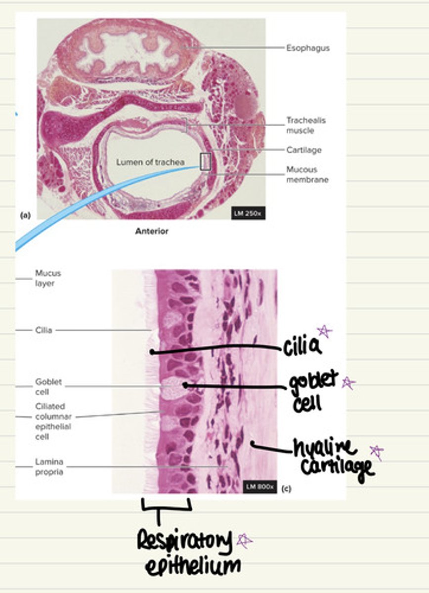
trachea histology
i. Respiratory epithelium - pseudostratified columnar
epithelium
ii. Cilia
iii. Goblet cells
iv. Hyaline cartilage
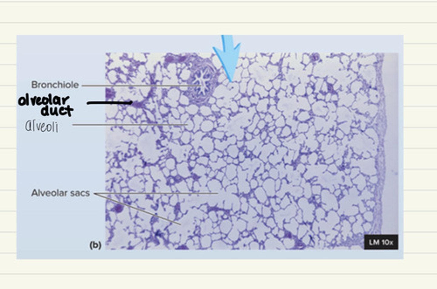
lung histology
i. Bronchioles
ii. Alveolar duct
iii. Alveolar sac
iv. Alveoli
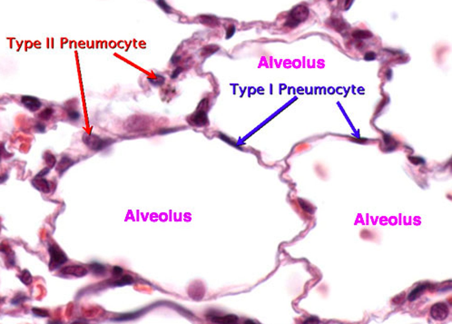
v. Simple squamous epithelium
vi. Type I pneumocytes
vii. Type II pneumocytes
type I pneumocytes
type II pneymocyte histology
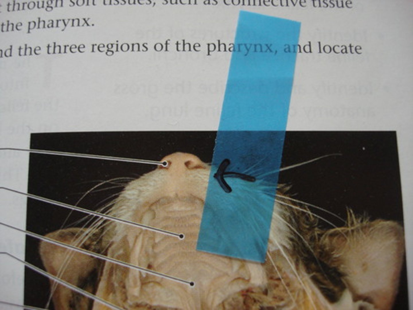
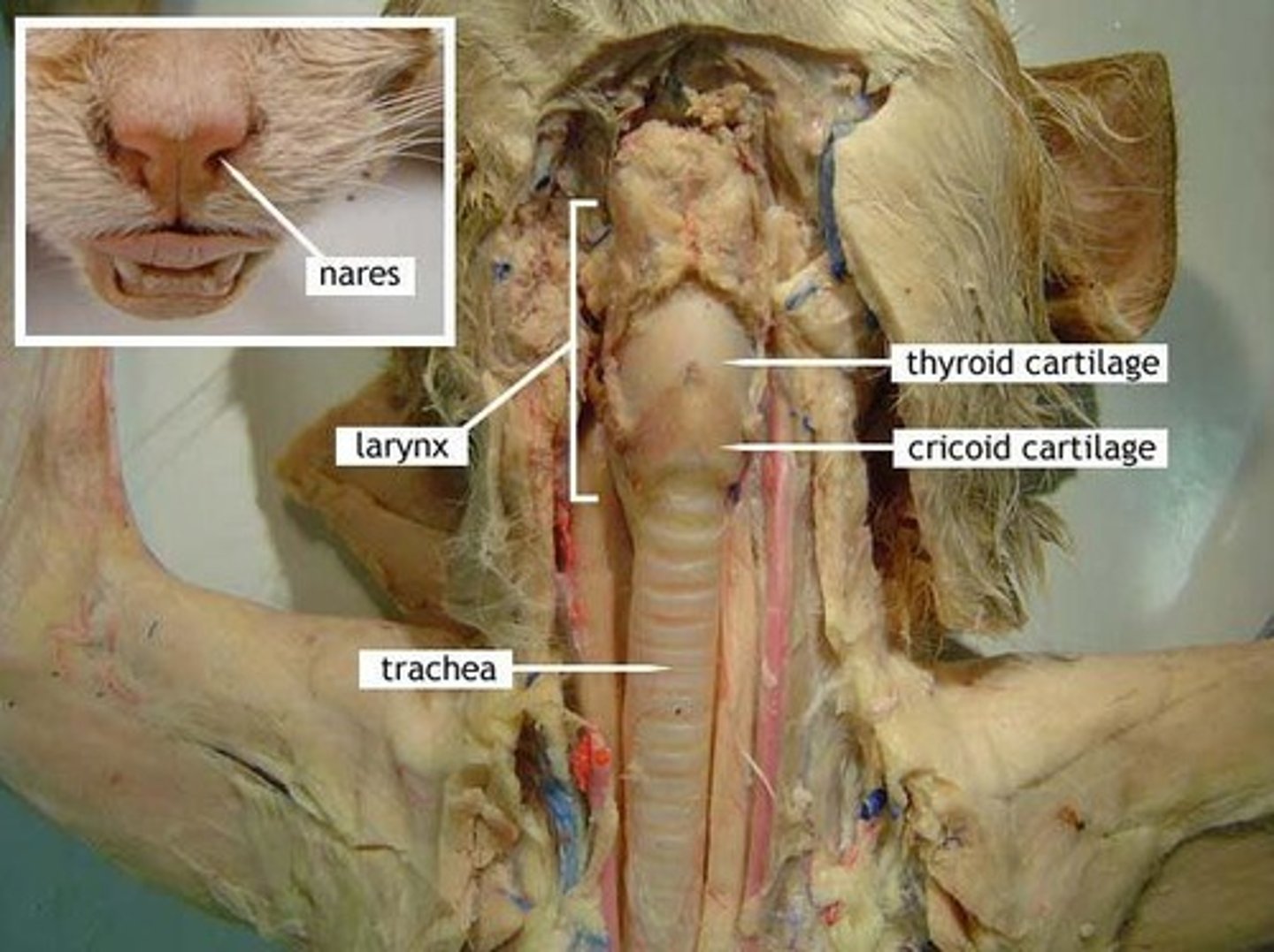
nares cat
hyoid bone (cat)
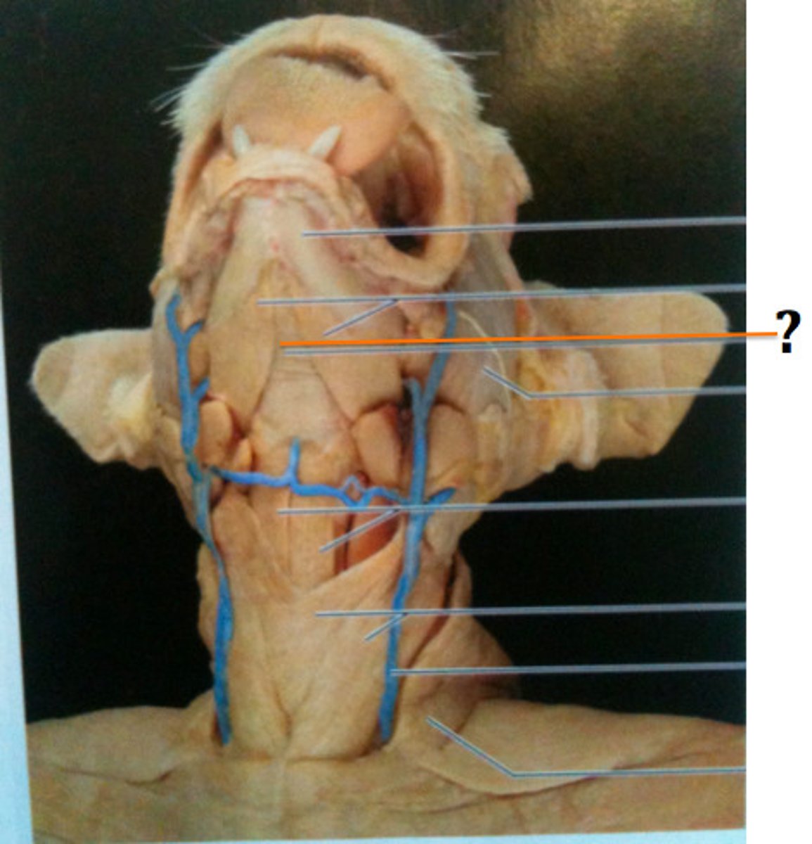
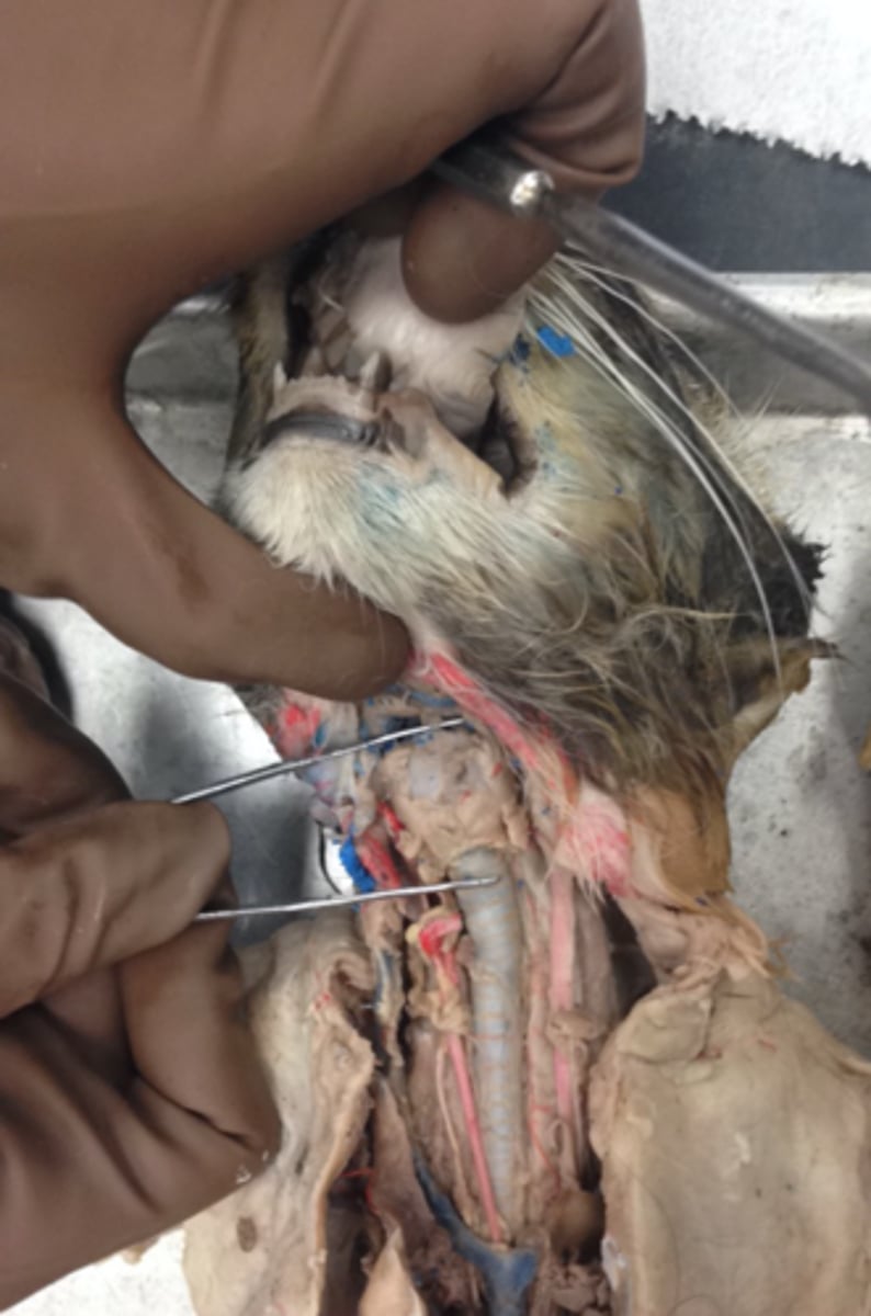
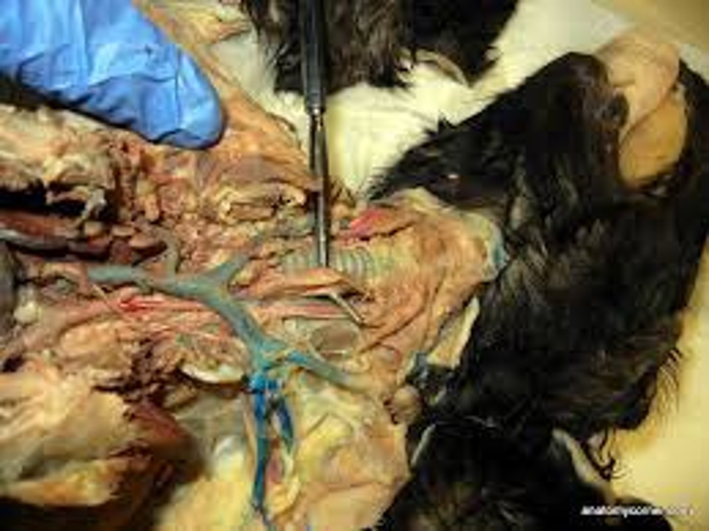
larynx (cat)
voice box
thyroid cartilage (cat)
cricoid cartilage (cat)
thyroid cartilage is superior to cricoid cartilage
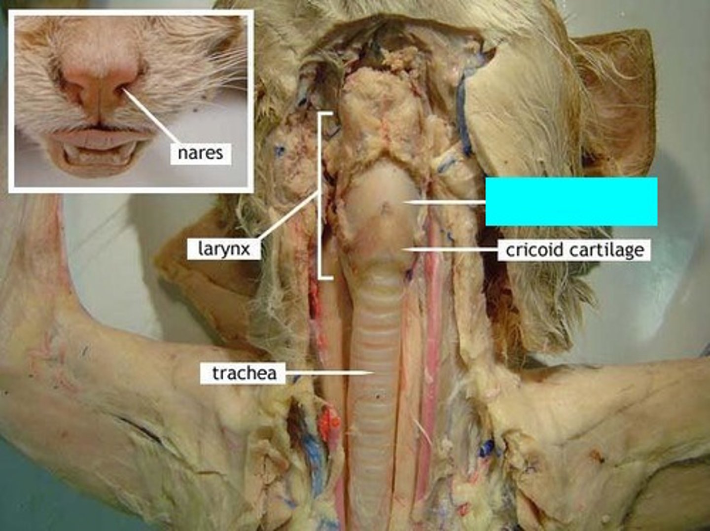
trachea (cat)
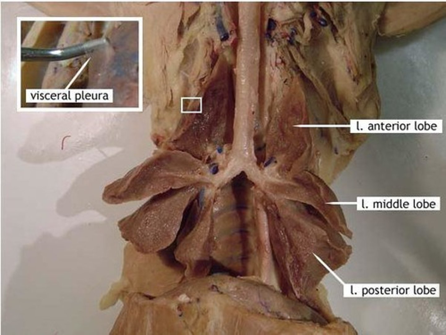
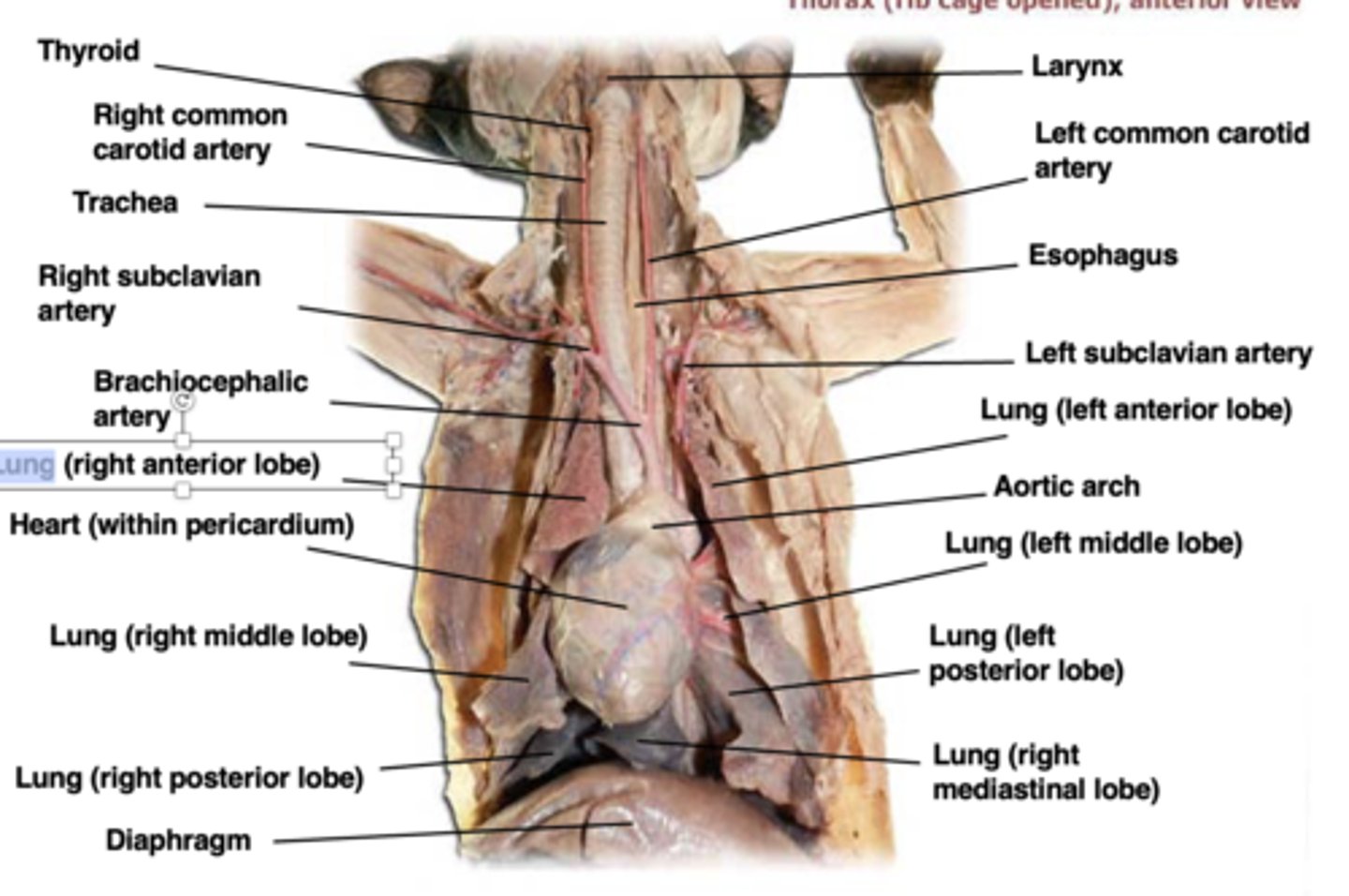
left lung (cat)
3 lobes (anterior, middle, posterior)
right lung (cat)
4 lobes: anterior, middle, posterior, and mediastinal
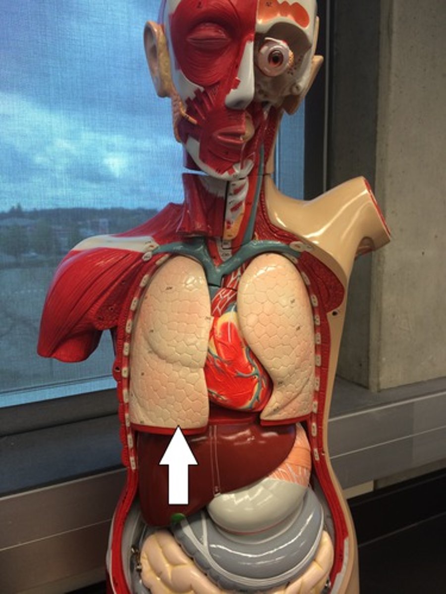
** know diaphragm as well
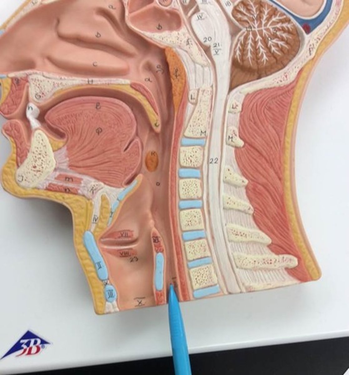
nares (model)
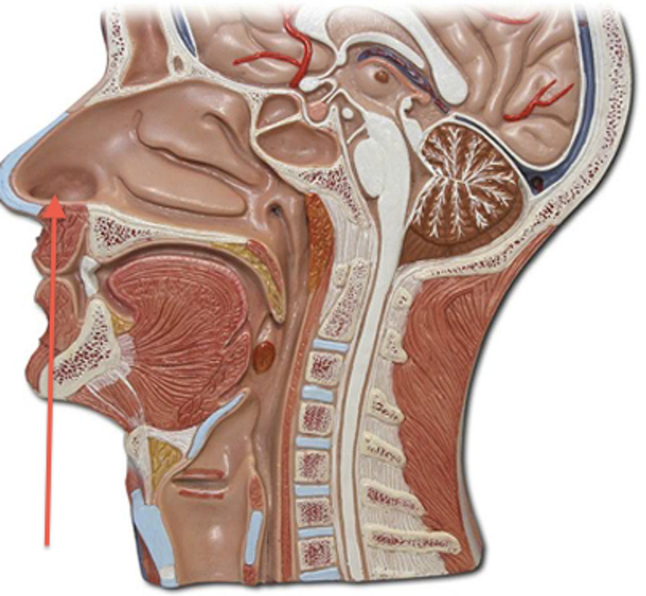
nasal cavity (model)
superior concha (model)
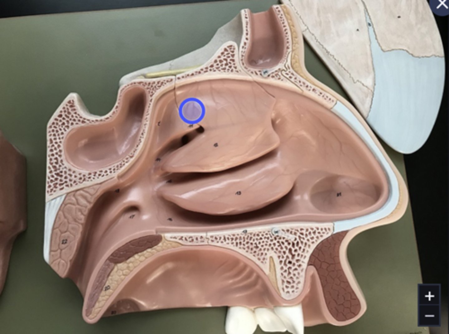
Middle concha (model)
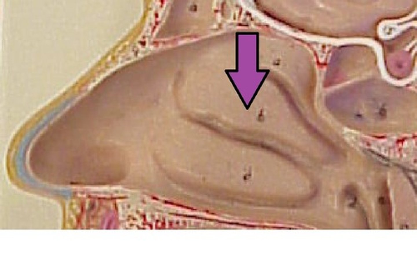
inferior concha (model)
superior, inferior, & middle meatus (model)
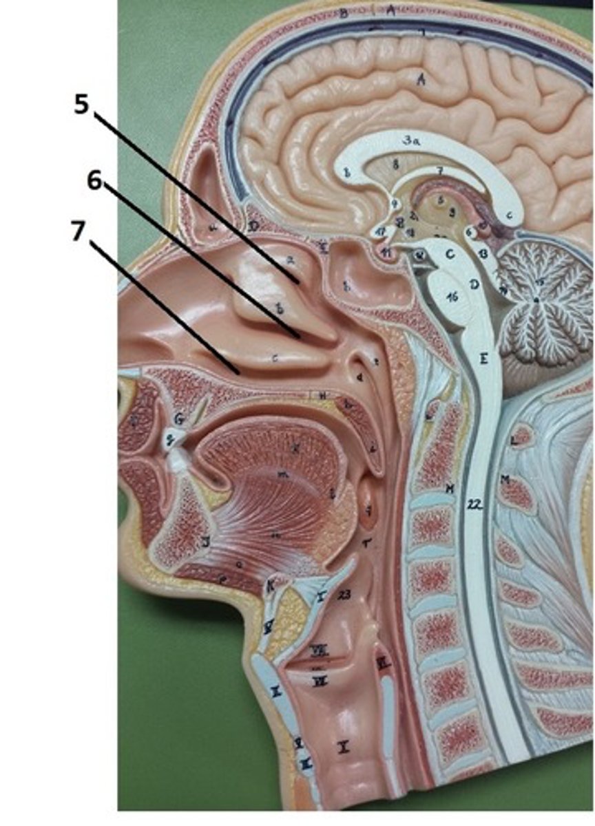
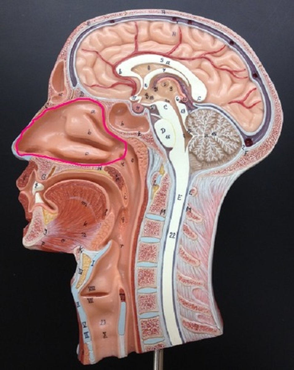
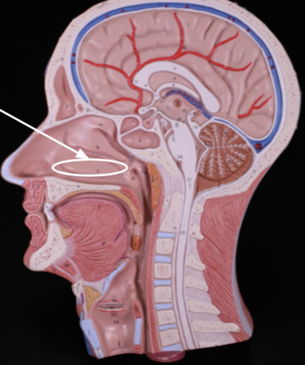
hard palate (model)
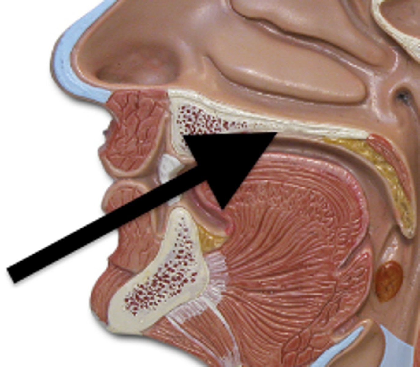
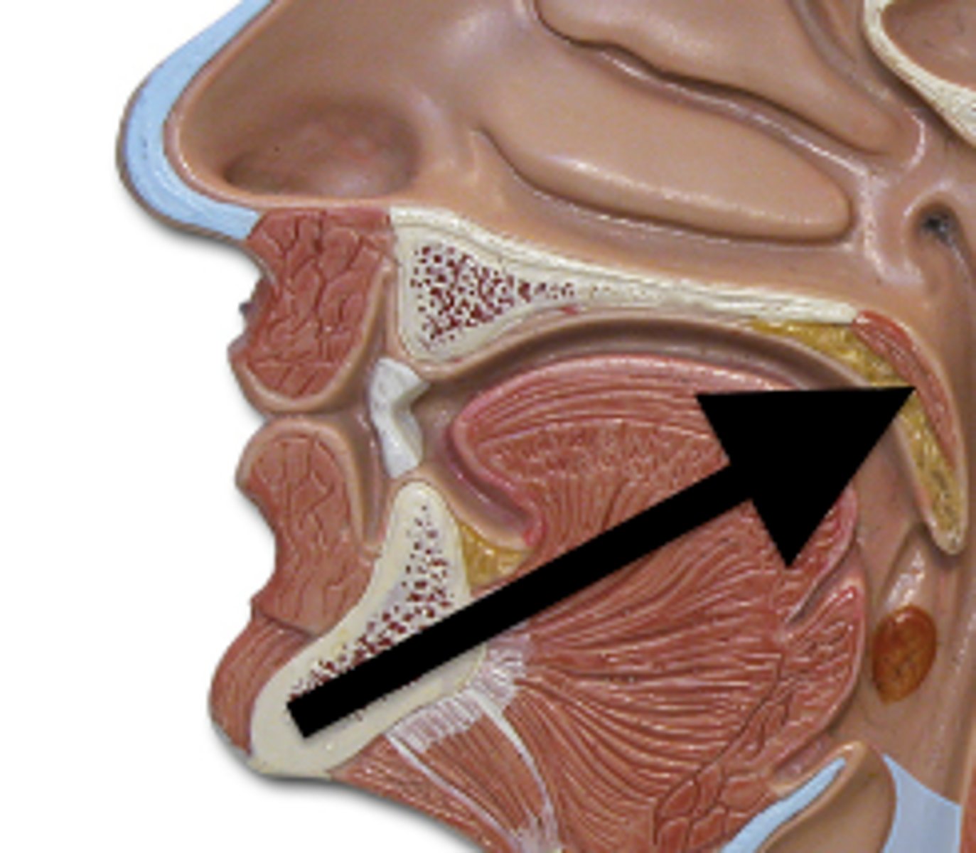
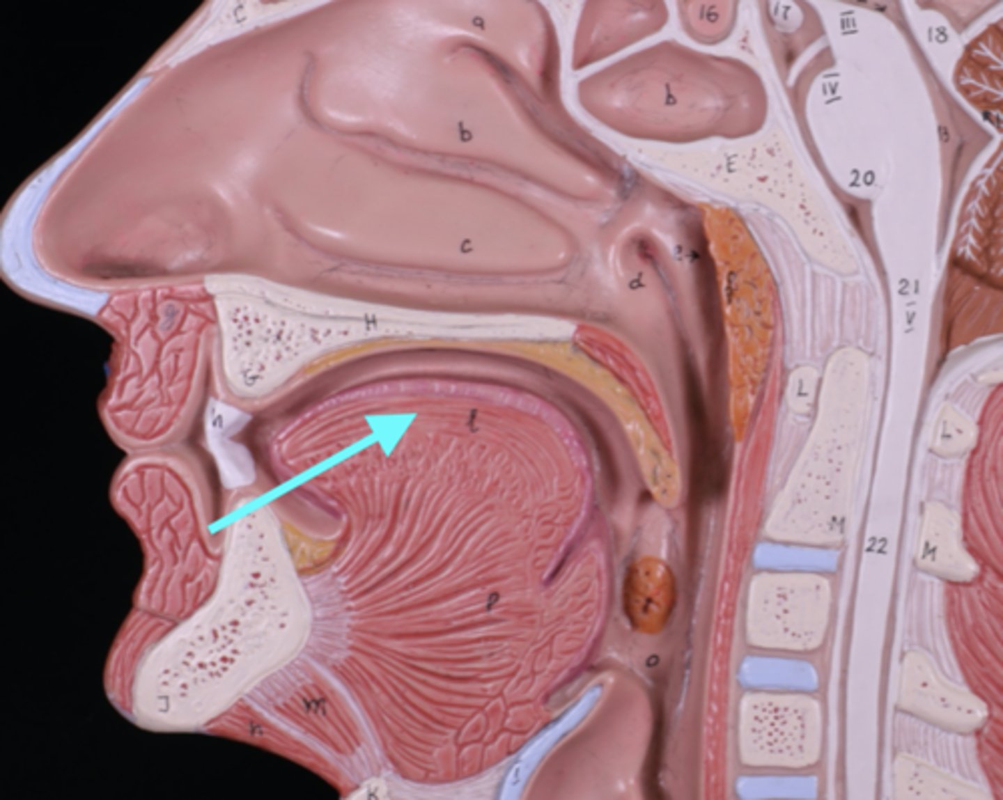
soft palate (model)
pharynx (model)
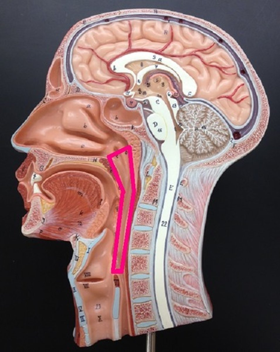
nasopharynx (model)

oropharynx (model)
laryngopharynx (model)
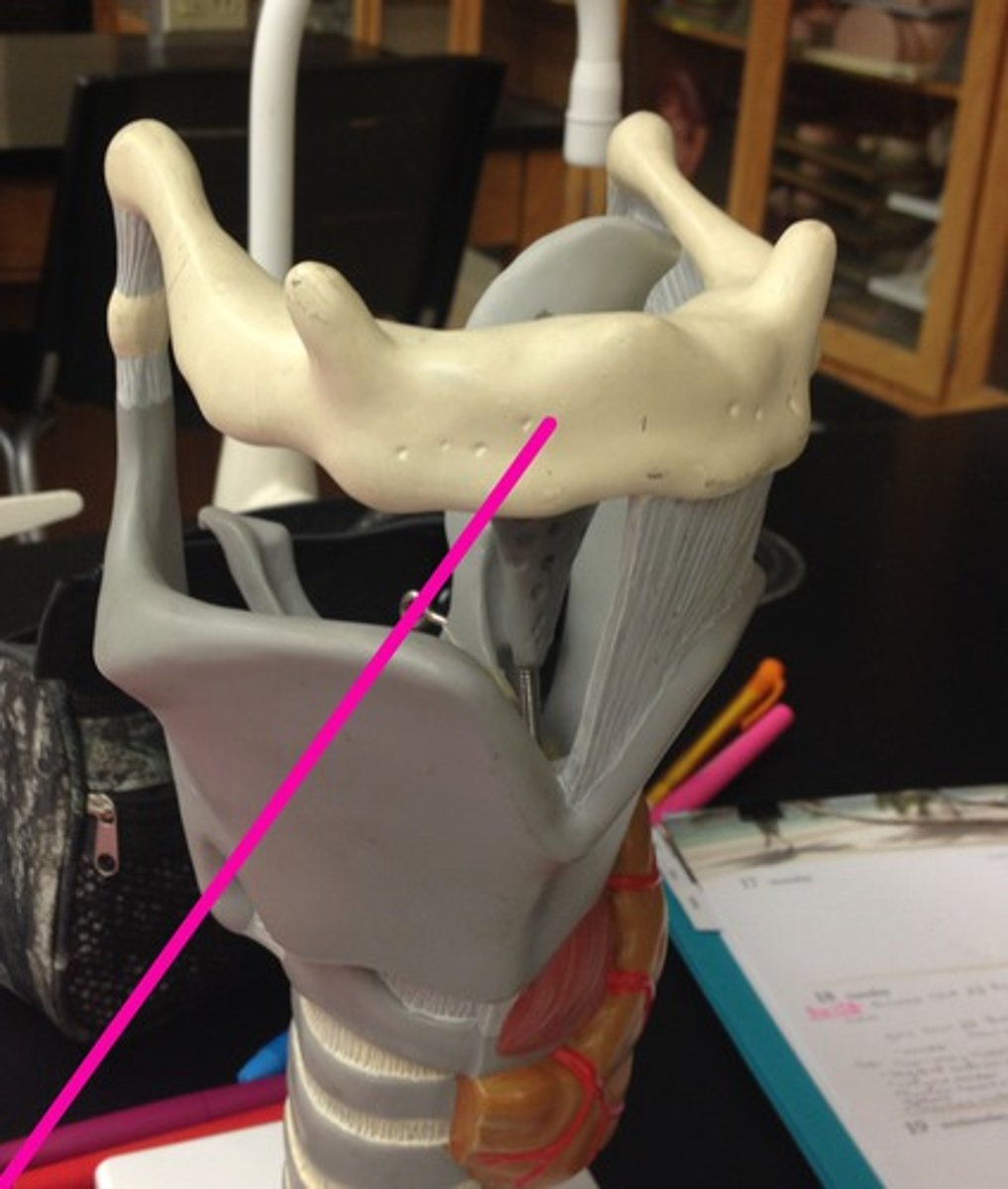
hyoid bone (model)
larynx (model)
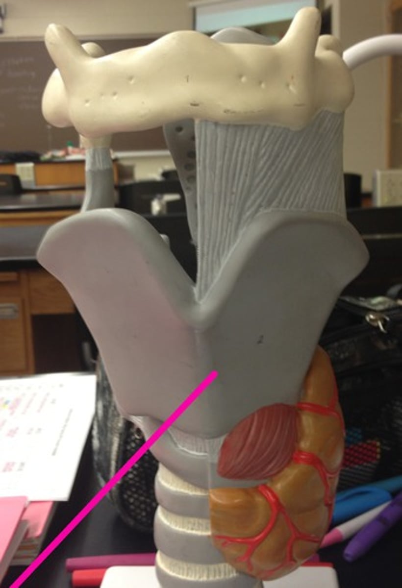
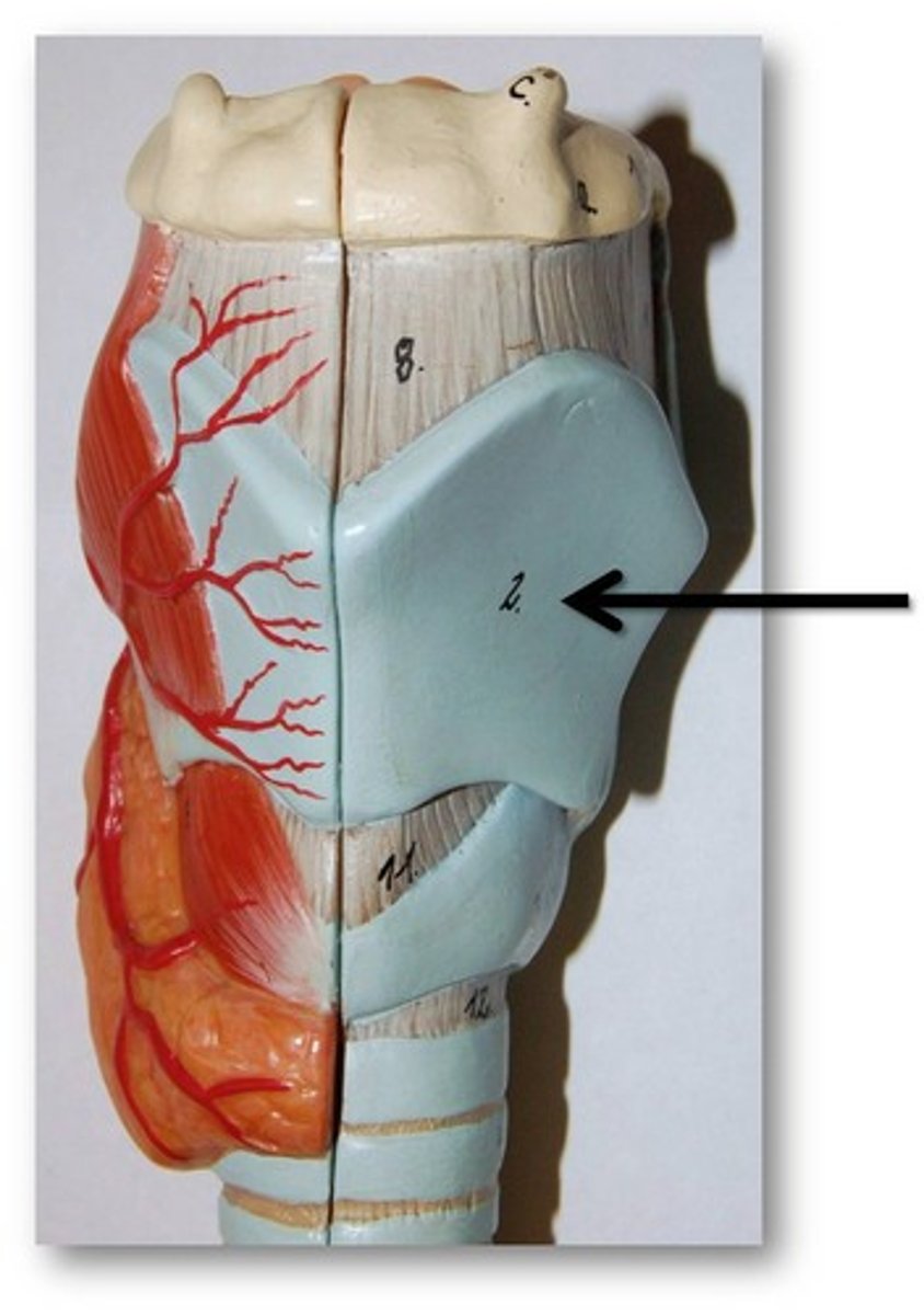
thyroid cartilage (model)
The big blue part
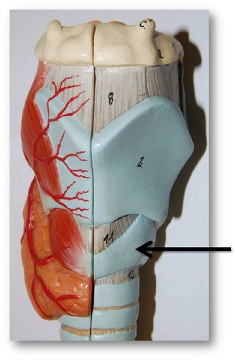
cricoid cartilage (model)
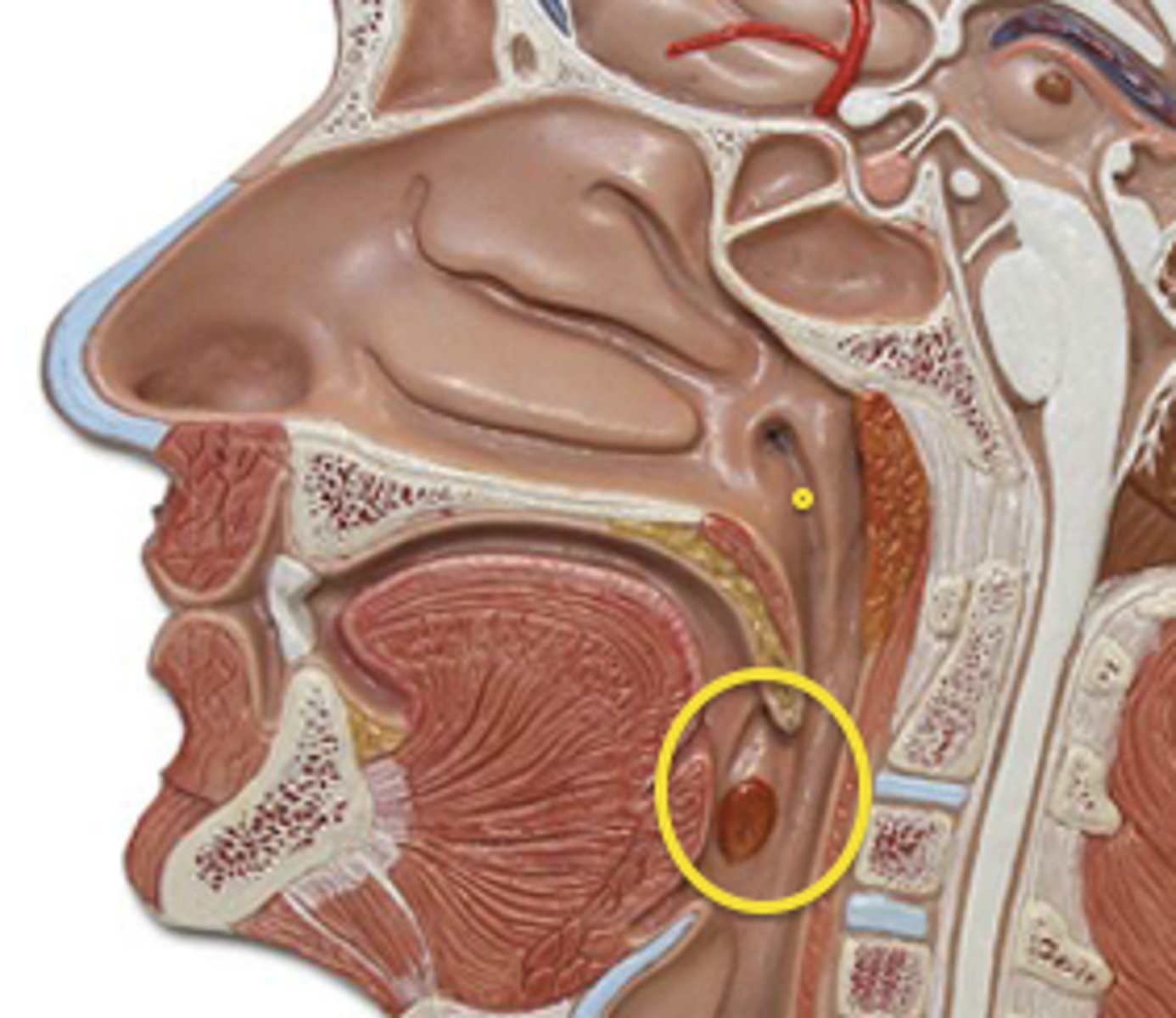
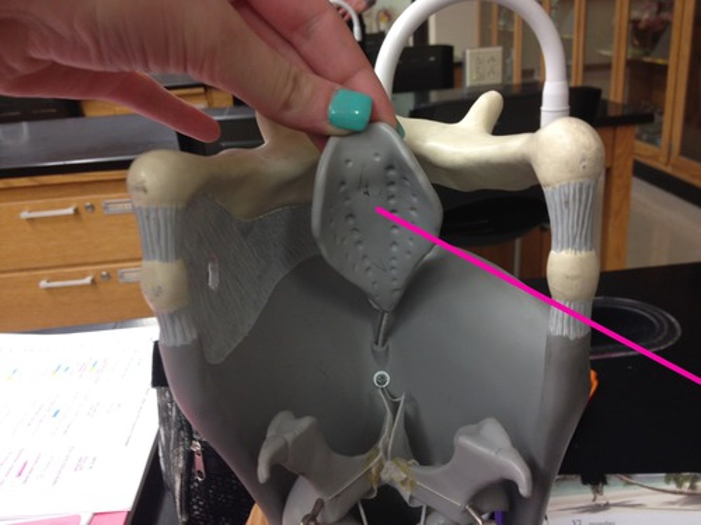
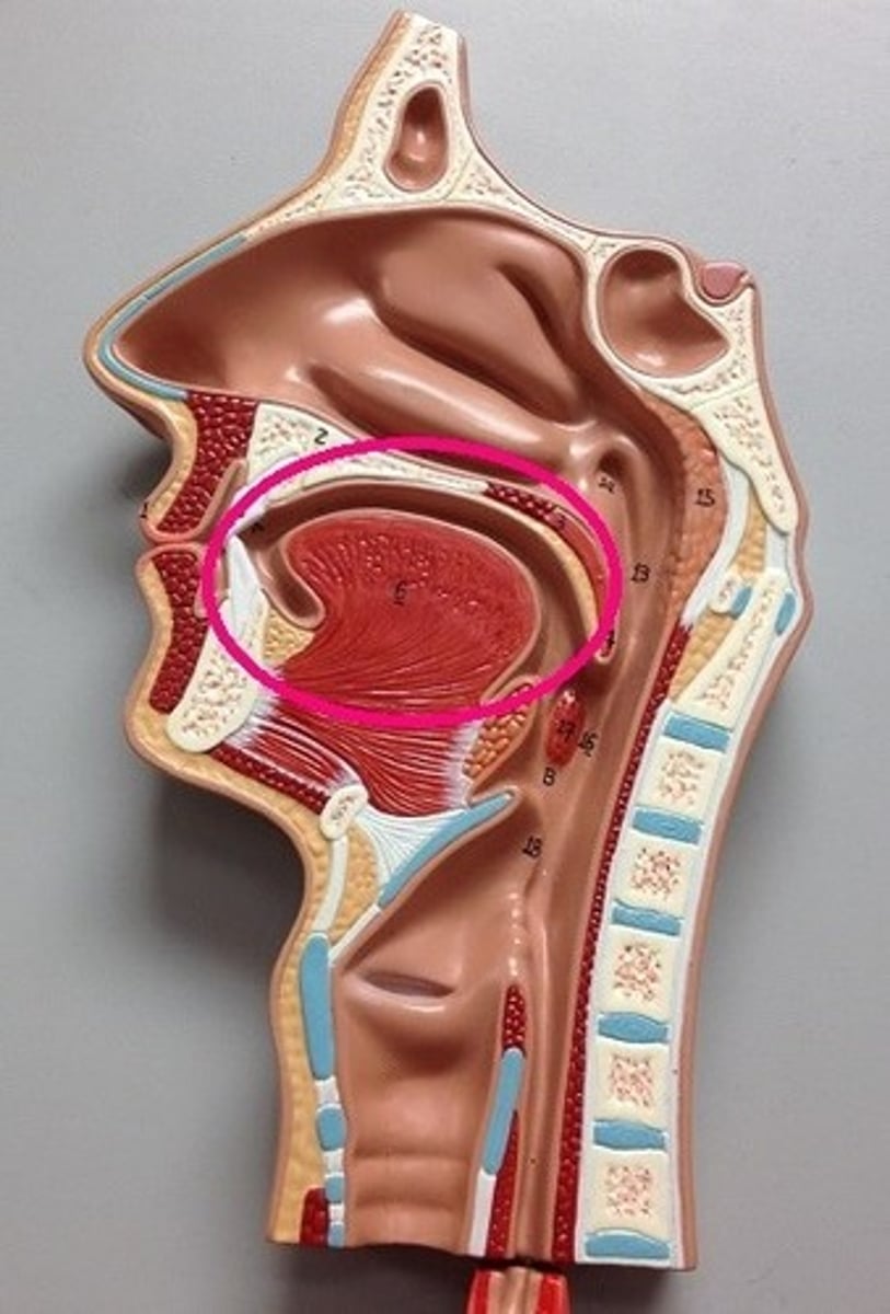
epiglottis (model)
flesh colored part; behind the hyoid bone
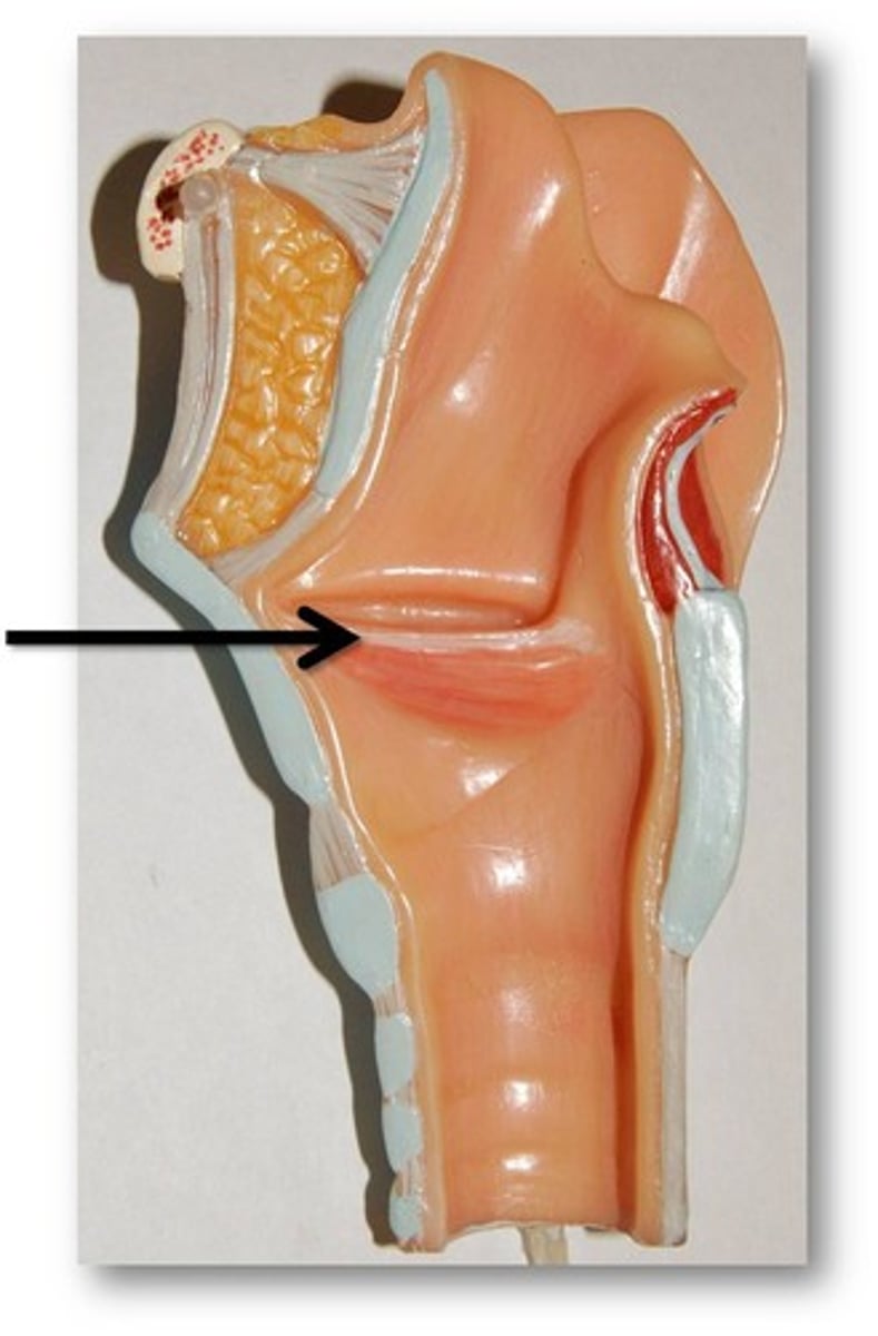
vestibular fold (model)
superior
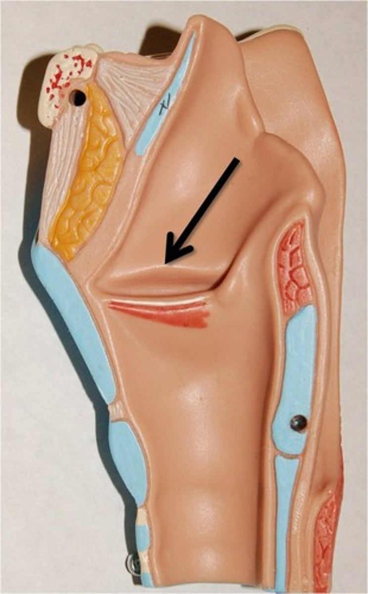
vocal fold (model)
inferior
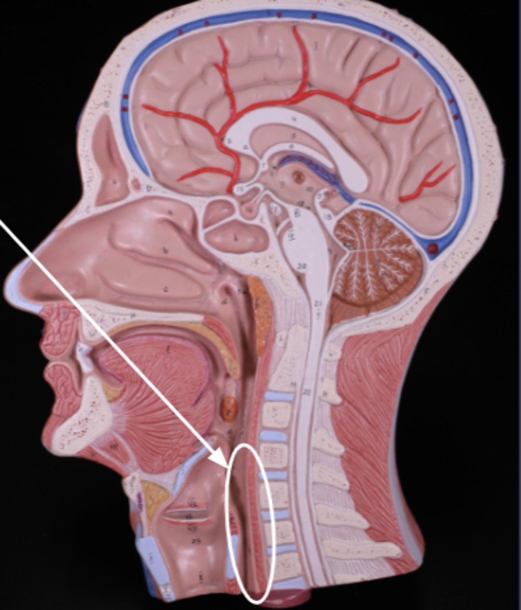
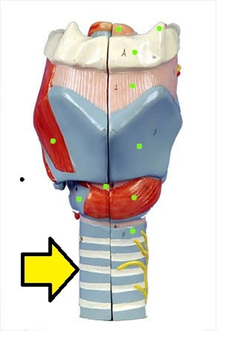
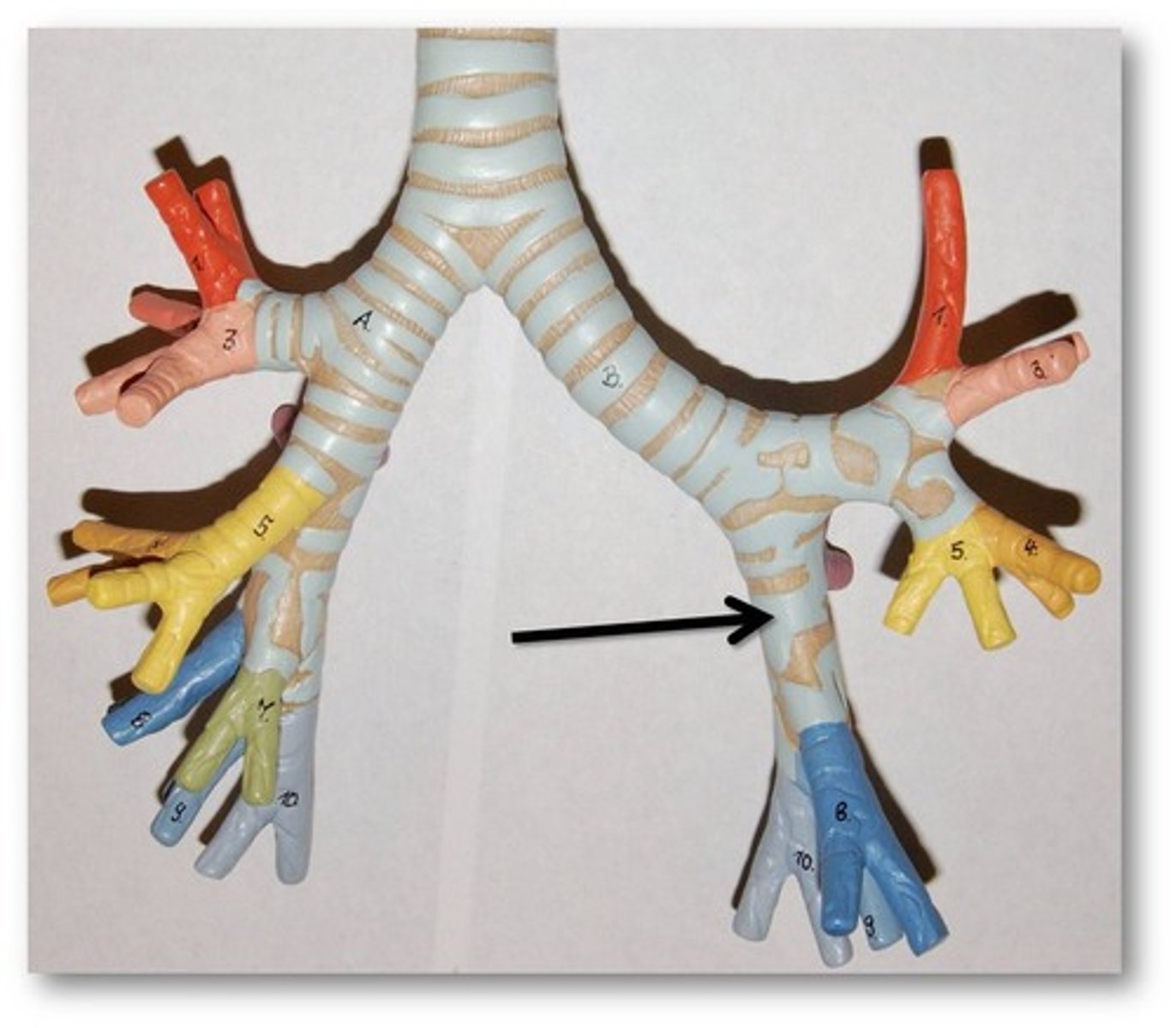
trachea (model)
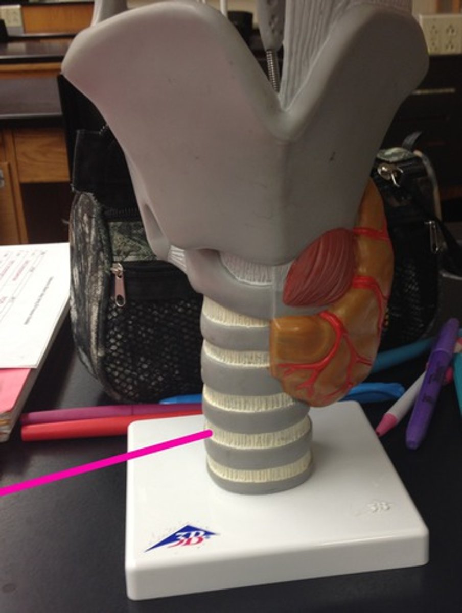
cartilaginous rings (model)
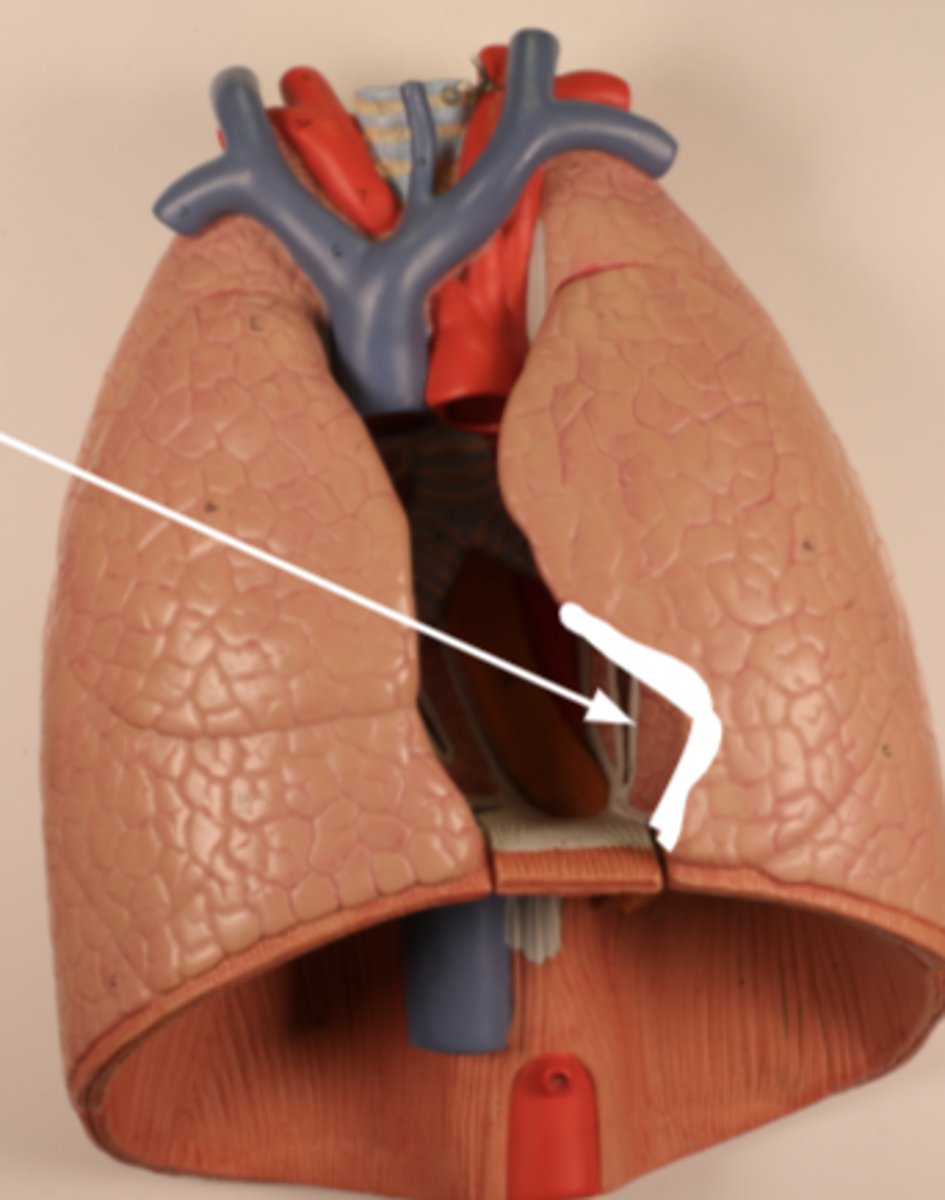
carina (model)
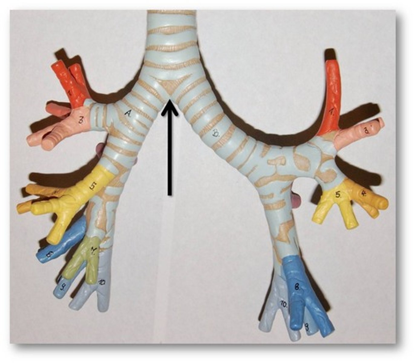
primary bronchi (model)
know R/L
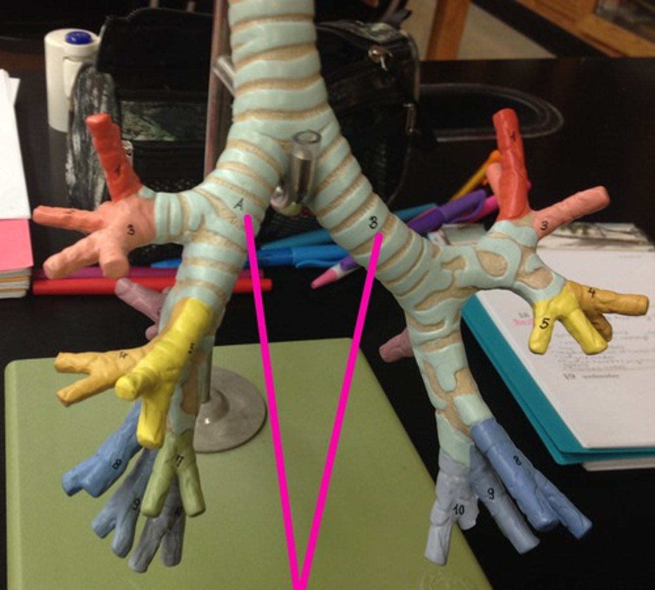
secondary bronchi (lobar) (model)
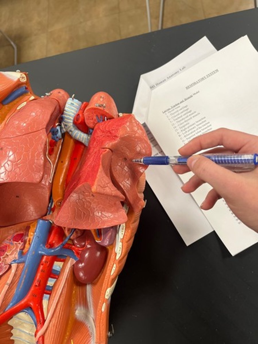
right lung (model)
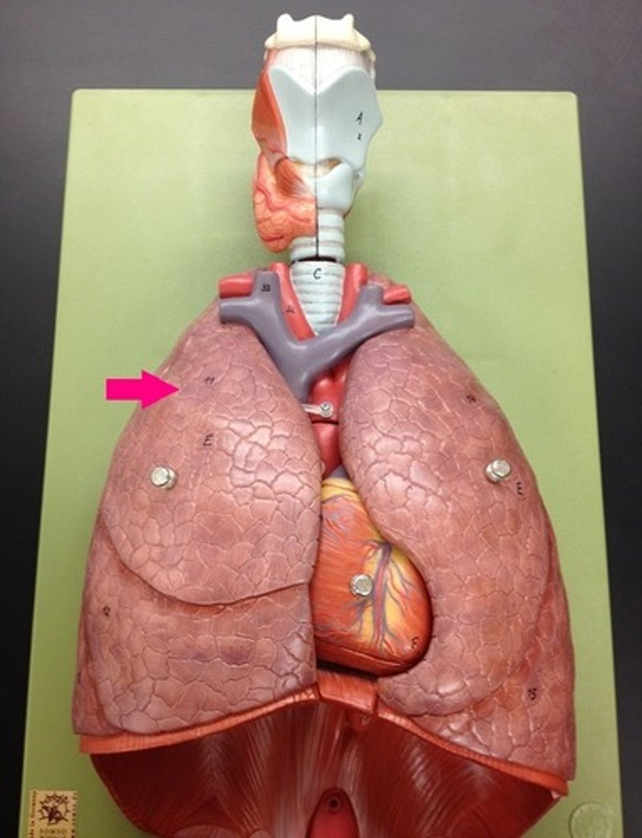
lung base (model)
apex of the lung
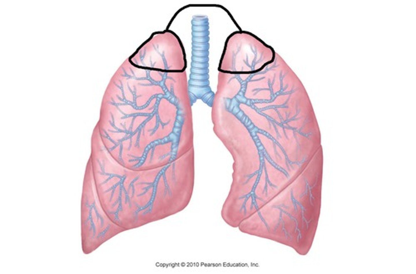
lobes of right lung (model)
R. lung has
superior lobe
middle lobe
inferior lobe
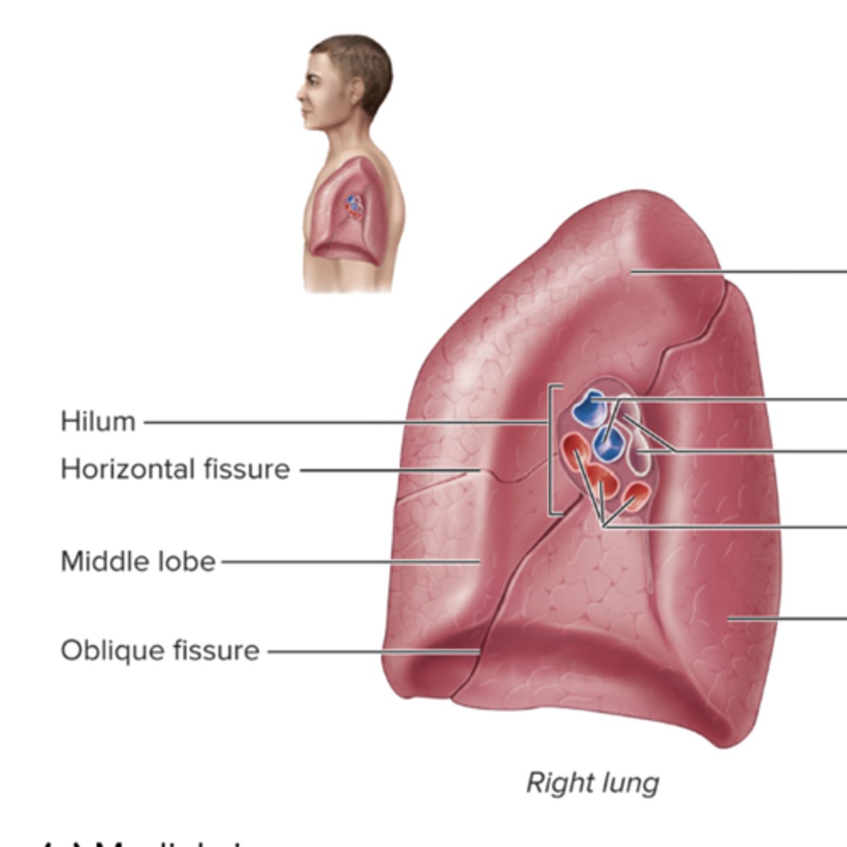
oblique fissure (model)
horizontal fissure (model)
** only on right lung
lobes of left lung (model)
Left lung has only superior and inferior lobes
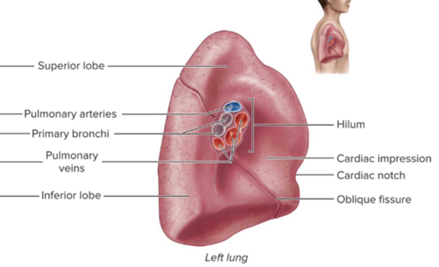
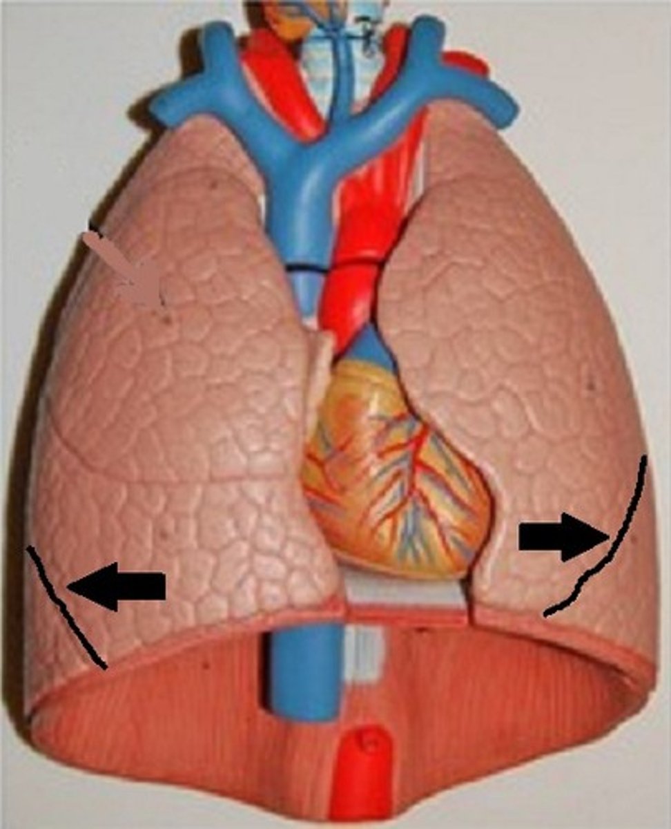
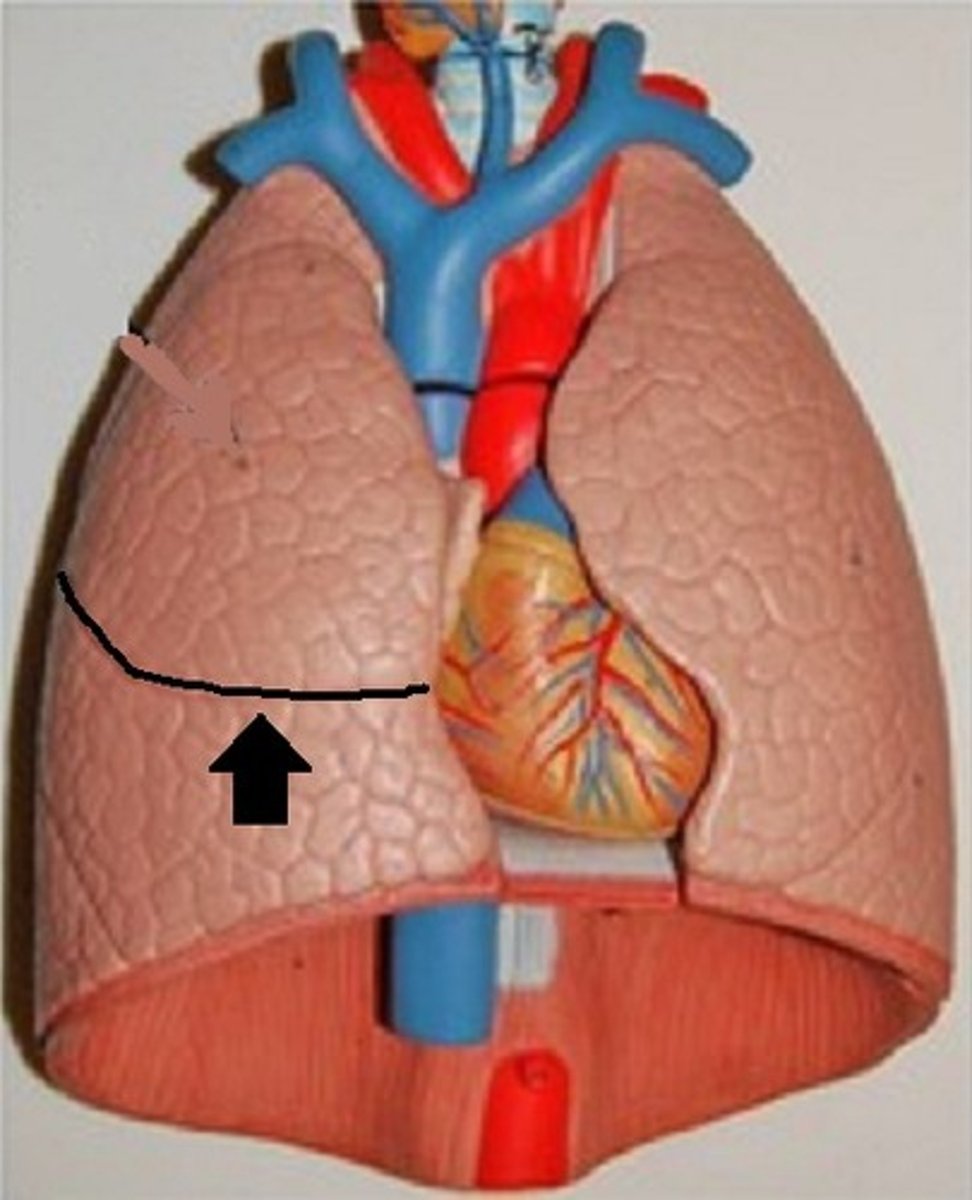
cardiac impression (model)
** left lung
cardiac notch (model)
** left lung
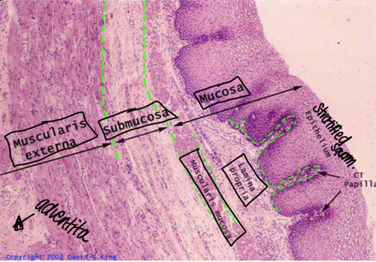
Esophagus Histology
Mucosa:
- stratified squamous epithelium
- lamina propria
- muscularis mucosae
Submucosa
Muscularis Externa
Adventita
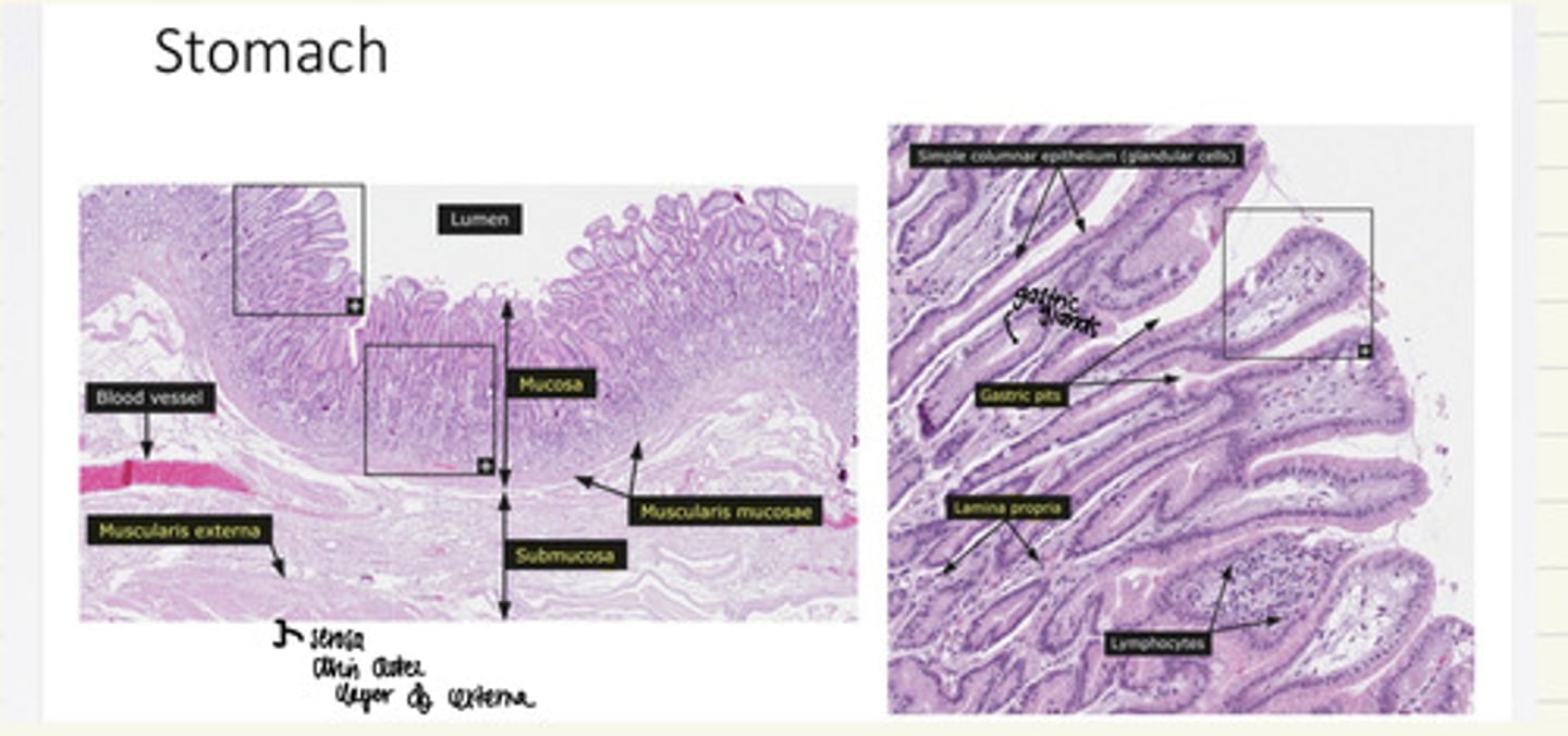
Stomach Histology
Mucosa
- gastric pits
- gastric glands
- lamina propria
Submucosa
Muscularis Externa
Serosa
Rugae **
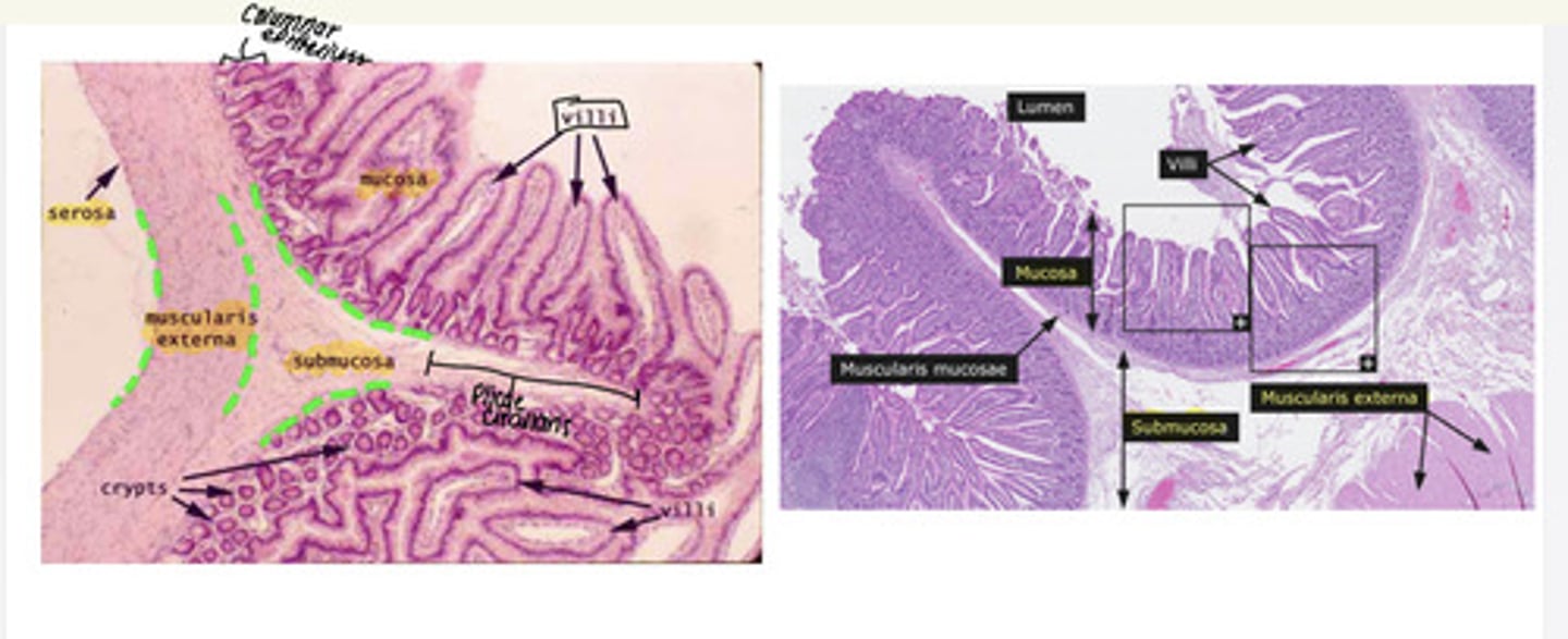
Small Intestine Histology
i. Plicae circulares
ii. Columnar epithelium
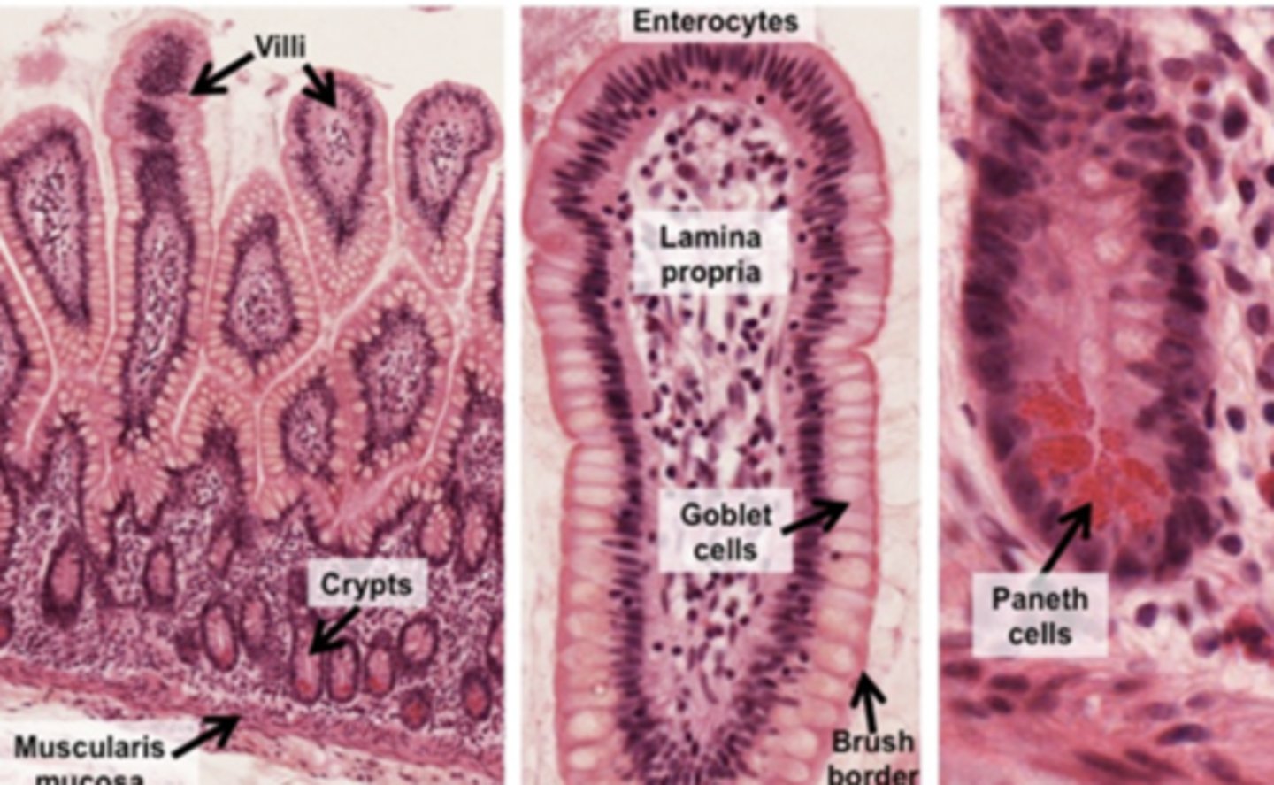
iii. Villi
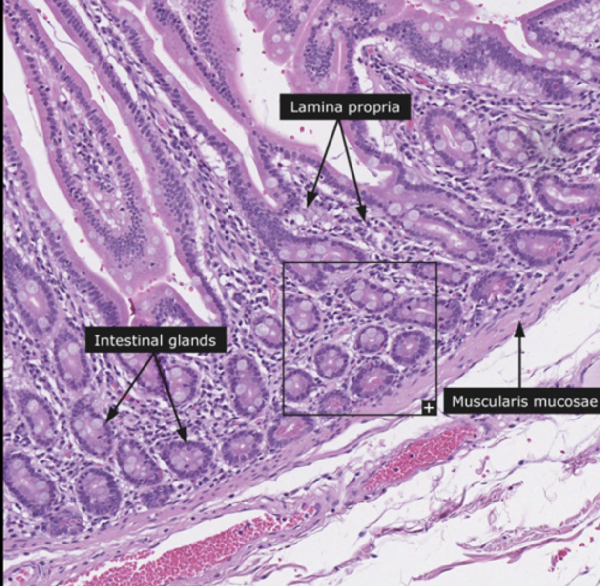
iv. Intestinal glands
v. Mucosa 1. Absorptive cells 2. Goblet cells 3. Brush boarder vi. Submucosa
vii. Muscularis externa
viii. Serosa
Intestinal Gland (small intestine)
Mucosa of small intestine (3)
absorptive cells
goblet cells
bush boarder
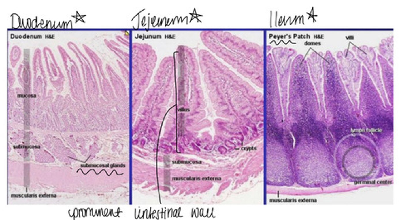
duodenum, jejunum, ileum histology
Duodenum: duodenal glands
Jejunum: prominent intestinal villi
Ileum: peyer's patches
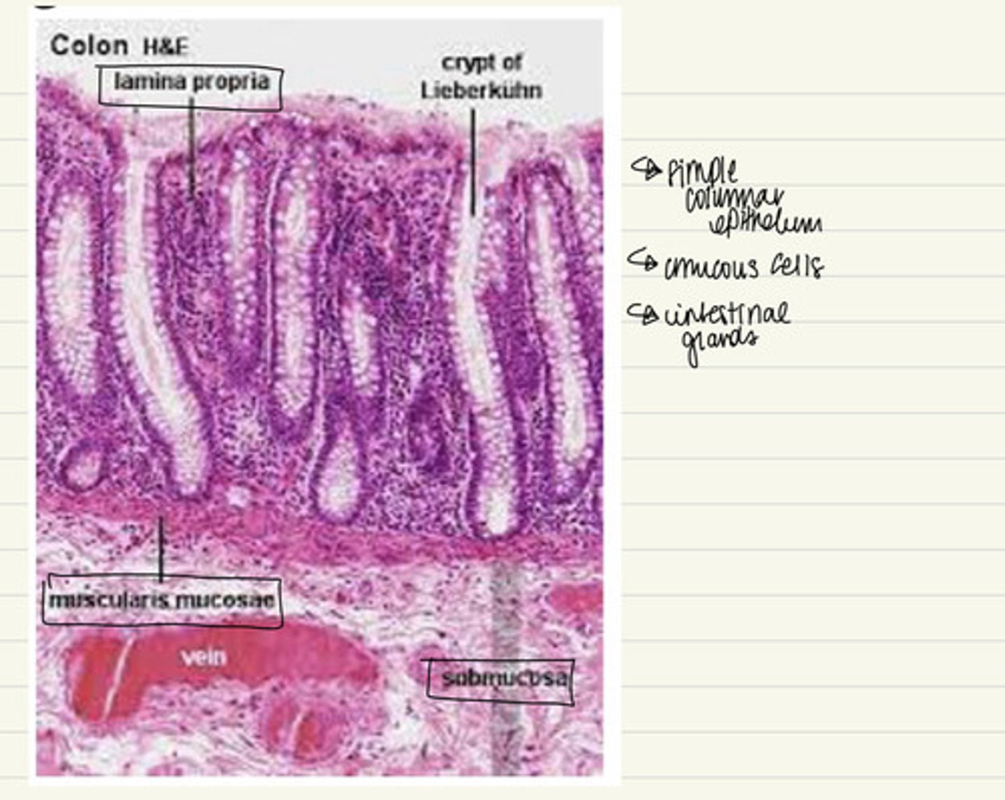
Large intestine Histology
i. Mucosa 1. Simple columnar epithelium 2. Mucous cells 3. Intestinal glands 4. Lamina propria 5. Muscularis mucosae
ii. Submucosa
iii. Muscularis externa
iv. Serosa
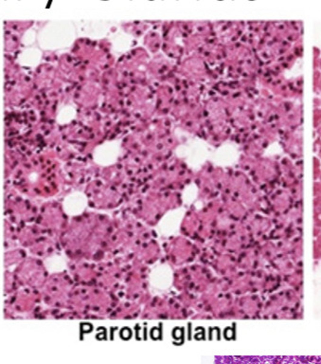
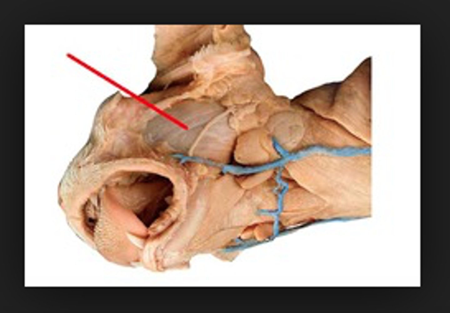
parotid gland
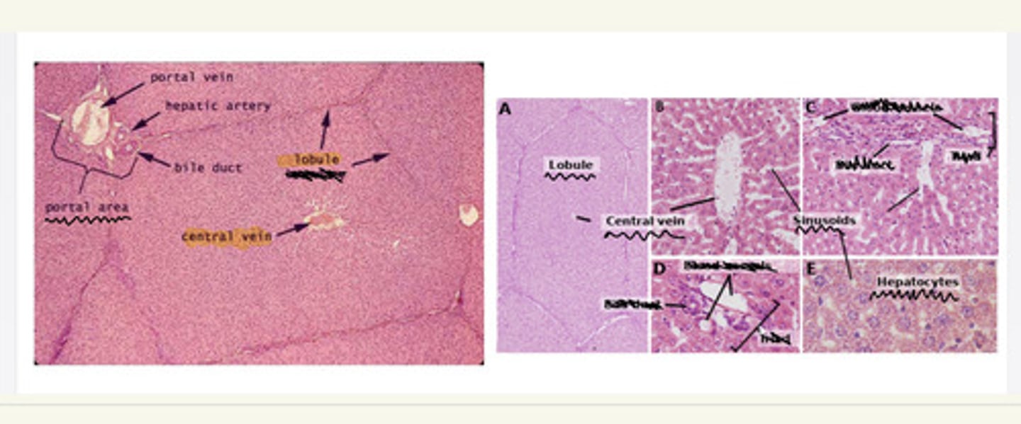
liver histology
i. Liver lobule
ii. Portal area
iii. Central vein i
v. Hepatocyte
v. Liver sinusoids
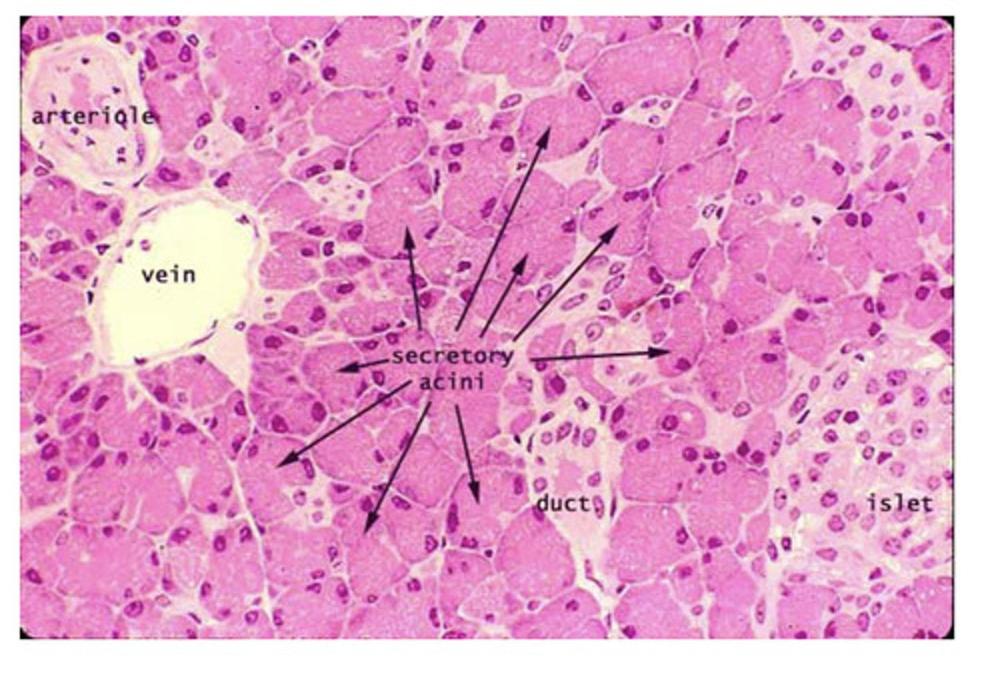
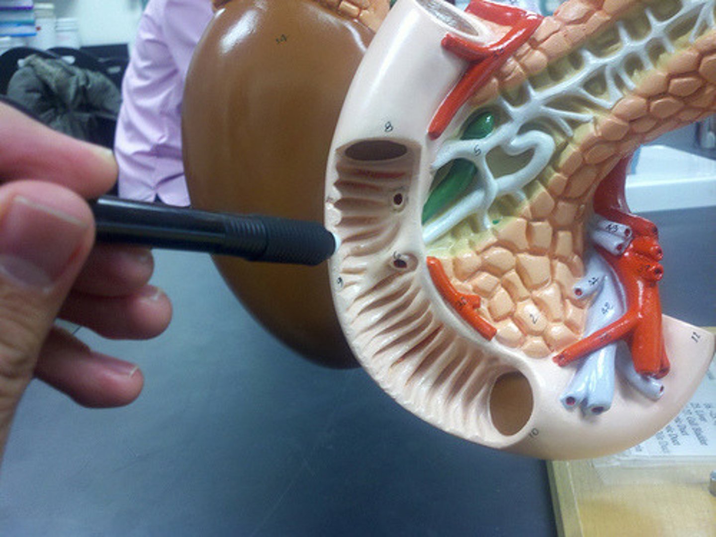
pancreas histology
pancreatic acini
pancreatic islets
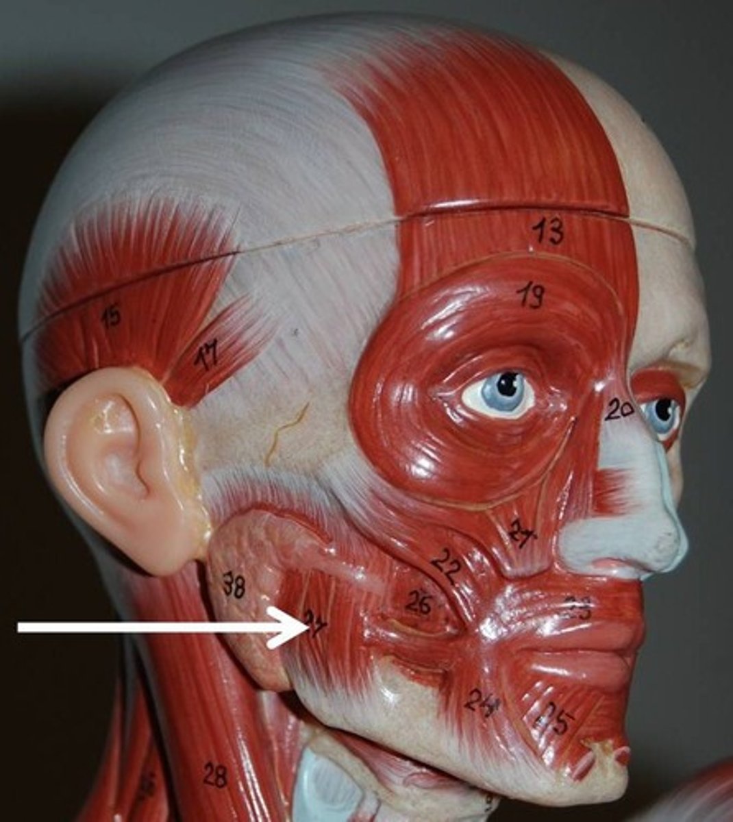
masseter (cat)

parotid gland (cat)
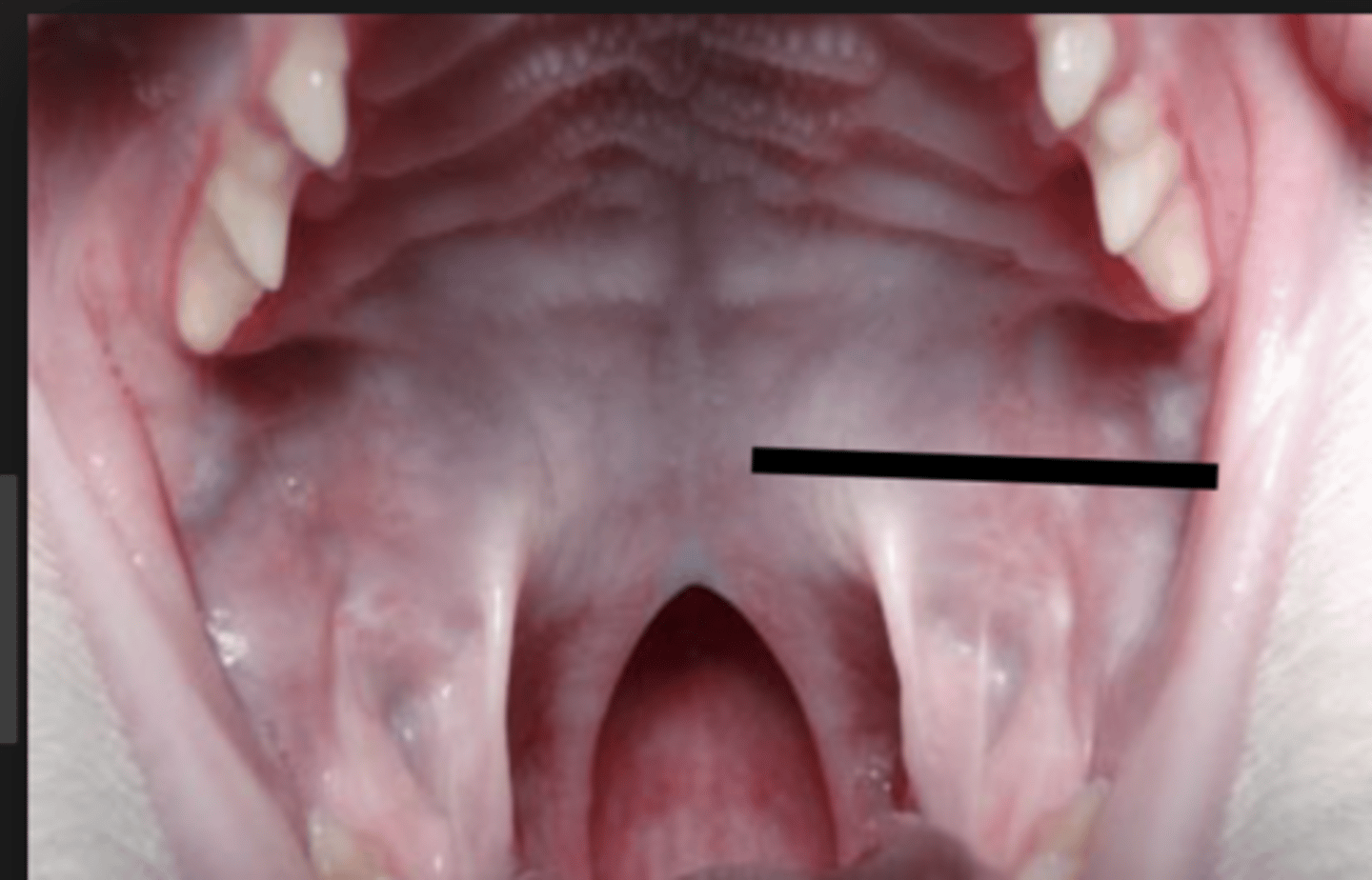
oral cavity (cat)
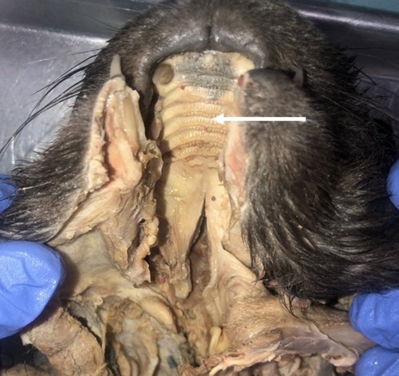
hard palate (cat)
roof of the mouth
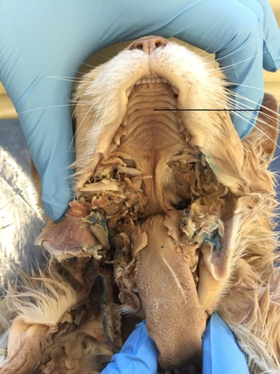
soft palate (cat)
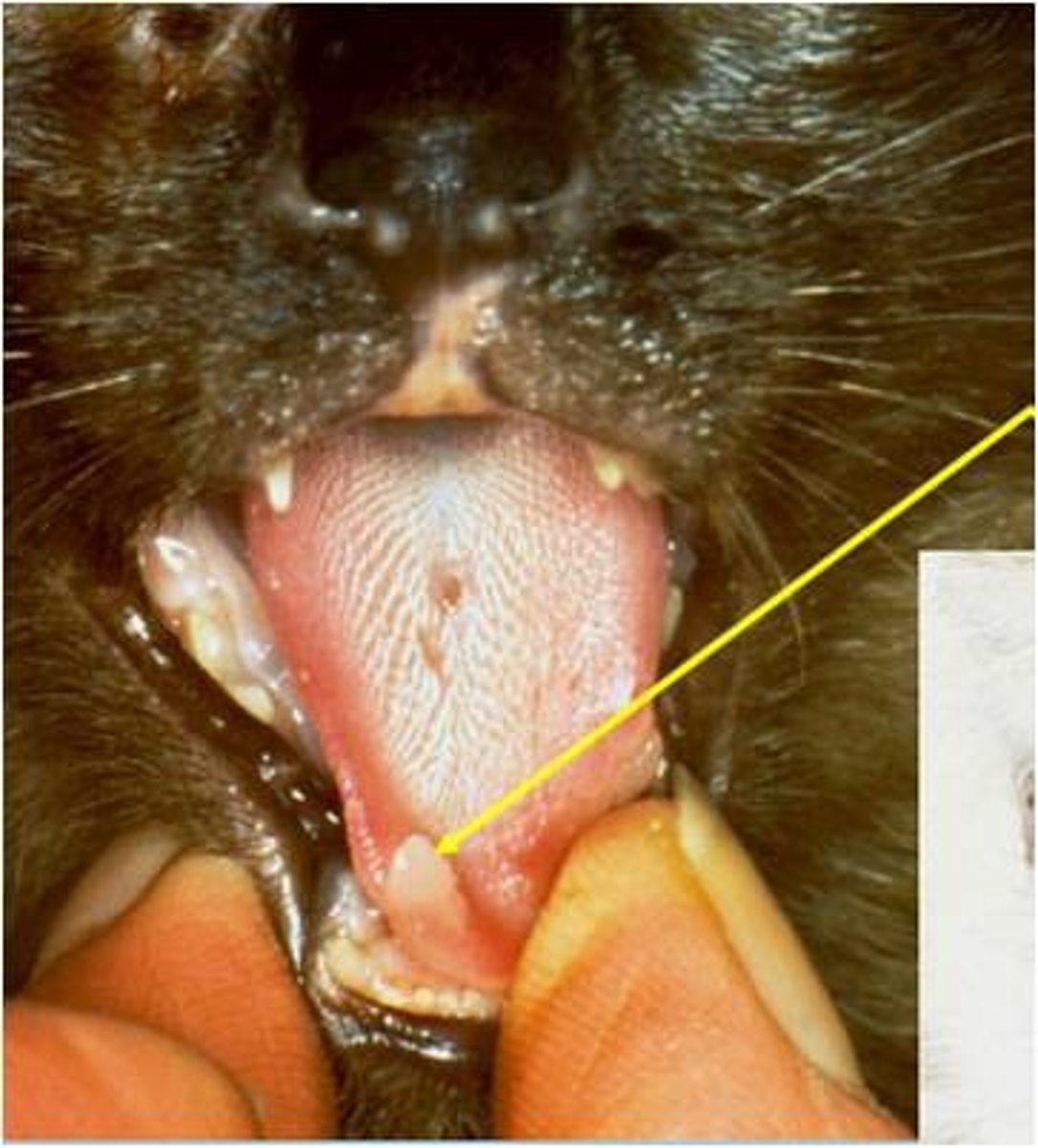
tongue (cat)
incisors (cat)
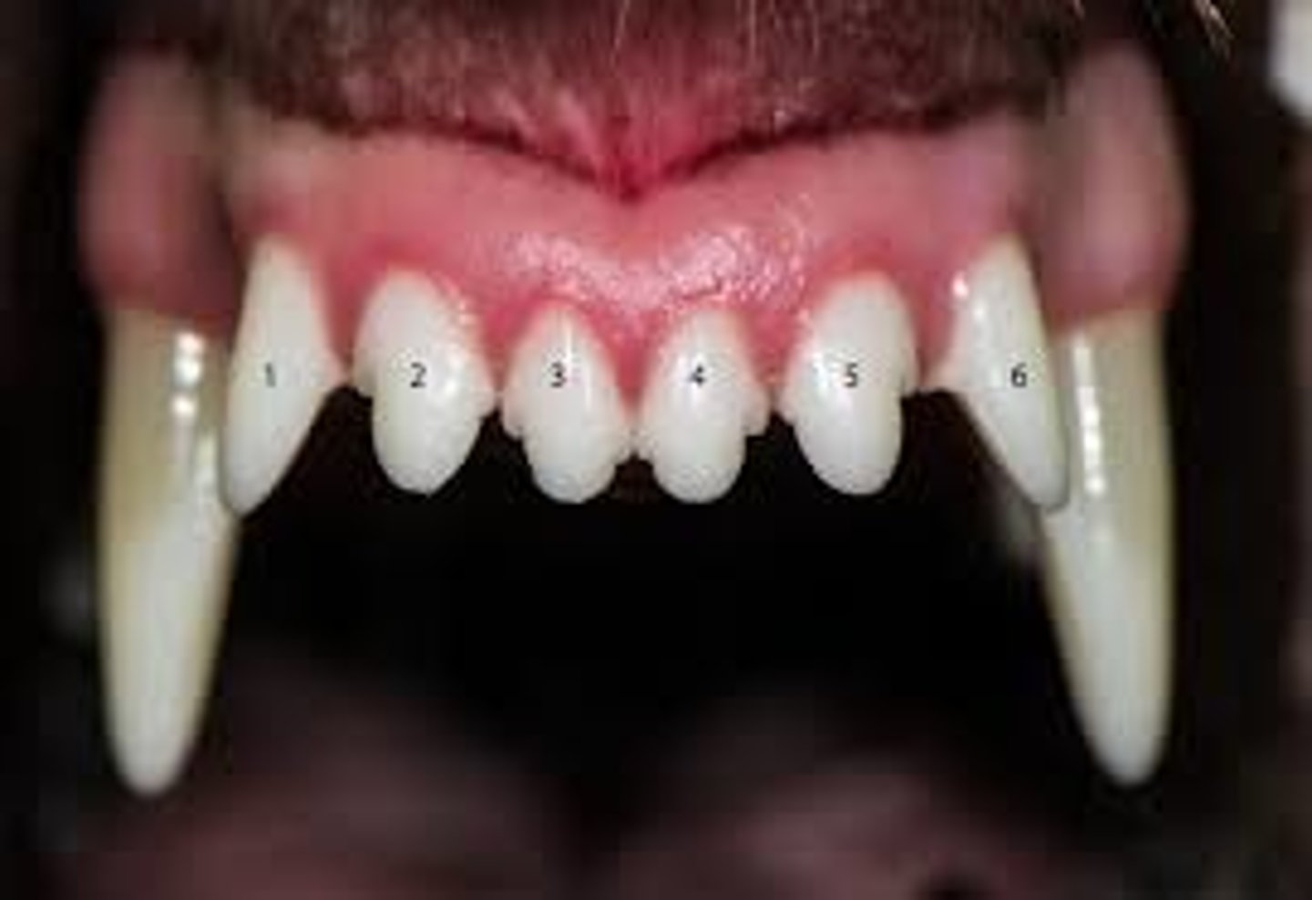
canines (cat)
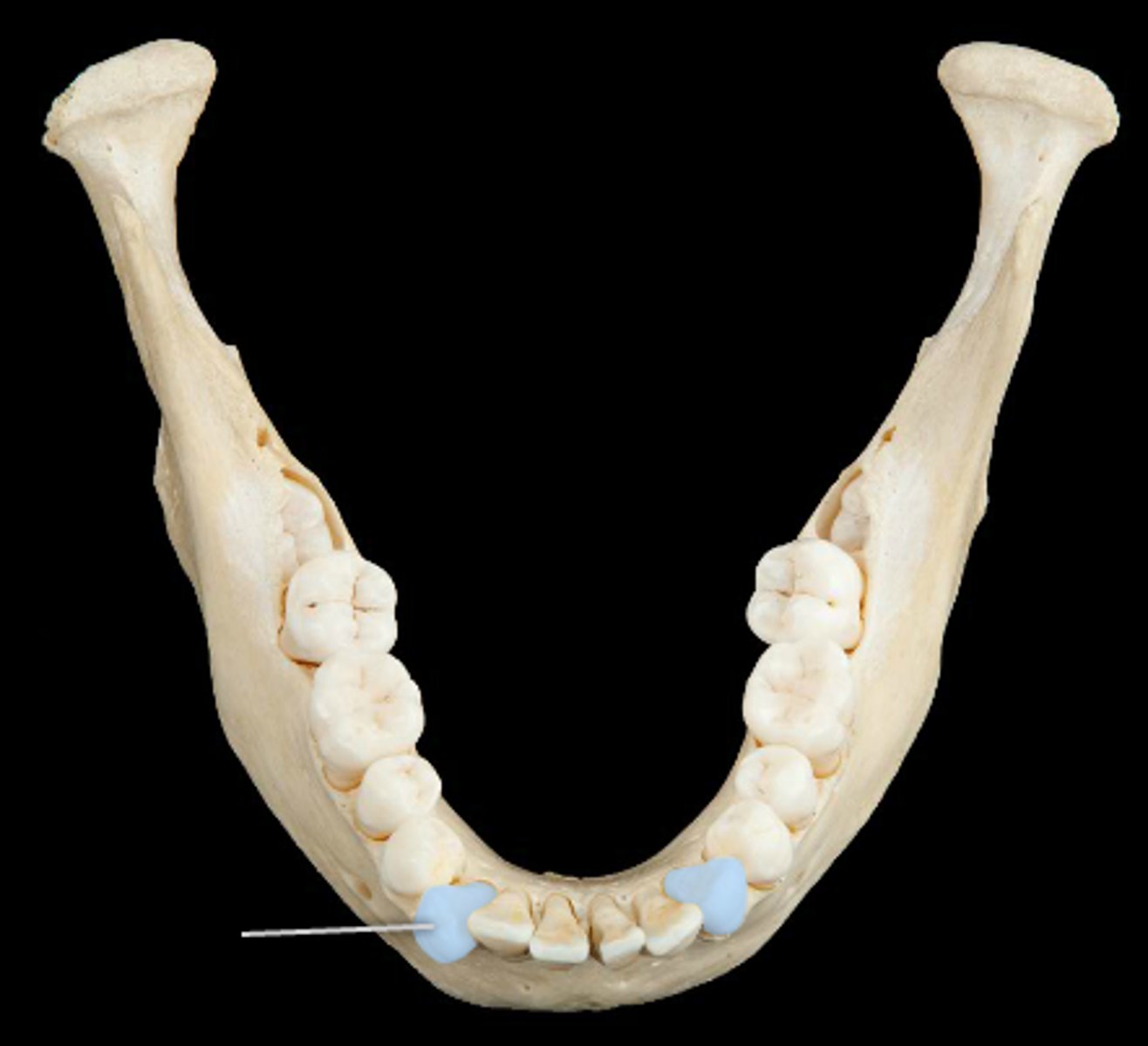
premolars (cat)

molars (cat)
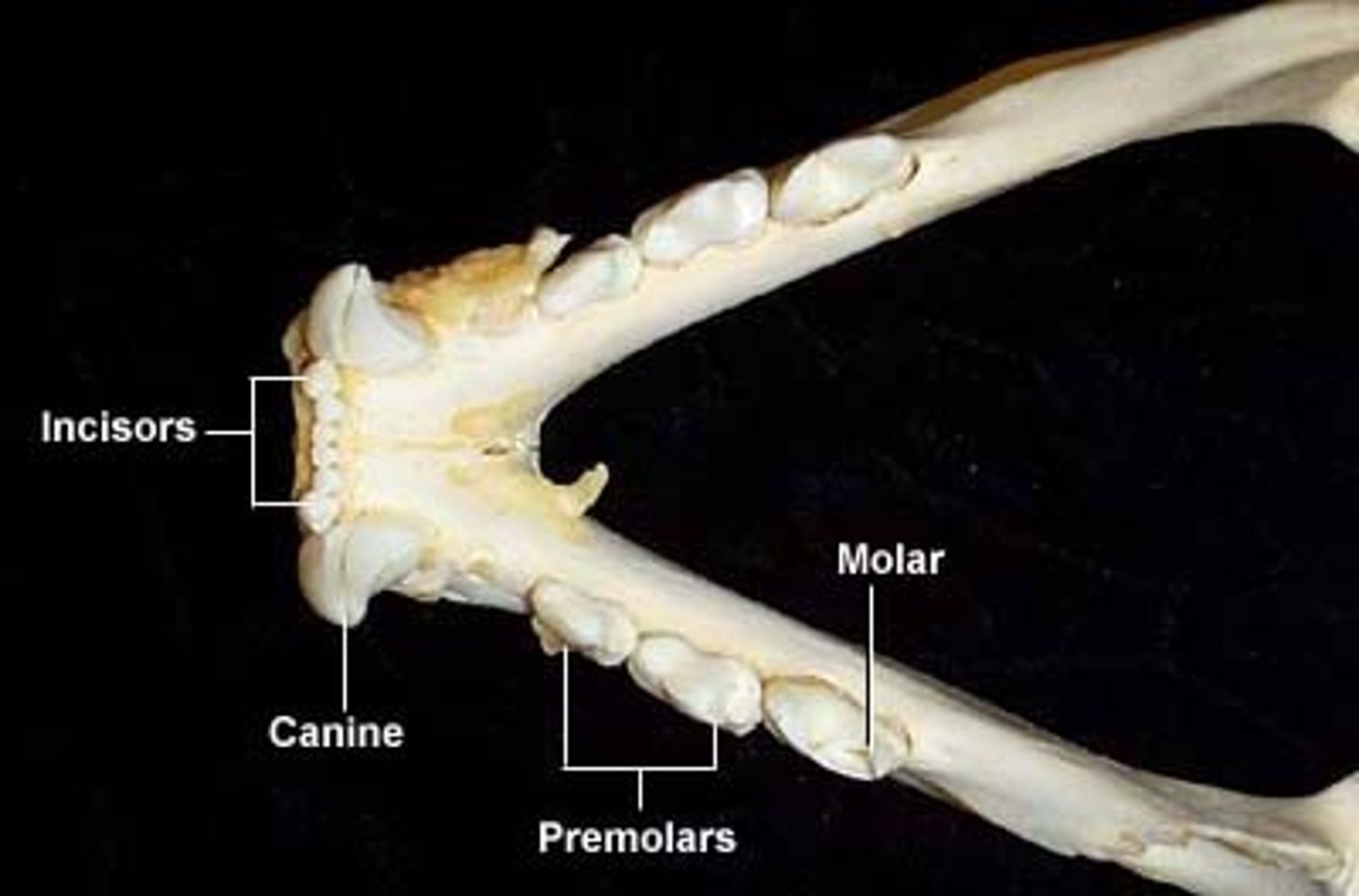
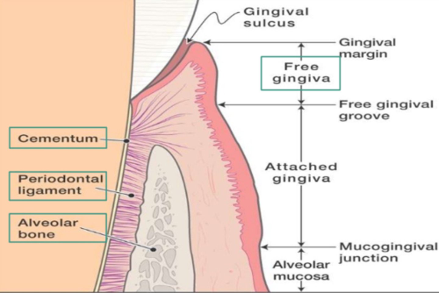
gingival (cat)
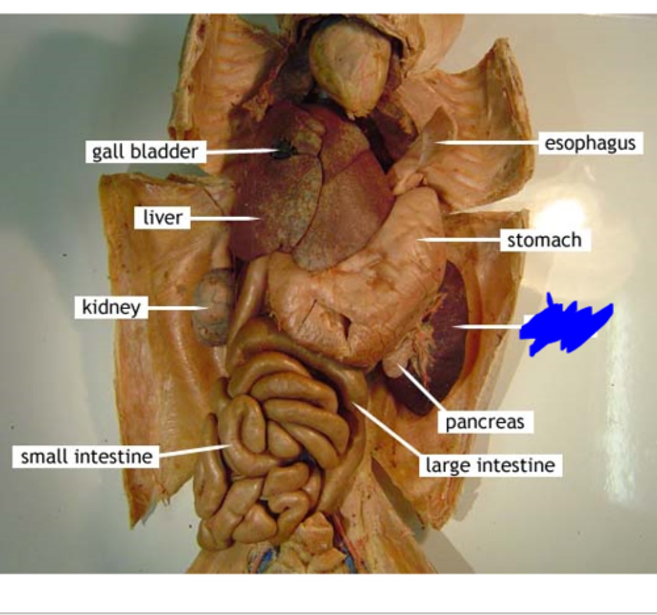
esophagus (cat)
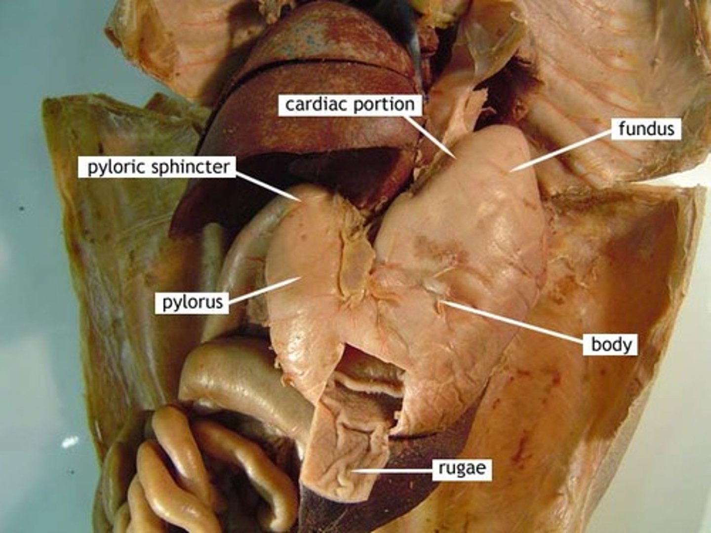
stomach (cat)
- cardiac region
- fundus
- body
- pyloric body
- pyloric sphincter
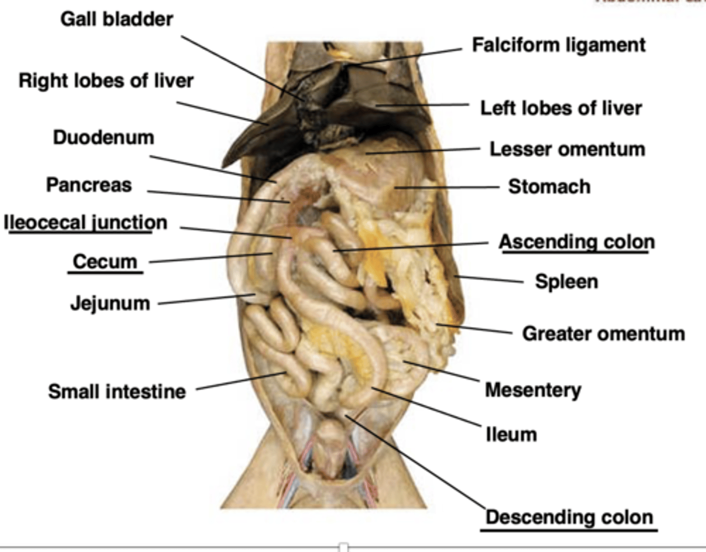
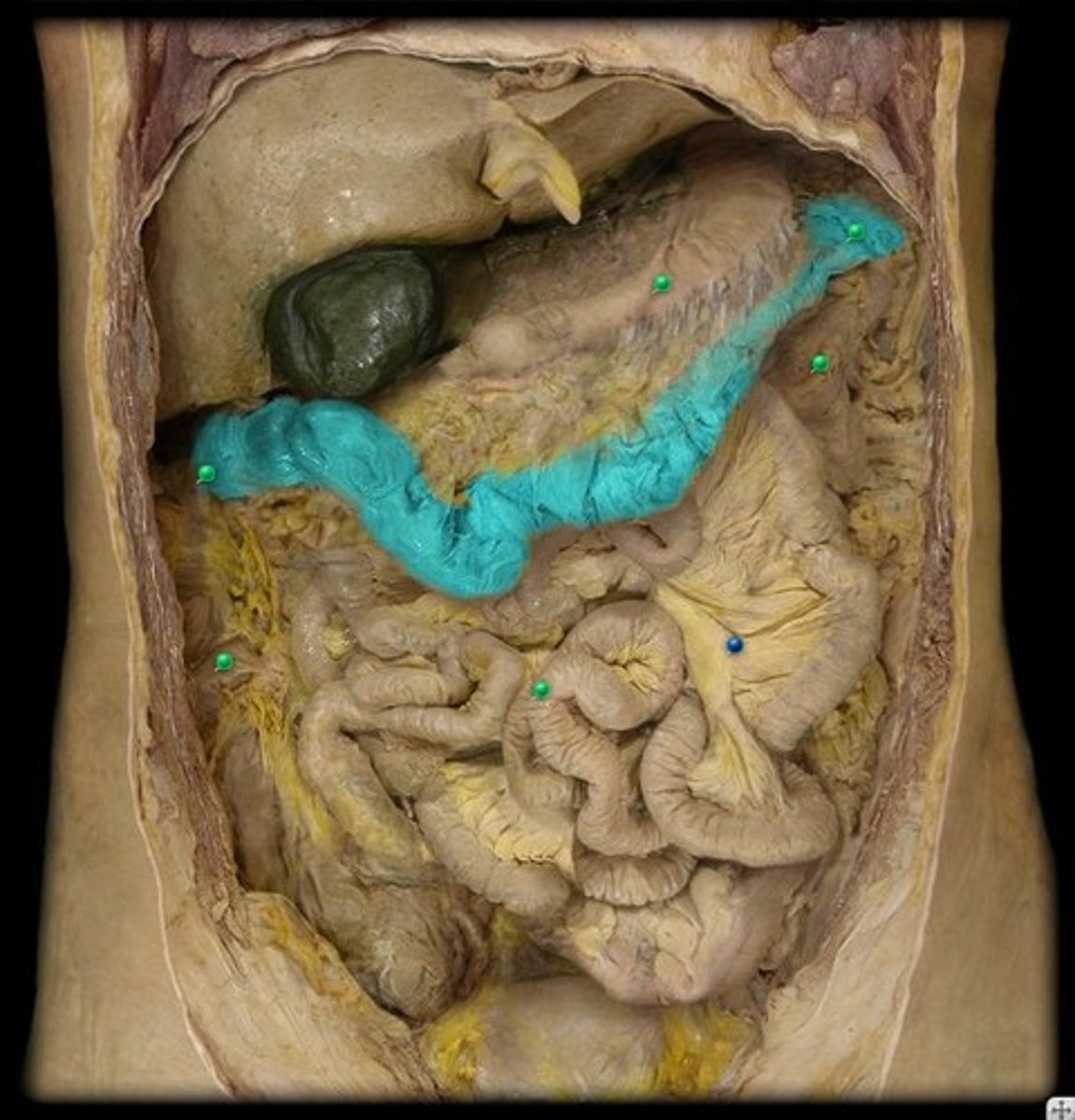
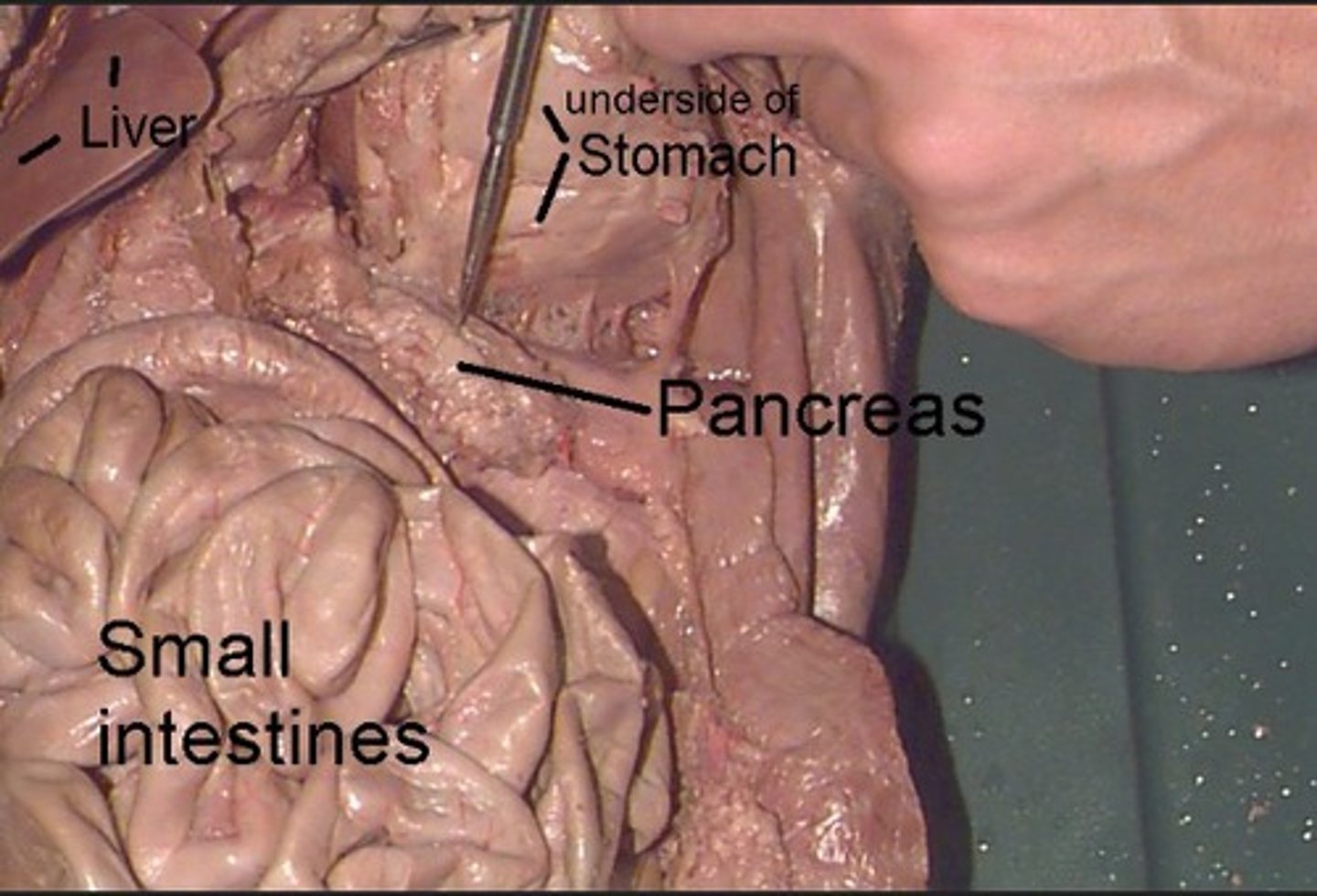
small intestine (cat)
duodenum (superior)
jejunum (middle)
ileum (inferior)
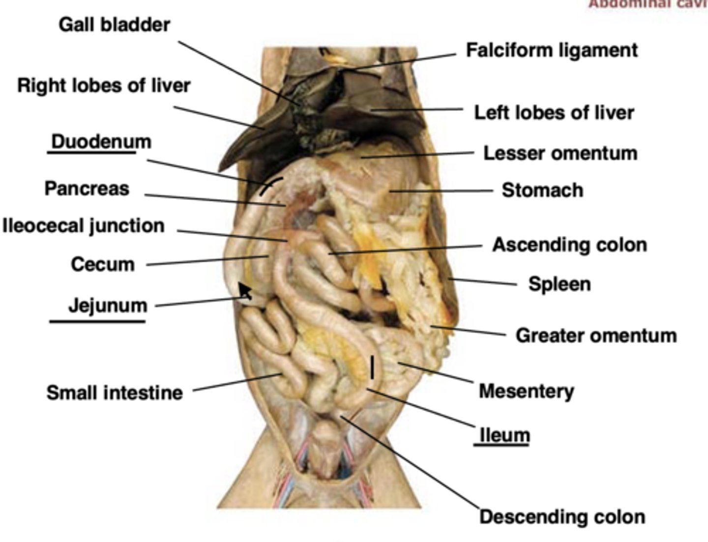
large intestine (cat)
ileocecal valve
cecum
ascending colon
transverse colon
descending colon
ileocecal valve (cat)
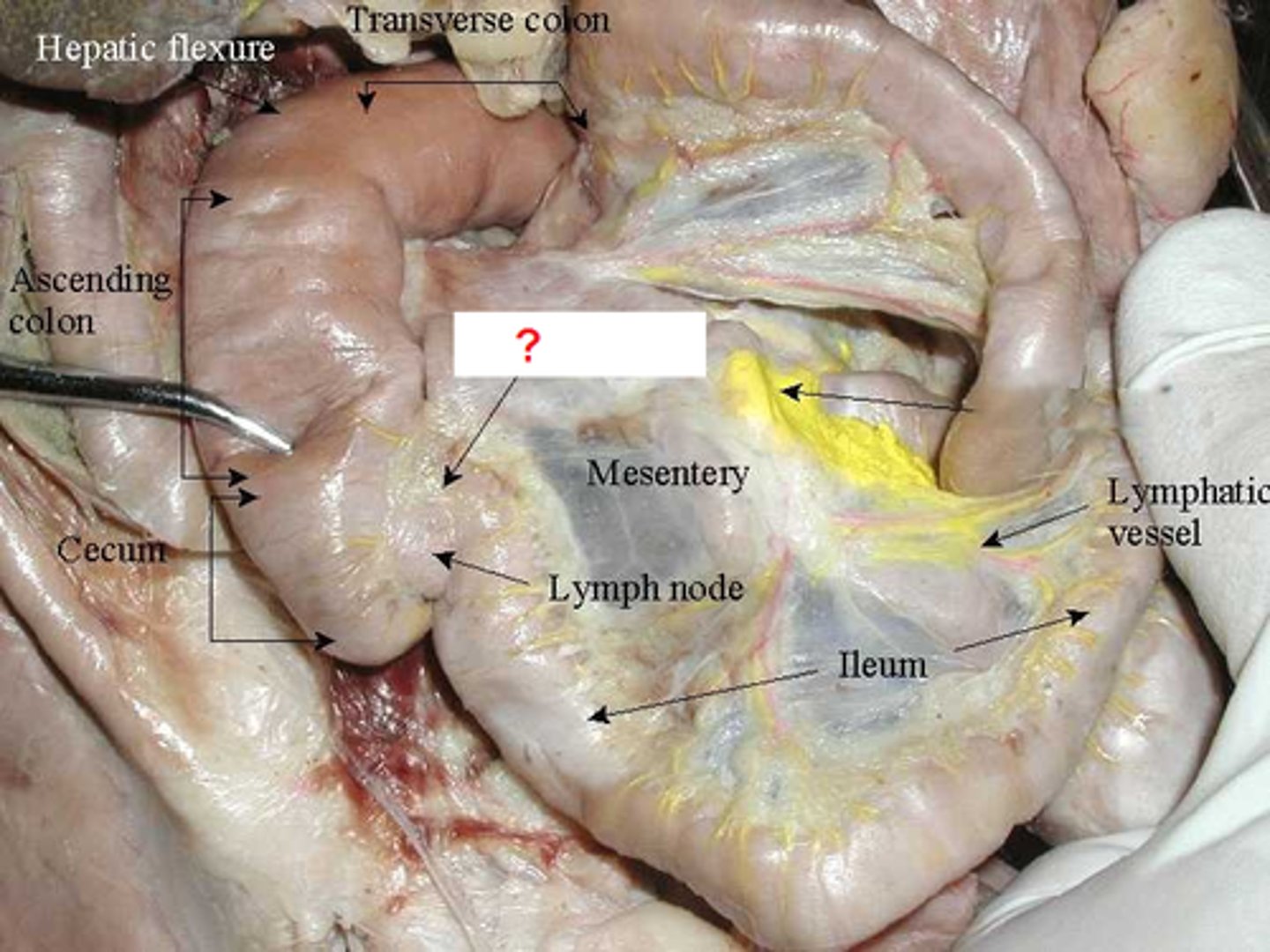
transverse colon (cat)
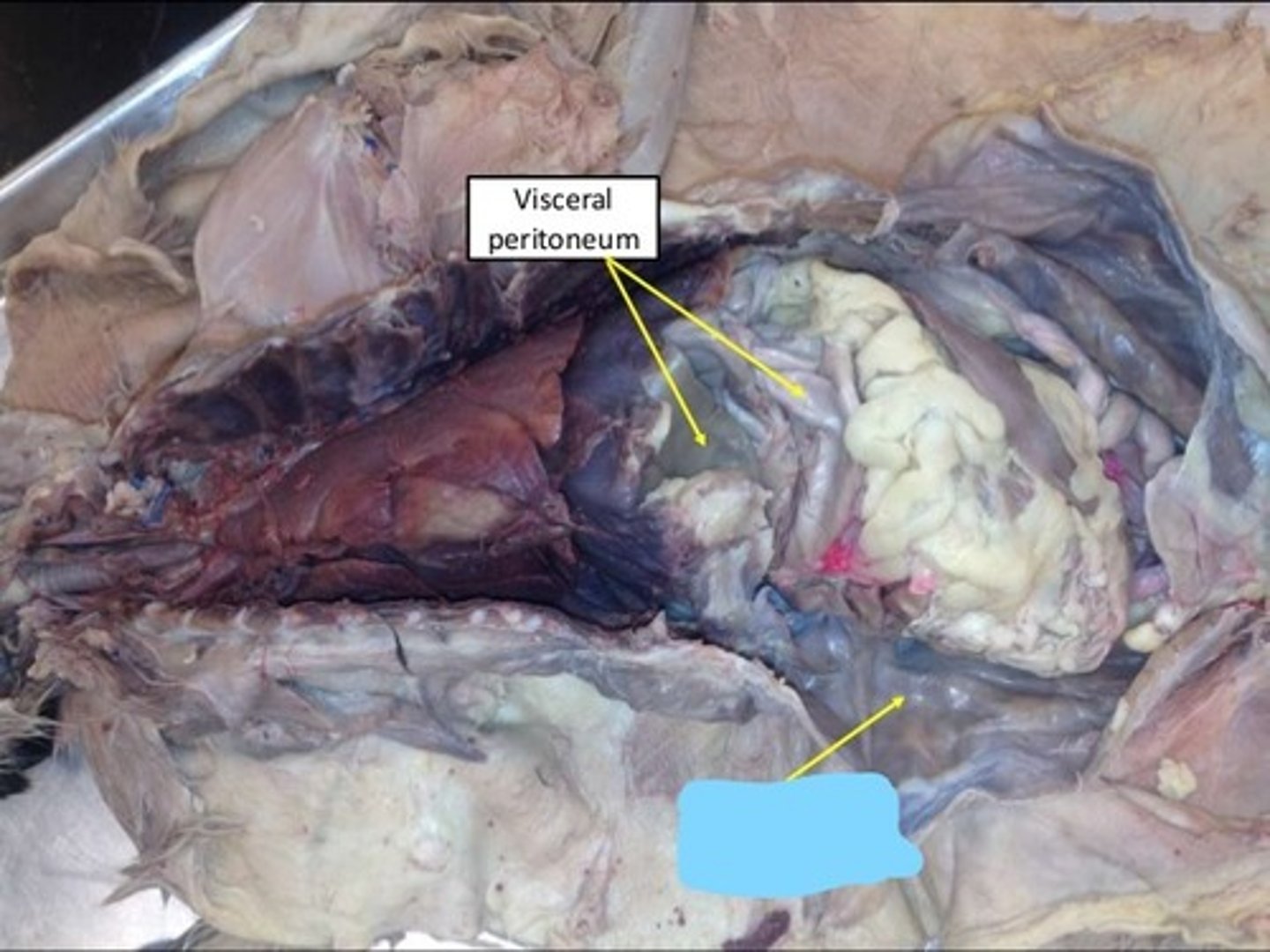
parietal peritoneum (cat)
shiny layer
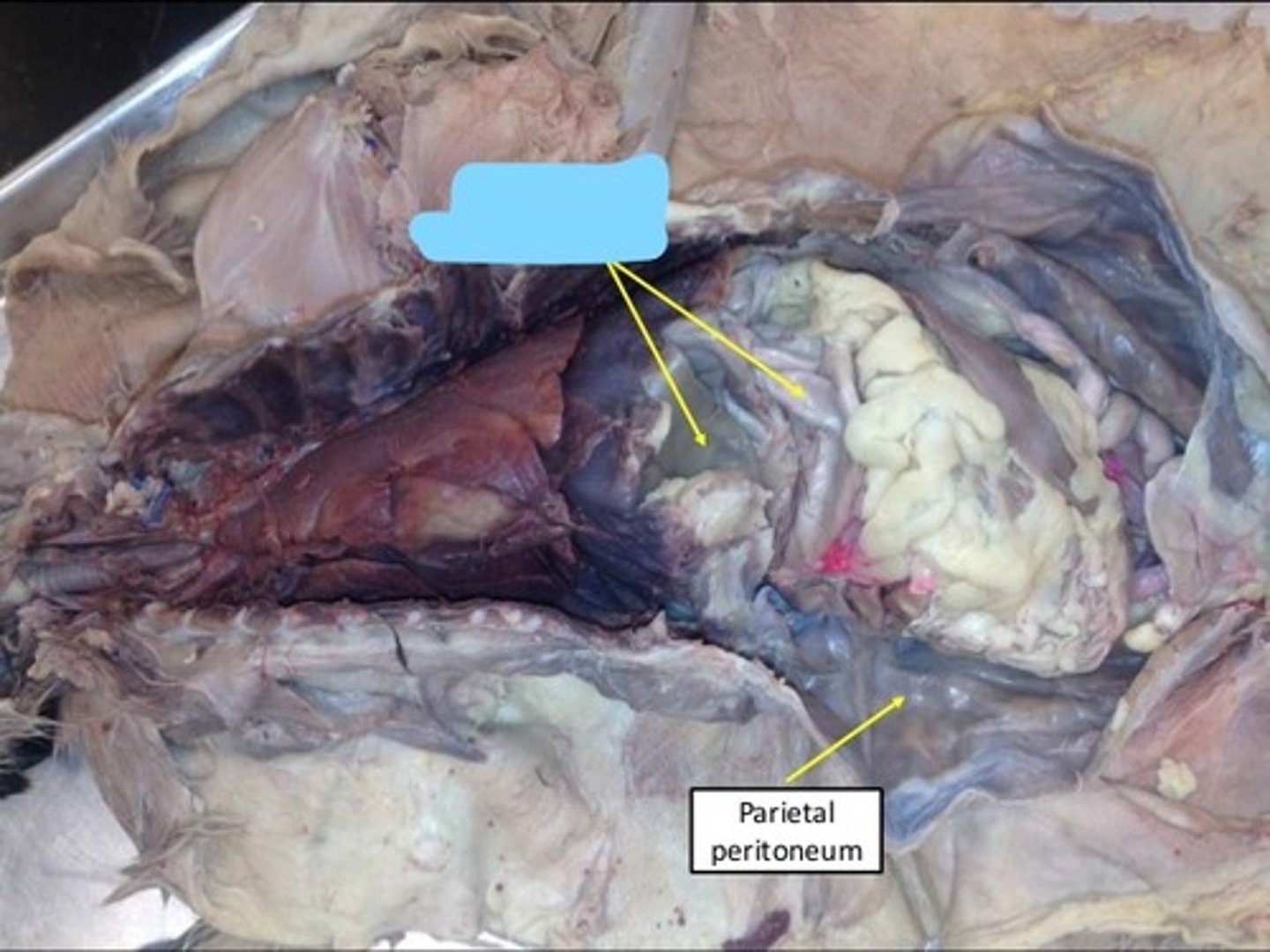
visceral peritoneum (cat)
deeper to parietal
greater omentum (cat)
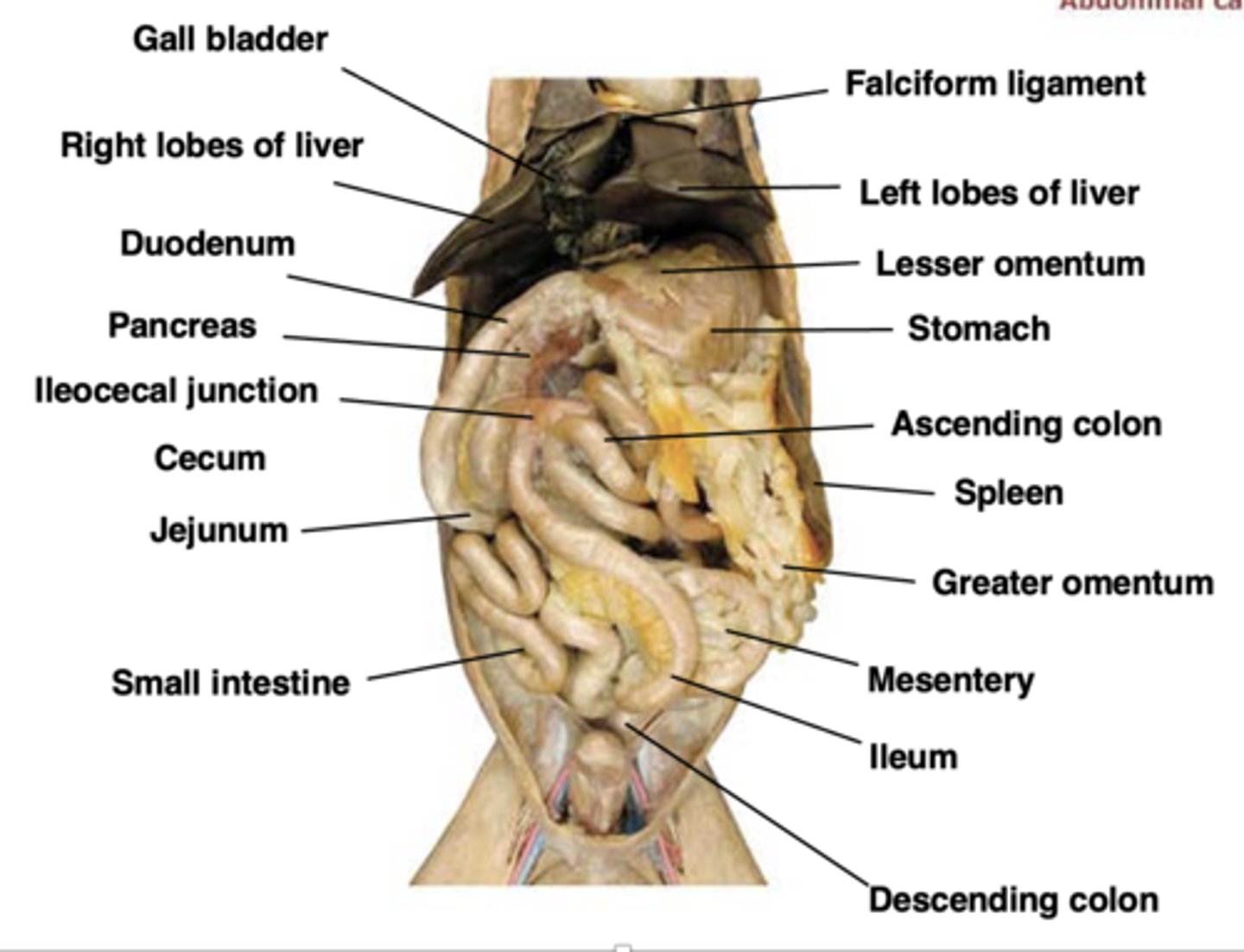
lesser omentum (cat)
attaches stomach to liver
liver (cat)
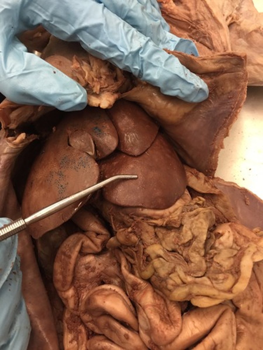
pancreas (cat)
gall bladder (cat)
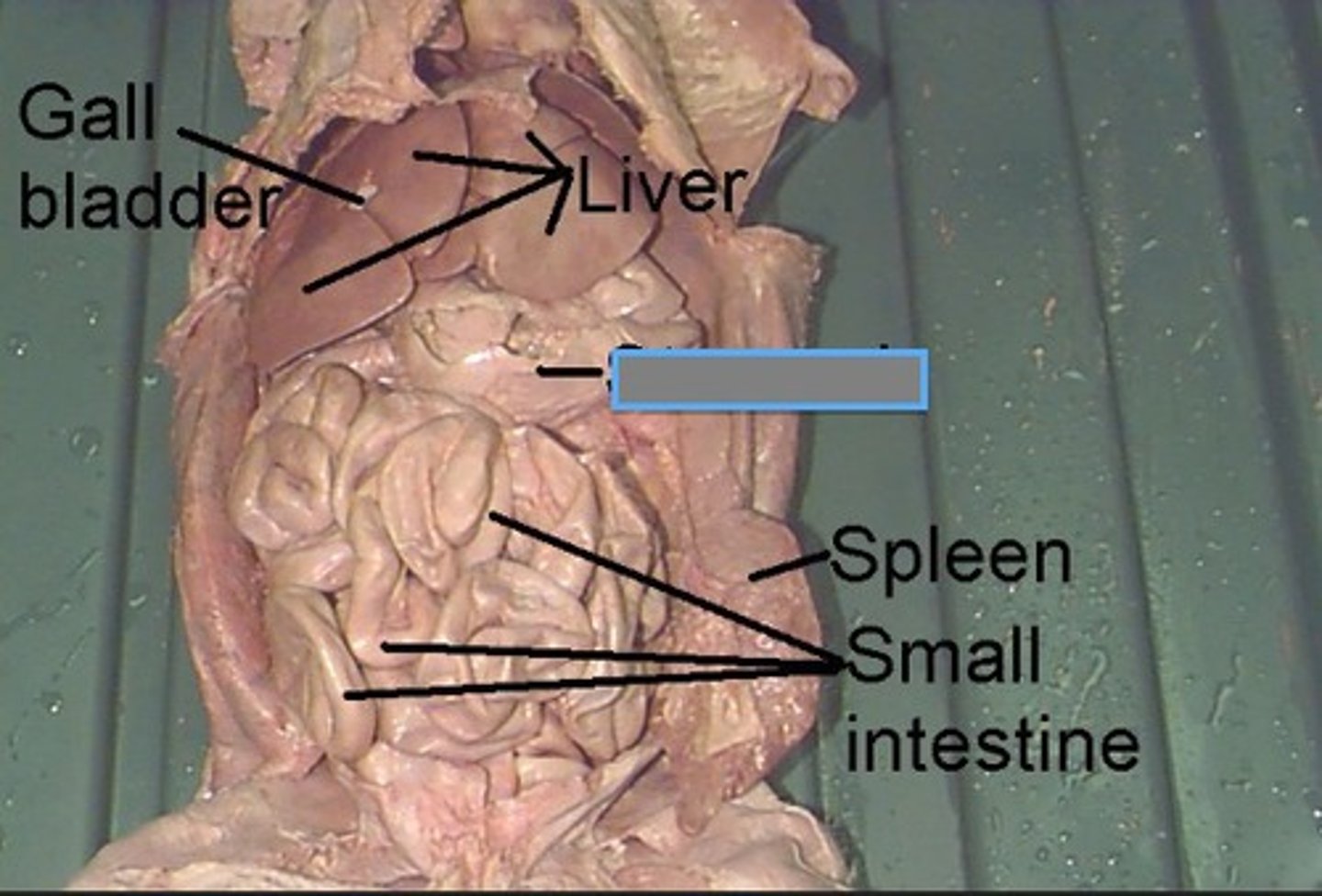
spleen (cat)
oral cavity (model)
cheek (model)
tongue (model)
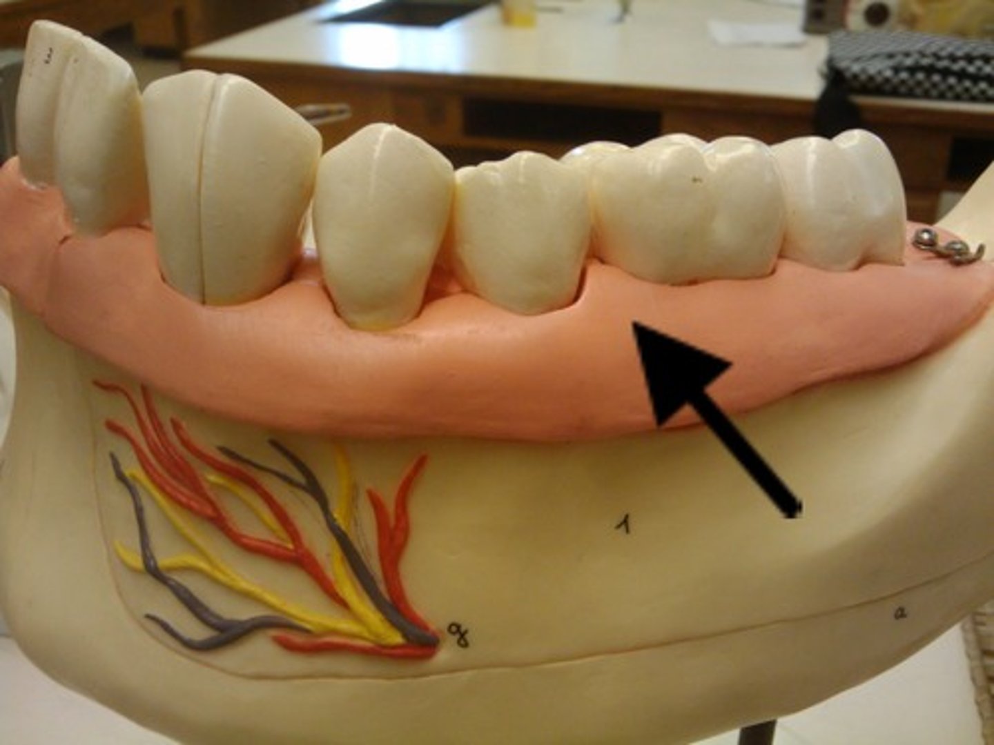
gingiva (model)
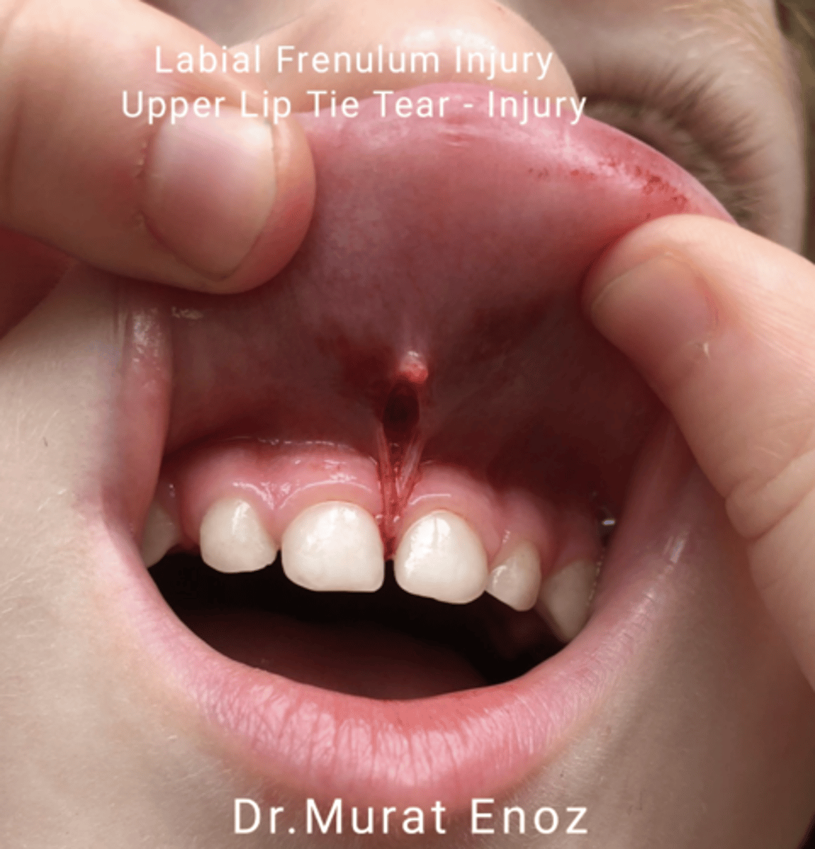
labial frenula (model)
vestibule (model)
** anterior region just inside nose?
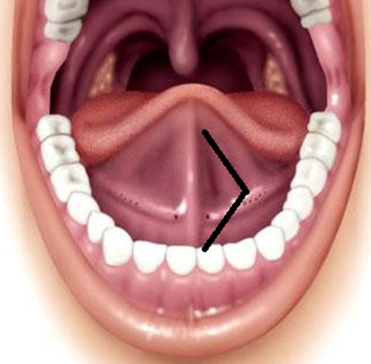
uvula lingual frenulum
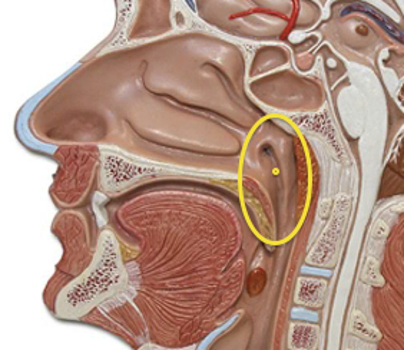
pharynx (model)
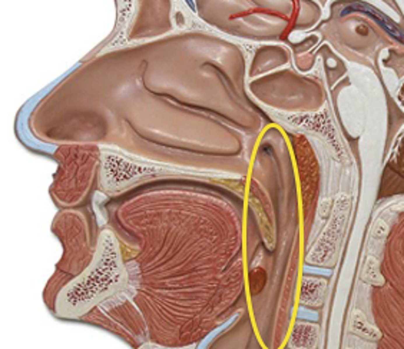
nasopharynx (model)
esophagus (model)
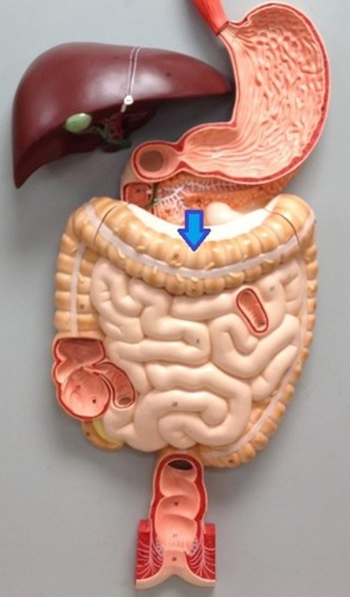
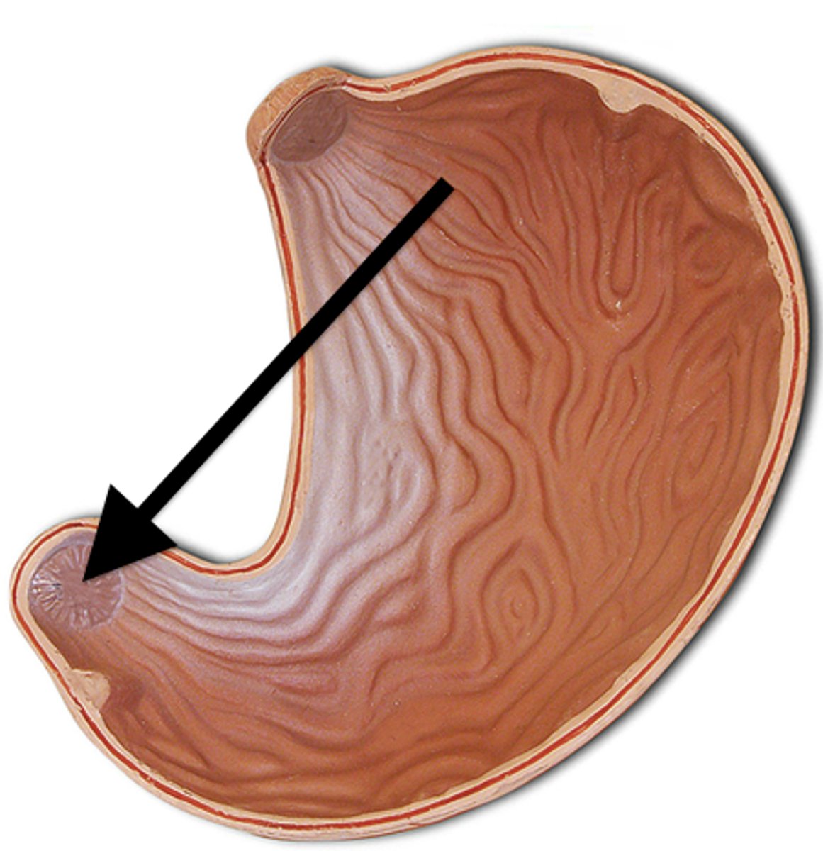
stomach (model)
large muscular sac that continues the mechanical and chemical digestion of food
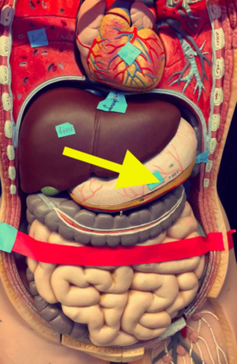
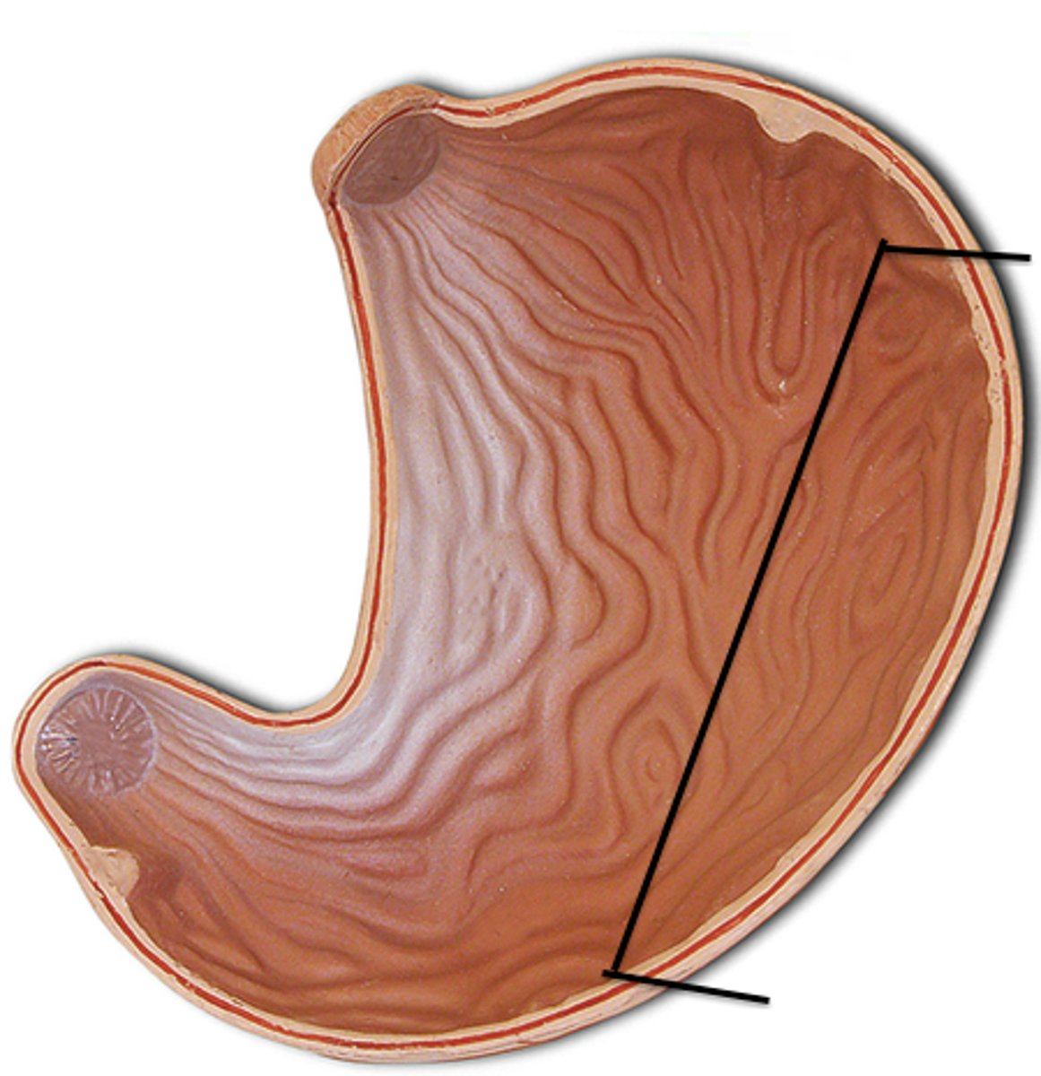
fundus (model)
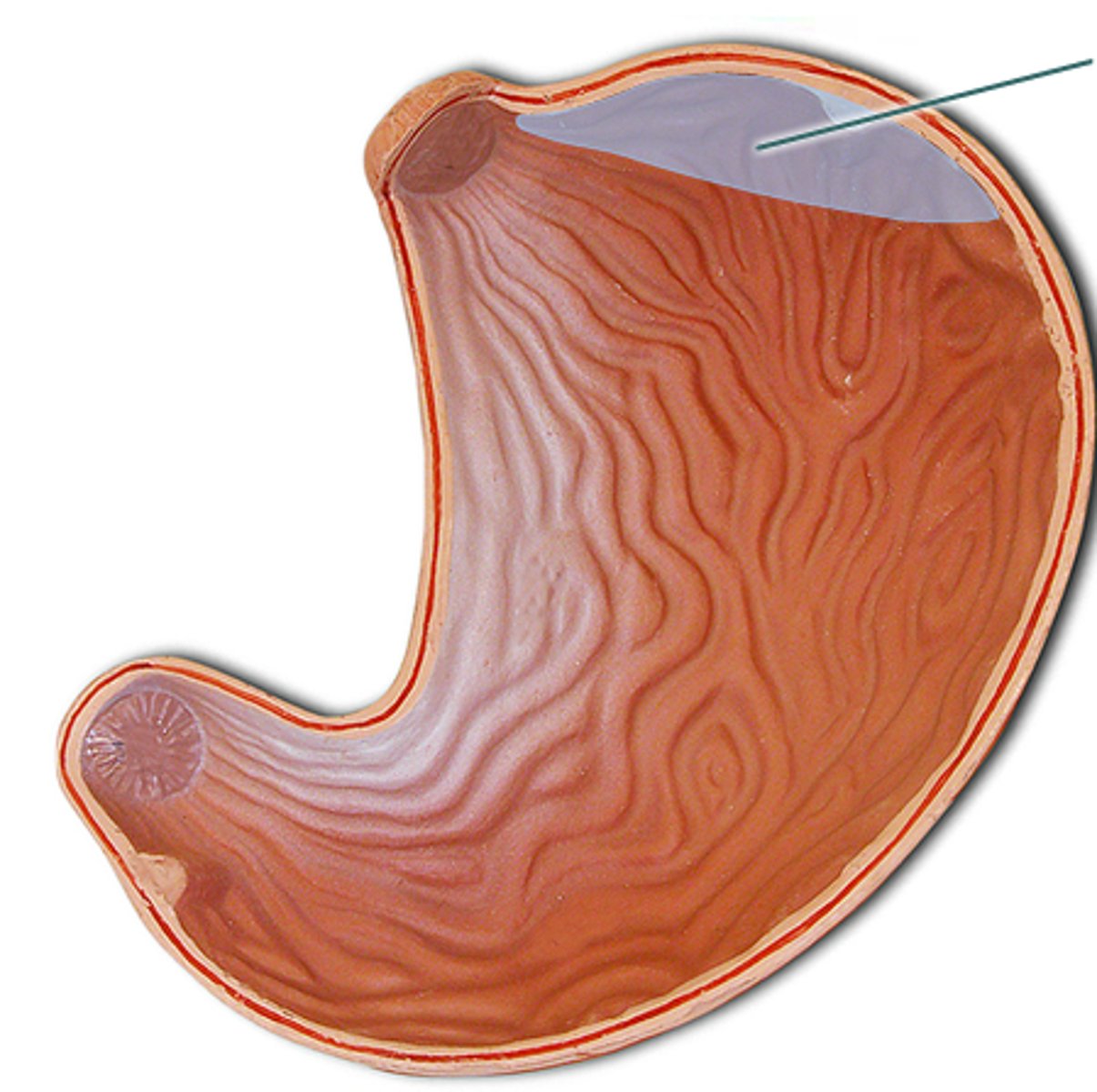
cardiac region (model)
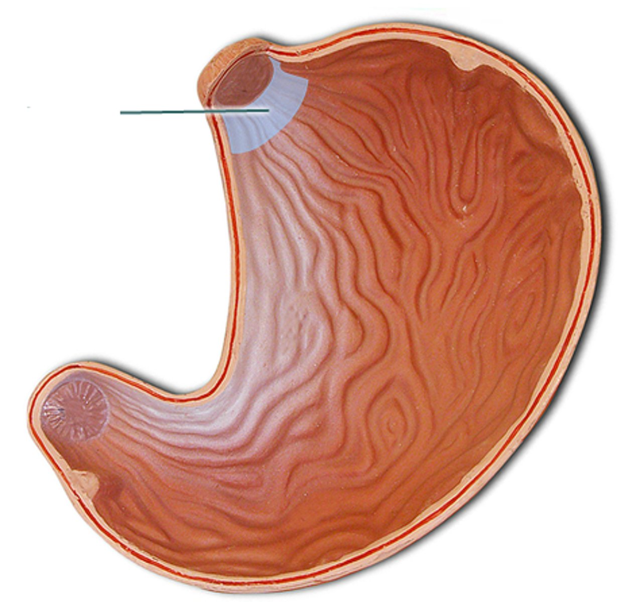
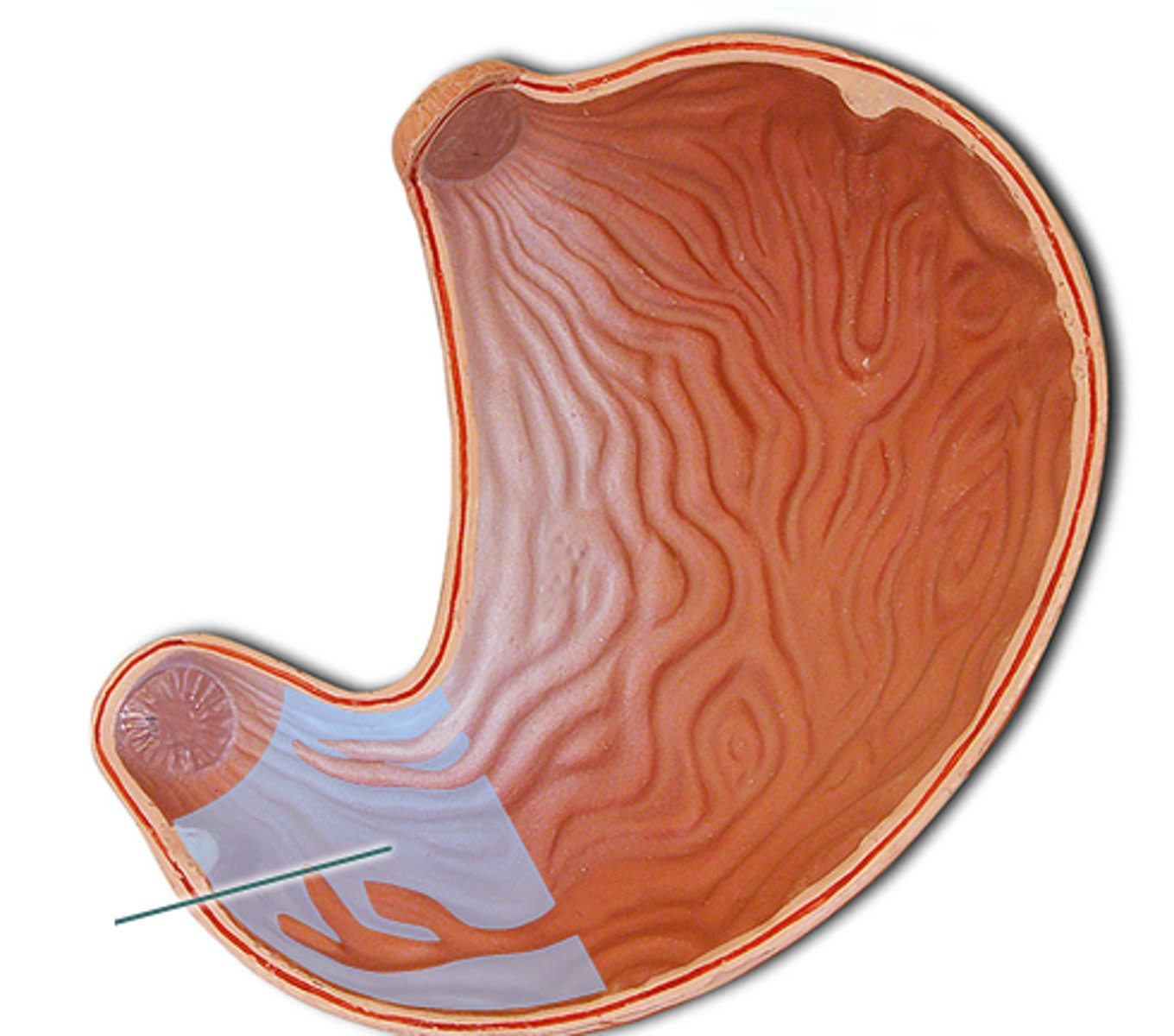
pyloric region (model)
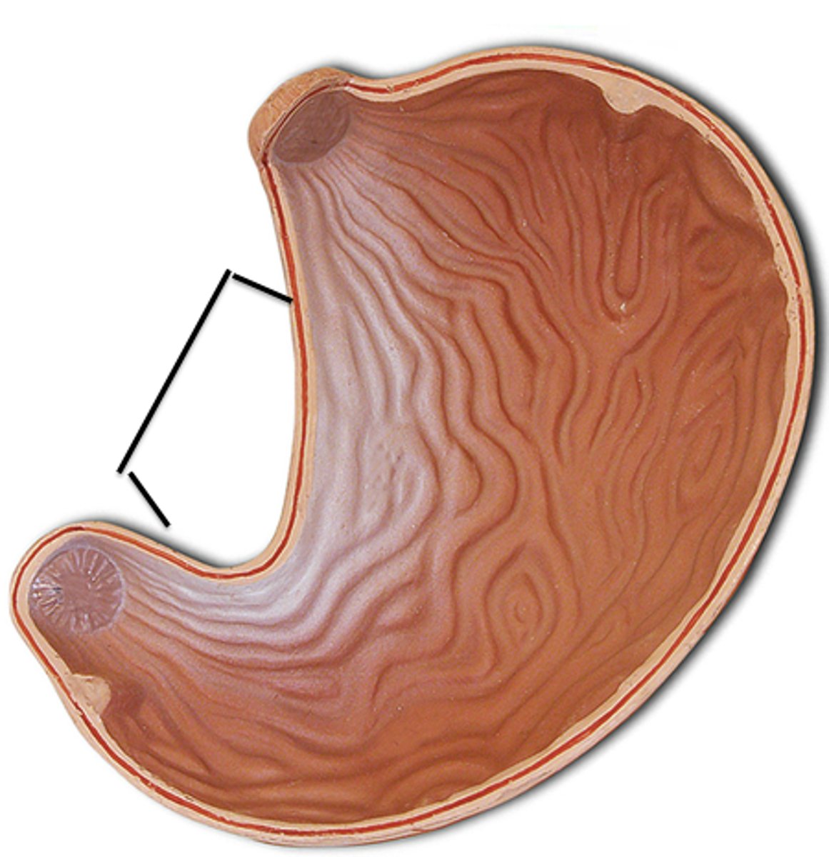
lesser curvature (model)
greater curvature (model)
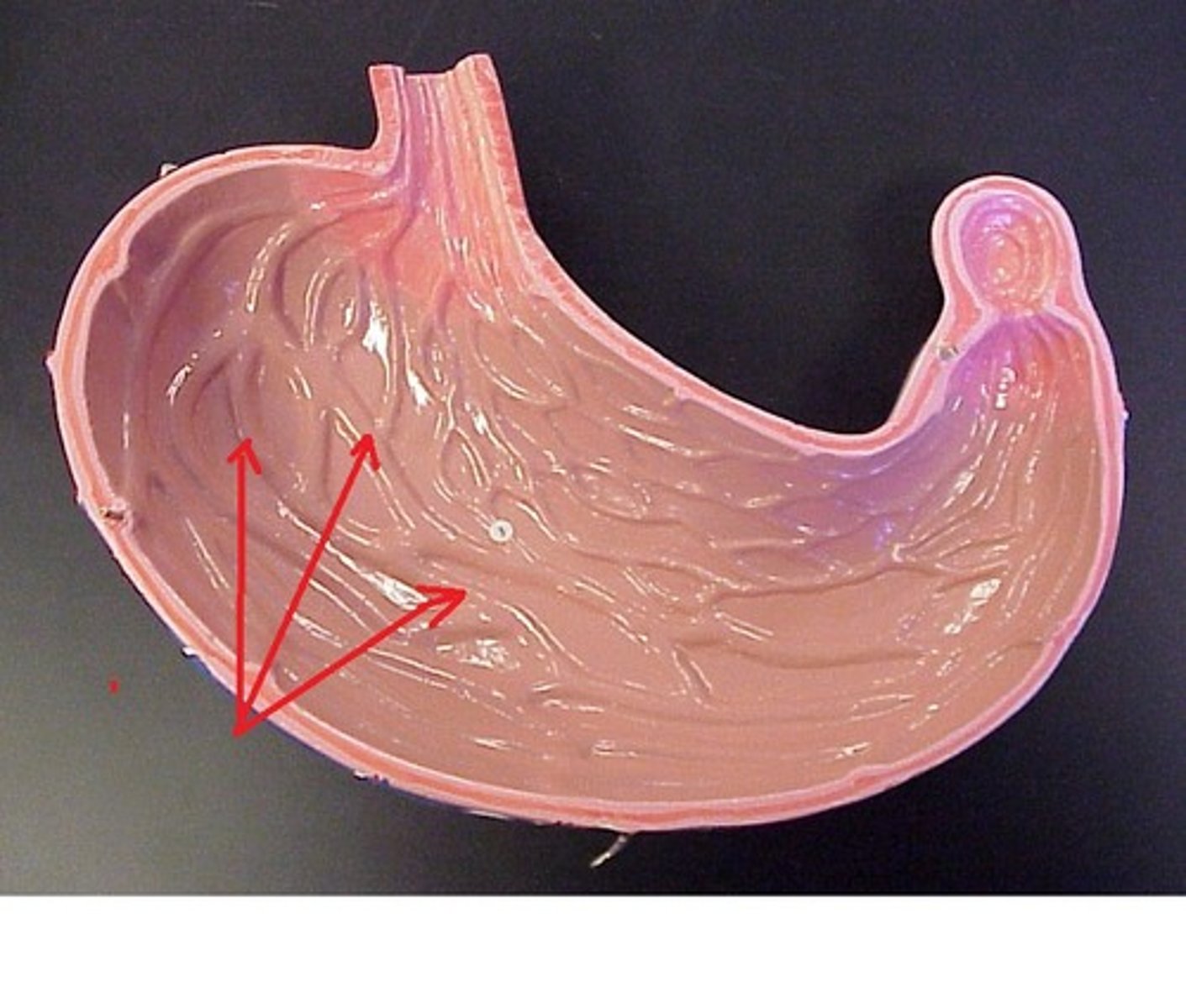
rugae (model)
inside
pyloric sphincter
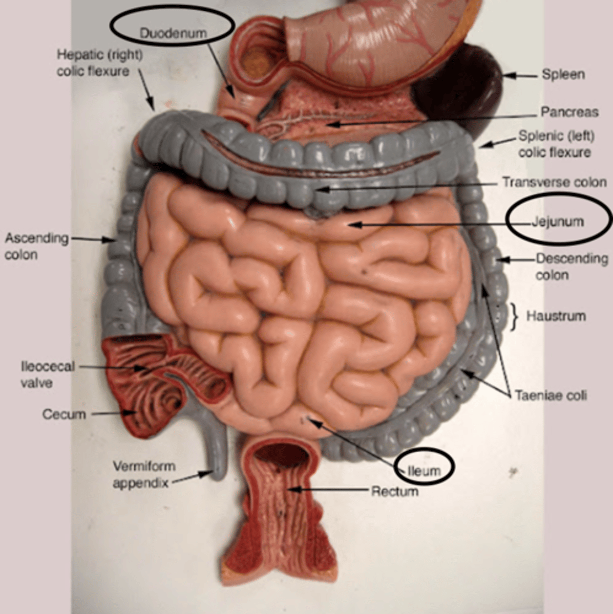
small intestine (model)
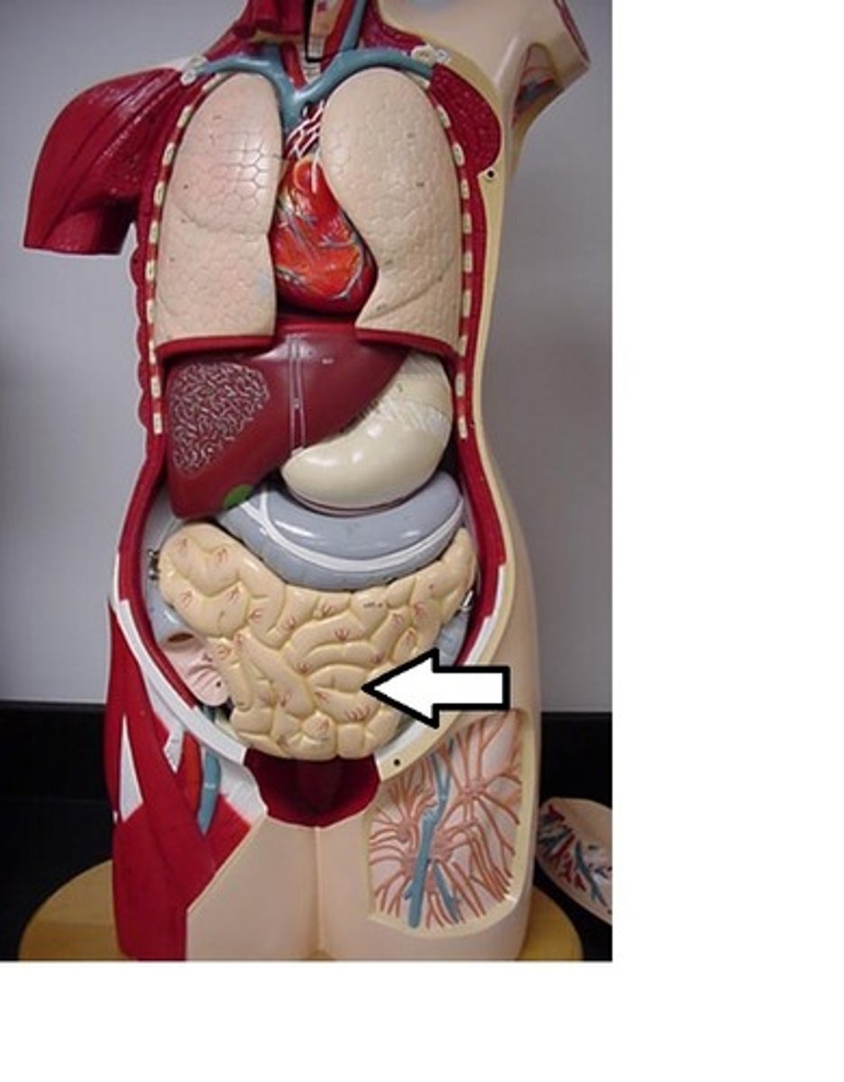
duodenom, jejunum, ileum (model)
plicae circulares
large intestine (model)