Kin 411 Lecture 23: Vascular Supply
1/40
There's no tags or description
Looks like no tags are added yet.
Name | Mastery | Learn | Test | Matching | Spaced | Call with Kai |
|---|
No analytics yet
Send a link to your students to track their progress
41 Terms
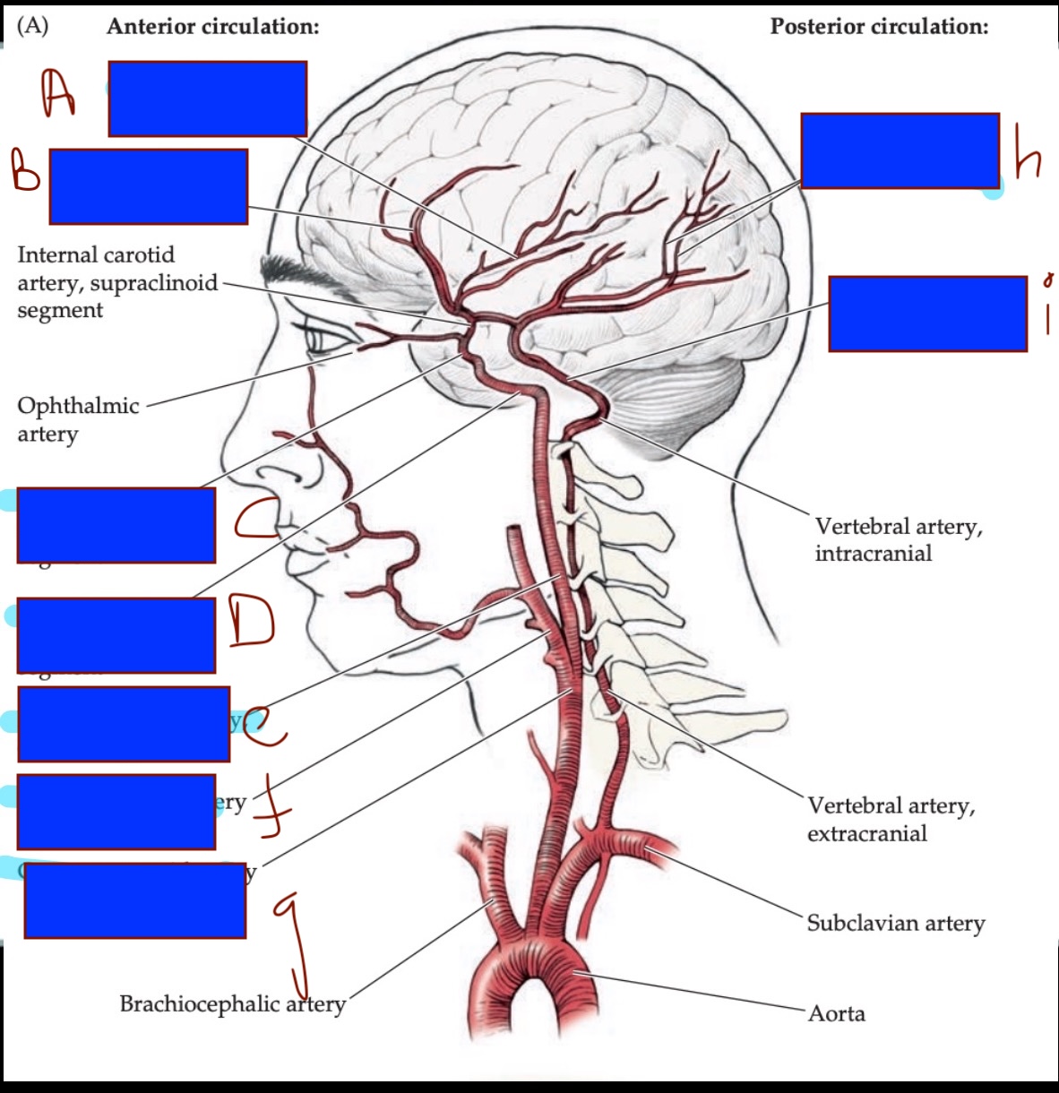
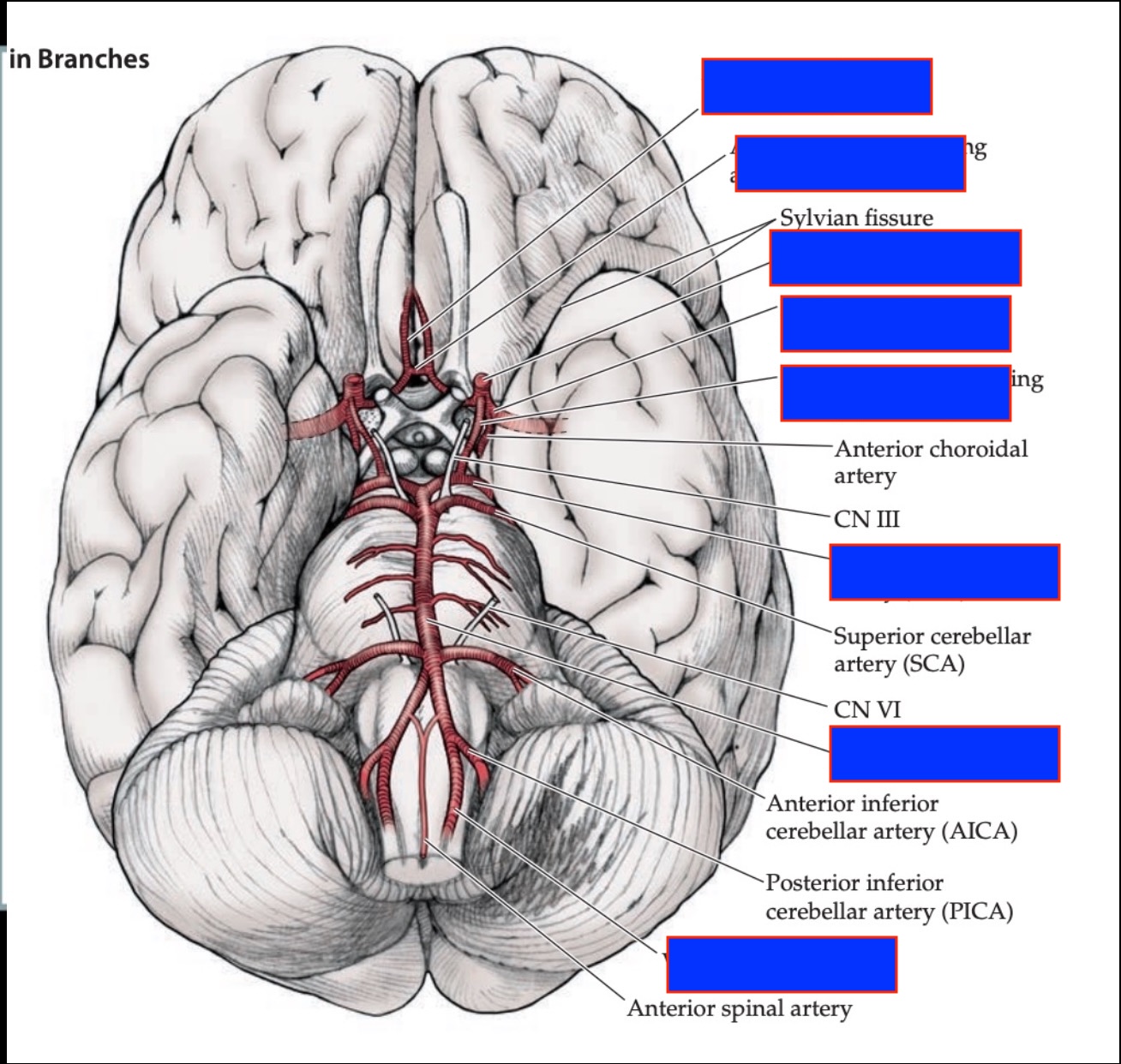
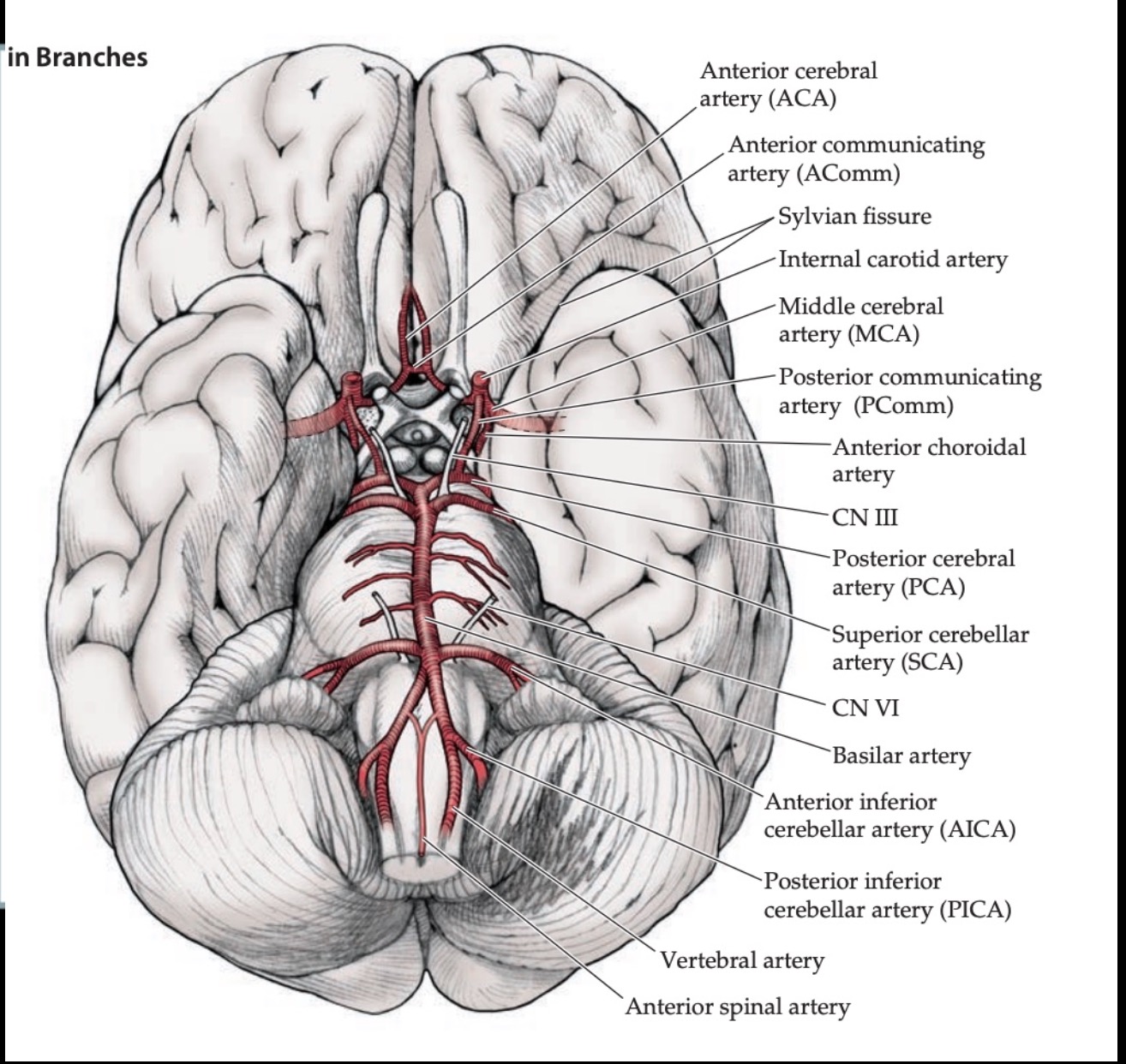
Label A-I
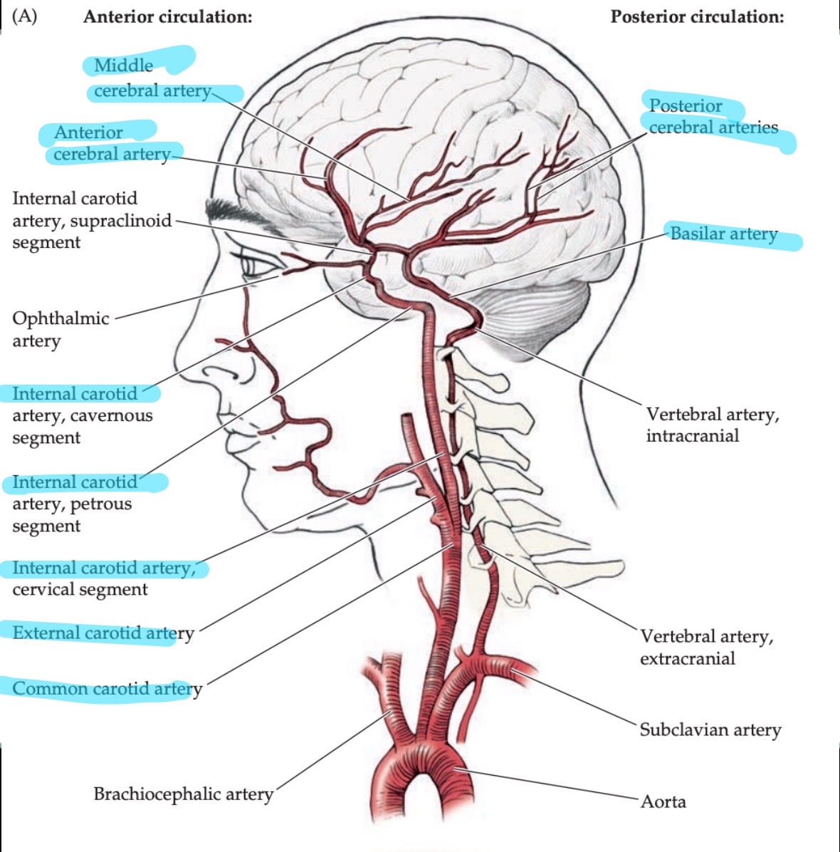
Two main sources of blood supply to brain?
Internal Carotid (anterior brain)
Vertebral-basilar (posterior brain)
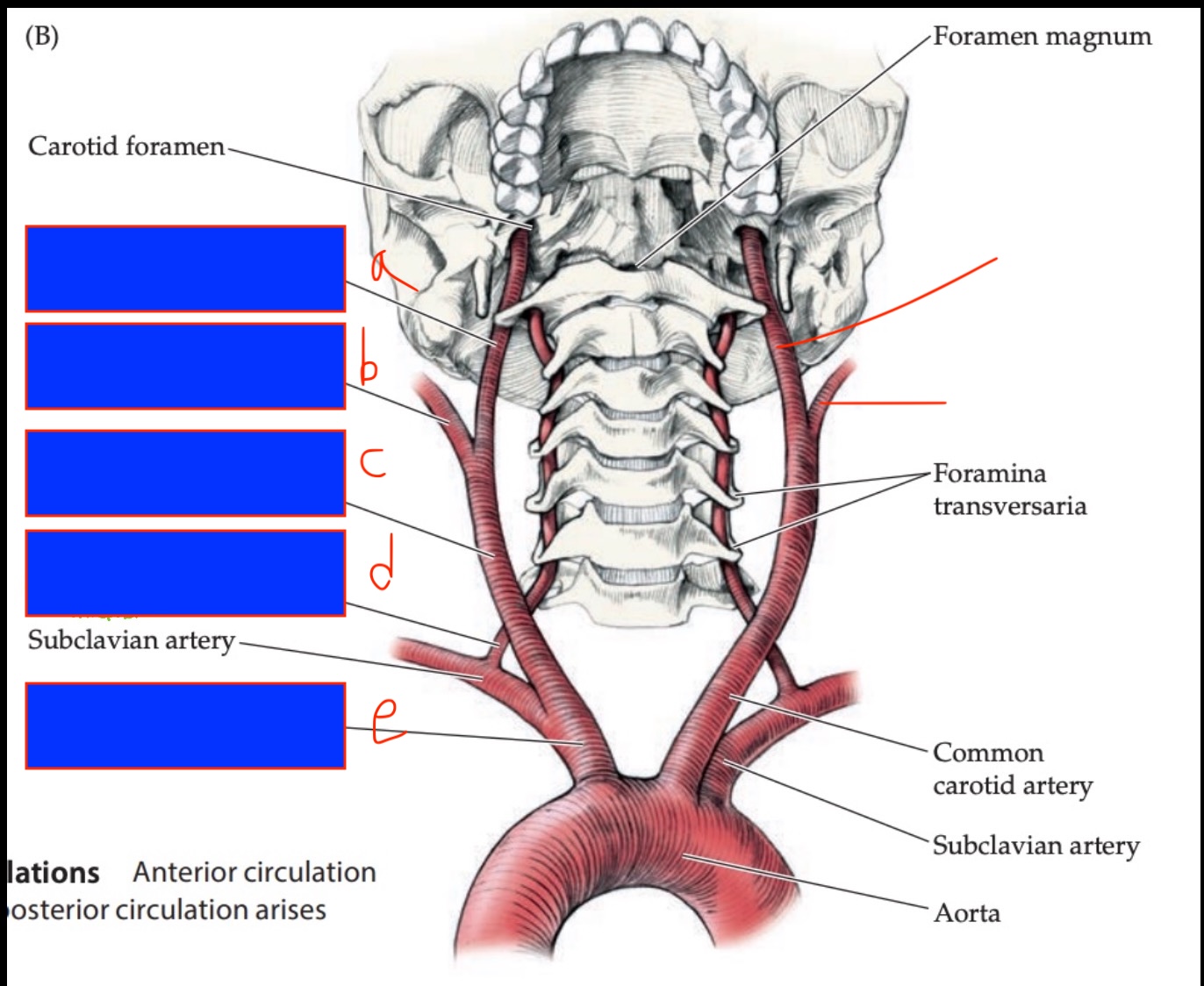
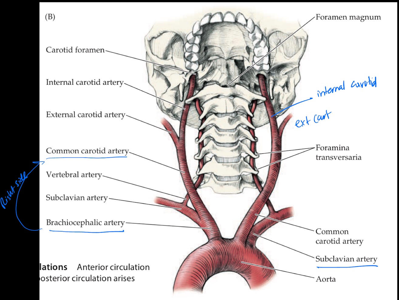
What does the common carotid artery divided into?
External carotid artery
Internal Carotid
What does the external carotid artery supply?
Outside of the head (face, scalp)
What does internal carotid artery supply?
Brain (anterior circulation)
Two main branches of the internal carotid artery?
Anterior Cerebral Artery (ACA)
Middle Cerebral Artery (MCA)
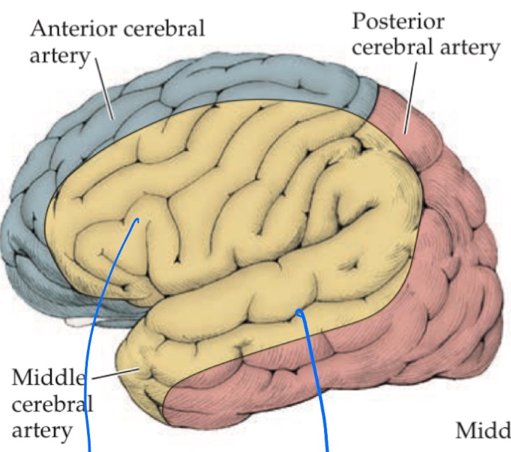
ACA vs MCA: What general area do they supply?
ACA: Medial surface of the brain
MCA: Lateral surface of the brain
Difference in left and right common carotid artery
Left: Common carotid comes right off the aorta
Right: Common carotid comes off a small branch of the aorta called Brachiocephalic artery
Through what structures do vertebral arteries travel?
Foramina transversaria of cervical vertebrae
What is Basilar artery? what arises from it?
Bilateral vertebral arteries join to form Basilar artery.
Basilar artery gives rise to Posterior Cerebral Artery (PCA)
Which arteries are part of anterior circulation vs posterior circulation?
Anterior circulation: ACA and MCA
Posterior circulation: PCA and Basilar system
Summary of cerebral blood supply
Internal carotids supply anterior brain (ACA, MCA), while vertebral arteries join to form basilar supply, which give rise to PCA that supplies the posterior brain
Vertebral artery path summary path
Subclavian artery → bilateral vertebral artery thru foramina transversaria → join + enter skull via foramen magnum → basilar artery → PCA
Carotid system path summary
Aorta splits → Common carotid → enters skull via carotid foramen → internal carotid = brain, external carotid= face
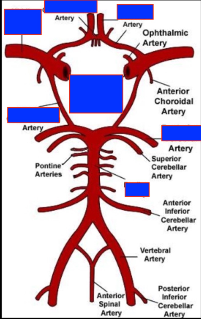
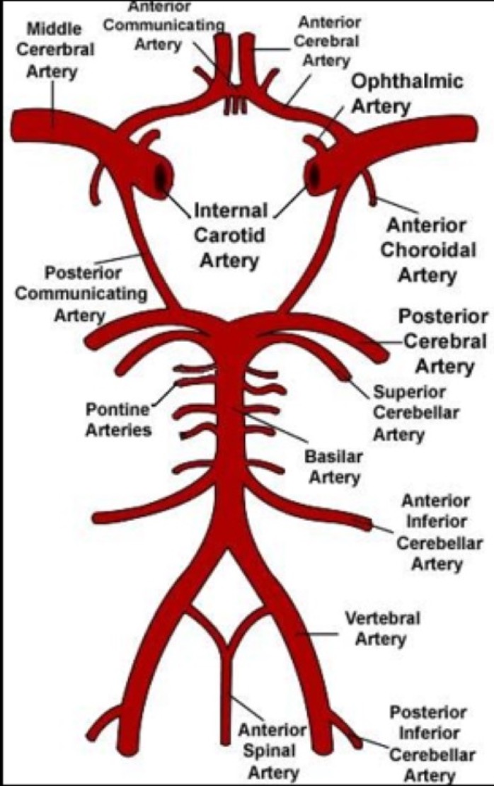
Label
Circle of Willis
Reflects connections between different cerebral arteries and the communicating arteries
Where are the cerebral arteries traveling?
Once ACA, MCA and PCA branch off, they enter into the sub-arachnoid space (CSF filled space under arachnoid mater and above pia mater)
There they run into smaller arteries and feed all the cortical areas of the brain
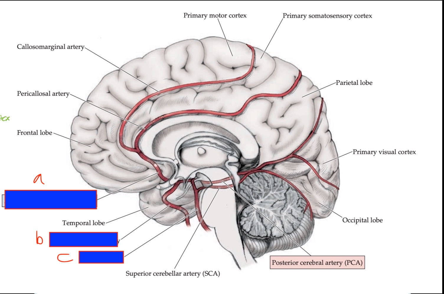
ACA blood supply
ACA supplies superior and medial cortex:
from frontal lobes to anterior parietal lobes
MCA path
Comes off the internal carotid artery → Goes out laterally into the sylvian fissure
Posterior Cerebral Artery blood supply
PCA supplies:
Inferior and medial temporal lobe and occipital lobe
Basilar artery gives rise to PCA → Goes into interhemispheric fissure → running along the midline, but goes to the posterior parts of the brain
Label A-C
A- Anterior Cerebral Artery (ACA): goes up interhemispheric fissure
B- Internal Carotid Artery
C- Basilar Artery: Goes up interhemispheric fissure along the midline to PCA → posterior brain
Label
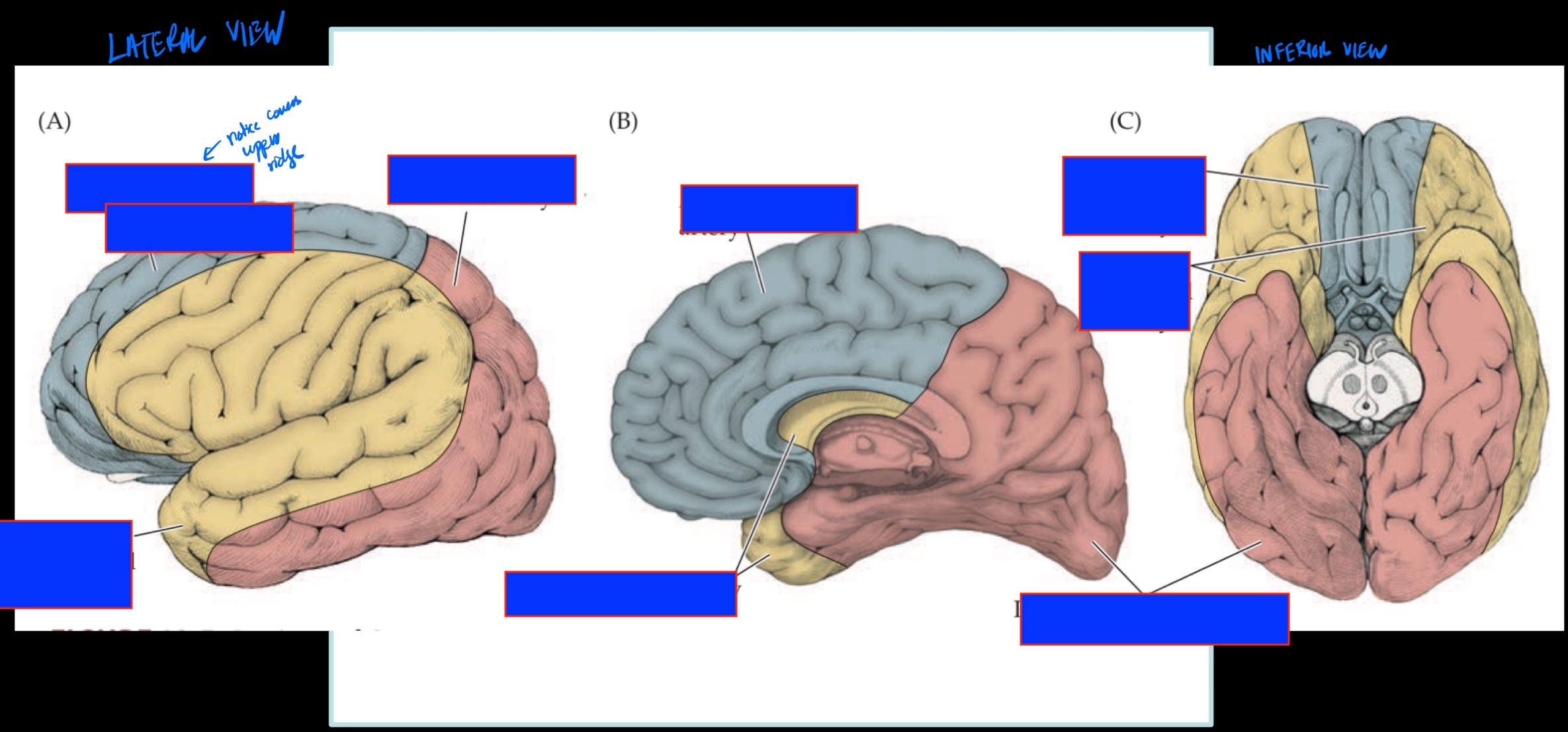
Blue: ACA
Pink: PCA
MCA: Yellow
ACA:
Medial surface of cortex as well as strip of superior cortex
From frontal lobe → anterior parietal lobe
PCA:
Inferior + medial temporal lobe, posterior parietal lobe, occipital lobe
MCA divisions
3 divisions:
MCA Superior division:
Emerge from Sylvian fissure → wraps UP → cover lateral frontal lobe to anterior parietal lobe
MCA Inferior Division:
Emerge from Sylvian fissure → wraps DOWN → anterolateral (superior) surface of temporal lobe and posterior parietal cortex
MCA Deep Branch:
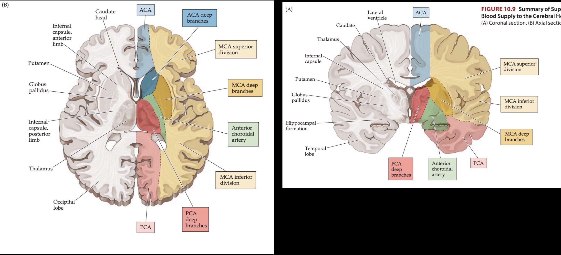
To deep nuclei within center of brain (body of caudate + lentiform nucleus)
Describe MCA supply in the lateral view
Superior MCA: Lateral surface of cortex anterior frontal lobe to anterior parietal lobe
Inferior MCA: Anterolateral (Superior surface) of temporal lobe
What does MCA superior division supply?
Lateral frontal lobe
Anterior parietal lobe
What does MCA inferior division supply?
Superior (anterolateral) temporal lobe
Posterior parietal cortex
Through what fissure does MCA travel?
Sylvia fissure
Memory trick for MCA blood supply?
MCA = Most of the lateral cortex
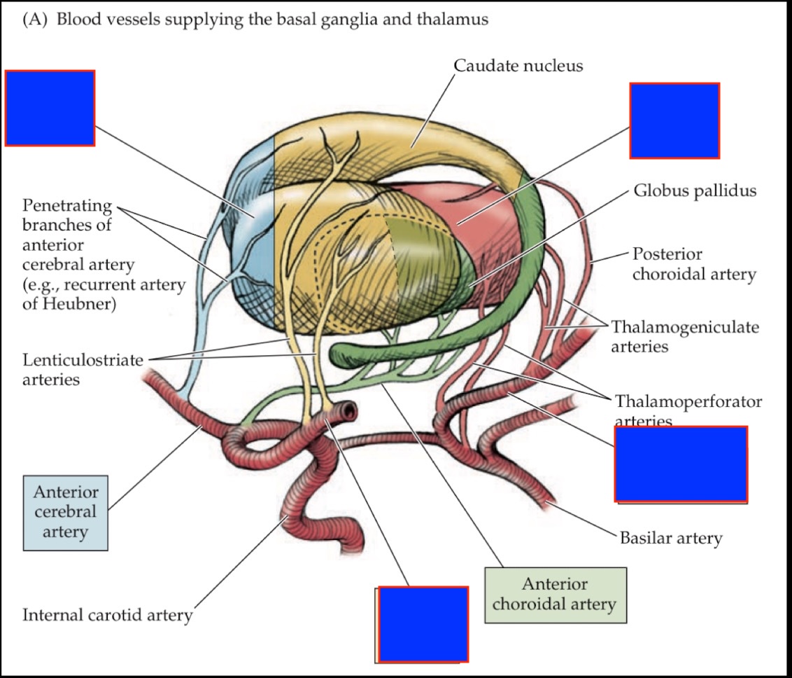
What structures do MCA deep division supply?
body of caudate
Lentiform nucleus (GPi + putamen)
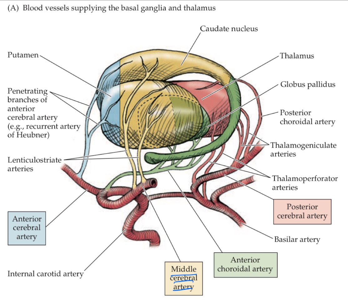
What do deep branches of the PCA supply?
Thalamus
What do the major sinuses do? Describe the path
Allow the venous blood to drain out of head and back into circulatory system
Superior saggital sinus runs along the superior part of interhemispheric fissure. Enters into interhemispheric fissure into the falx cerebri.
Superior drains into transverse sinus (made by tentorium cerebelli)
From Transverse sinus it drains out internal jugular vein and into the circulatory system
Vascular injury types
Blockages/occlusion of arteries: blood not flowing becuase blocked
Hemorrhage: artery is burst or torn open
Vascular injury: Hemorrhage
Associated with ruptured aneurism (weakening of arterial wall)
Arterial blood spills out into subarachnoid space for subarachnoid hematoma
Vascular injury: Blockage/occlusion
2 different mechanisms:
Thrombus: blood clot forms within arteries around artherosclerotic clot. Narrows/prevent flow of blood thru artery
Embolism: blockage by lodged material (embolus) within the vessel. Embolus is blood clot that has formed somewhere else in the circulatory system and becomes a plug in that artery
What dictates symptoms of vascular injury?
Depends on where the block or bleed occurs
e.g. block ACA → superior + medial frontal lobe and anterior parietal cortex = UMN for feet and legs probs