Opthalmology OSCE rev PART 1
1/330
There's no tags or description
Looks like no tags are added yet.
Name | Mastery | Learn | Test | Matching | Spaced | Call with Kai |
|---|
No analytics yet
Send a link to your students to track their progress
331 Terms
Conjuctival injection: Dilation of the conjuctival vasculture.
Conjuctival injection: Dilation of the conjuctival vasculture.
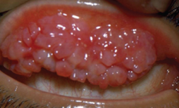
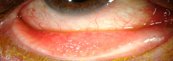
Giant papillae are typical of allergic eye disease , and they may result as a result of contact lens wear.
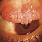
papillae: raised lesions in the upper tarsal conjunctiva, they are non-specific sign of chronic inflammation.
Follicles
Found usually in the lower tarsal conjunctiva and upper tarsal border and occasionally at the limbus.
-each follicle represents a lymphoid collection
-it’s a specific sign for viral and chlamydial infections
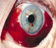
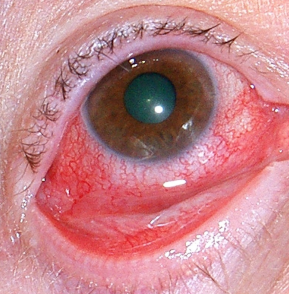
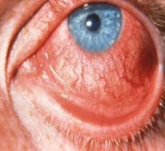
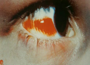
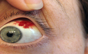
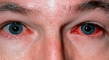
subconjuctival hemorrhage:
Bright red in colour because it’s fully oxygenated through the ambient air through the conjunctiva.
Ddx: high blood pressure, trauma to the eye most common cause, or a base of skull fracture if you can’t see the posterior border, warfarin or aspirin side effect.
subconjuctival hemorrhage
subconjuctival hemorrhage
subconjuctival hemorrhage
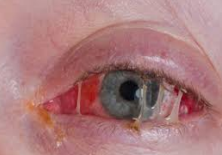
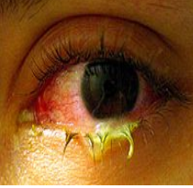
bacterial conjunctivitis
bacterial conjunctivitis
Bacterial conjunctivitis symptoms
Redness (conjunctival injection).
Purulunt discharge.
Ocular irritation (NOT PAIN).
Bacterial conjunctivitis causative organism:
Staphylococcus, streptococcus, Pneumococcus, and haemophilus.
Bacterial conjunctivitis treatment
usually it’s self limiting
-topical Broad spectrum antibiotic eye drop (chloramphenicol)
- Conjunctival swabs for culture is indicated In severe cases or if there is no resolution.
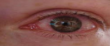
viral conjunctivitis
viral conjunctivitis
viral conjunctivitis characterized by:
-Redness (conjunctival injection).
-Ocular irritation (NOT PAIN).
-watery discharge
-Conjuctival follicles(follicular conjunctivitis).
-preauricular lymph nodes. -Lid edema and excessive lacrimation.
-can cause corneal ulceration
Viral conjunctivitis causative agents
Adenovirus,it’s self limiting 4-7 days and highly Contagious.
Viral conjunctivitis treatment
Self limiting, Avoid contact with others, wash your hands, If you wear contact lenses, you should throw away contacts worn
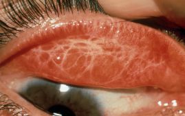
Subconjunctival scarring in trachomas
Entropion (turned in eyelashes rubbing the cornea)
typical micro pannus of trachoma
follicles of trachoma
intense inflammation of trachoma
conjunctival scarring of trachoma
trichiasis of trachoma
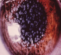
Allergic conjunctivitis:
Acute: itchiness, chemosis, lacrimation vernal conjunctivitis: itchiness, photophopia, lacrimation, papillary conjunctivitis may form giant cobblestones , limbal follicles and white spots, punctate lesions on the cornea,
Allergic conjunctivitis treatment
Initially: Mast cell stabilizers or antihistamines,
In severe cases: Topical steroids (avoid long uses)

Giant cobblestone papillae in vernal conjuctivitis
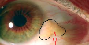
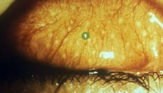
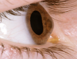
Pingueculae, yellow raised patch
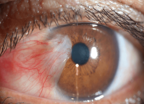
Pterygia
Pterygia
Pterygia and pingueculae
Forms of corneal degeneration found on the interpalpebral bulbar conjunctiva.
Result from the excessive exposure to the reflected or the direct ultraviolet component of the sunlight.
Pingueculae
are small, elevated yellowish paralimbal lesion and never impinge on the cornea, may cause discomfort.
Tx: Artificial tears for discomfort and for cosmetic reason, surgical excision
Pterygia
wing-shaped lesions, they may extend into the cornea and cause irritation, FB sensation , itchy eyes , tearing and may encroach onto the visual axis if extensive.
Tx: they can be excised but may recur.
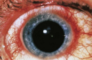
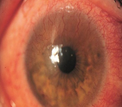
Ciliary flush (injection). Redness is localized to the limbus.
3 important Differential diagnosis:
- Keratitis – uveitis - acute glaucoma
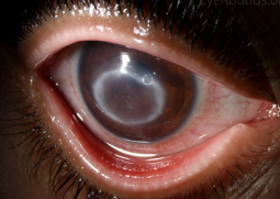
corneal edema (epithelial or sromal): It causes clouding of the cornea.
keratic precipitates KPs: Deposits of cells on the corneal endothelium.
-Fine KPs: neutrophils, lymphocytes.
keratic precipitates KPs
Coarse KPs(mutton fat): macrophages
keratic precipitates KPs
Fine KPs: neutrophils, lymphocytes
Corneal-pannus
Blood vessels formation under the corneal epithelium
Corneal-pannus caused by
Chronic keratitis
Ill fitting contact lenses
Alkali injury
Ocular insult due to infectious keratitis

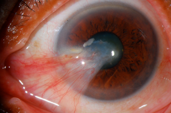
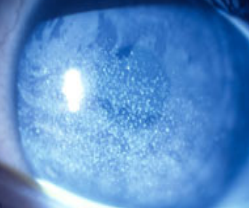
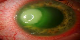
Hypopyon collection of WBCs in the anterior chamber.
causes : bacterial keratitis , ant. Uveitis
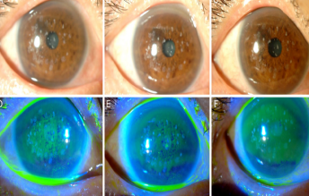
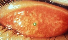
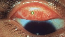
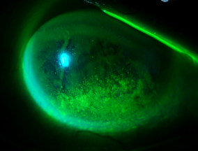
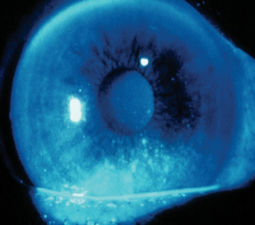
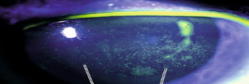
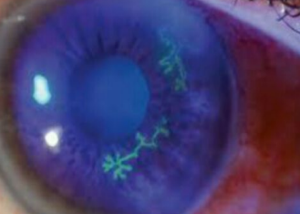
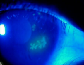
punctate epithelial erosions (PEE): Points of superficial epthelial cell loss or dysfunction.
punctate epithelial erosions (PEE): Points of superficial epthelial cell loss or dysfunction.
punctate epithelial erosions (PEE) may be on:
1 - in the cornea : they are best detected by fluorescein dye viewed with blue light
2- in the conjunctiva: they are best stained by lissamine green.
punctate epithelial erosions (PEE)
punctate epithelial erosions (PEE)
punctate epithelial erosions (PEE)
corneal abrasion with flourescein staining
corneal abrasion
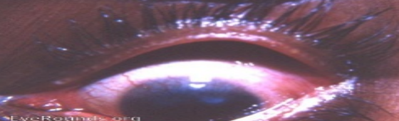
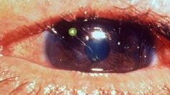
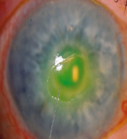
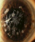
herpex simplex keratitis
Sign: Dendritic ulcer
herpex simplex keratitis
Sign: Dendritic ulcer
herpex simplex keratitis clinical info
-most common cause: herpes simplex virus type 1 (HSV1).
-primary infection it may causes conjunctivitis then followed by resolution and latency of the virus in the trigeminal ganglion.
-reactivation of the virus increases if the pt is debilitated (systemic illness,immunosuppression).
-It Causes Dendritic ulcer.
-Maybe associated with uveitis and glaucoma.
- Risk factors: contact lens, prolonged steroid treatment
herpex simplex keratitis Tx
topical antivirals (Aciclovir). Topical steroids is contraindicated, they may excacerbate the condition.
Dendritic ulcer may heal with scar which may need a corneal graft to restore vision in severe cases.
Disciform keratitis
Disciform keratitis
-another immune reaction to herpes simplex antigen in the stroma.
-cause : HSV
- presents as disc or ring-shaped stromal edema and clouding without ulceration, blurred vision , light sensitivity
-Tx: oral /topical steriods + oral antiviral
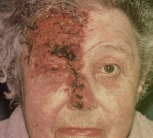
Herpes zoster ophthalmicus (ophthalmic shingles)
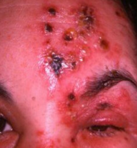
Herpes zoster ophthalmicus (ophthalmic shingles)
Herpes zoster ophthalmicus (ophthalmic shingles)
- cause: varicella zoster
- ocular manifestations : lid swelling , keratitis , iritis , secondary glaucoma , pain and vesicles at the ophthamic divion of the trigiminal nerve .
- there is usually a prodromal period where the pt is systemically unwell.
Herpes zoster ophthalmicus (ophthalmic shingles) Tx
-oral antivral (acyclovir, famciclovir) within 3 days of vesicles eruption for reducing the neuralgia.
-topical steroids for ocular disease
-antibacterials to cover secondary infection
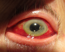
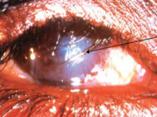
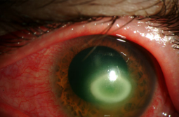
Bacterial keratitis
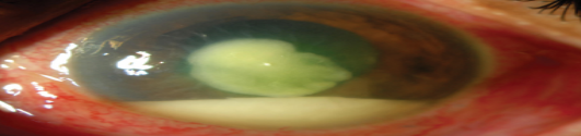
Bacterial keratitis
Bacterial keratitis Signs and symptoms
Severe pain, purulent discharge, ciliary injection (flush), visual loss( if the visual axis is involved), hypopyon(a mass of WBCs collected in the anterior chamber), white corneal opacity
Bacterial keratitis clincial info
-3 serious findings: hypopyon, ciliary flush, corneal cloudiness
-Microorganisms: staph epidermidis, staph aureus, strep pneumonia.
-RFs: keratoconjuctivitis sicca (dry eye),contact lens, breach in the epithelium by surgery or trauma, prolonged use of steroids.
Bacterial keratitis Tx
topical broad spectrum antibiotics.
Either fluroquinolones (ciprofloxacin, ofloxacin) as monotherapy, or combined therapy : cefuroxime against
acanthamoeba keratitis
acanthamoeba keratitis
acanthamoeba keratitis
-also named as infective keratitis.
-fresh water amoeba -associated highly with soft contact lenses
Extremly painful keratits with prominent infiltrated corneal nerves.
The amoeba can be isolated from the cornea or from the contact lens
acanthamoeba keratitis tx
Topical chlorhexidine, PHMB, propamidine.
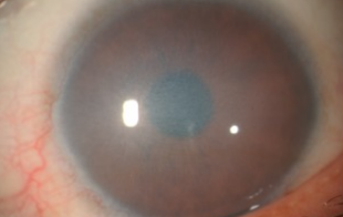
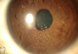
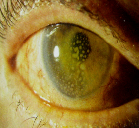
Corneal dystrophy
Corneal dystrophy
Corneal dystrophy
Corneal dystrophy
rare inherited disorder
sx: affect corneal transparency and can cause visual loss, it may cause pain if they cause corneal erosions.
Tx: corneal graft
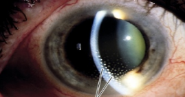
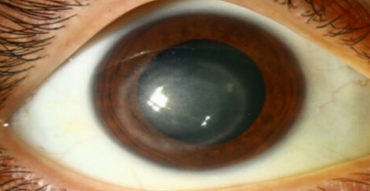
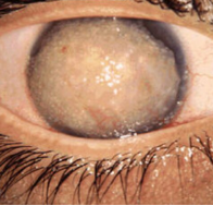
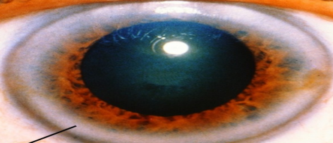
Keratoconus
Keratoconus
sporadic, occasionally inherited.
sx; marked myopia, irregular astigmatism , vision loss (painless).
signs; fleisher’s ring , apical scar , vogt’s stiae, prominent corneal nerve.
mgt : astigmatism corrected by; glasses , contact lenses , UVA radiation in the presence with riboflavin, corneal graft.
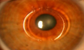
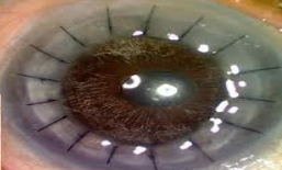
Double intrastromal corneal rings. Used for keratoconus.
Complications of surgery: Infection (Give TOPICAL antibiotic, not IV), corneal perforation, displacement,
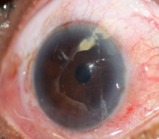
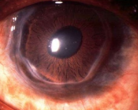
Band keratopathy
Band keratopathy
-subepithelial deposition of calcium phosphate . Occur with chronic uveitis or glaucoma.
-may cause visual loss and erosions and discomfort.
-it may be a sign of systemic hypercalcaemia as in hyperparathyroidism or renal failure. ttt: scraped off surgically or by excimer laser
More likely occupy the 3’oclock and 9’oclock position of the paralimbal cornea.
corneal thinning
corneal thinning
-rare cause: painful peripheral corneal thinning is (Mooren’s ulcer)
-melting, starts at limbus & spreads across cornea, immune basis. It may be seen in RA & Wegener’s granulomatosis
corneal thinning tx
systemic and topical immunosuppression (steroids or cytotoxic drugs) and antiproteases. Adequate corneal protection and wetting.
corneal thinning
corneal thinning with inflammation
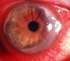
lipid arcus
lipid arcus
-peripheral white ring-shaped lipid deposits, separated by the limbus by clear interval.
-normally seen in elderly (above 50)=arcus senilis.
-in younger pts: maybe a sign of hyperlipoproteinaemia.
- doesn’t affect vision, no Tx is required.
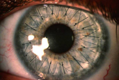
corneal transplantation, corneal grafting
corneal transplantation, corneal grafting
corneal transplantation, corneal grafting
corneal transplantation, corneal grafting Indications:
to restore corneal clarity or repair a perforation in these conditions: keratoconus, traumatic scar, herpes infxn, corneal dystrophy, interstitial keratitis with marked opacity and decreased visual acuity, decompensated cornea in old ages
corneal transplantation, corneal grafting Complications
rejection, astigmatism, endophthalmitis, recurrence of previos pathology, cataract.
postop :steroid eye drops to prevent graft rejection
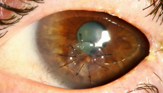
Corneal graft
-Penetrating keratoplasty: When the entire cornea is replaced.
-lamellar keratoplasty: when only part of the cornea is replaced.
Corneal graft rejection
-Any patient with: Red eye, pain or visual loss and had a corneal graft must be seen urgently.
- Examination: Graft edema,iritis, and a line of activated T-cells attacking the graft endothelium
-Tx: intensive topical steroid application can restore graft clarity
Indications for penetrating keratoplasty:
-pseudophakic bullous keratopathy(m.c.c in developed countries). -keratoconus (m.c.c in developing countries). -corneal degeneration. -keratoglobus. -Corneal dystrophy.
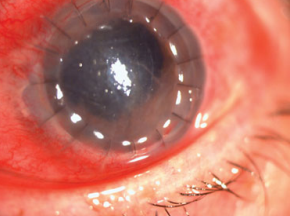
Corneal graft rejection
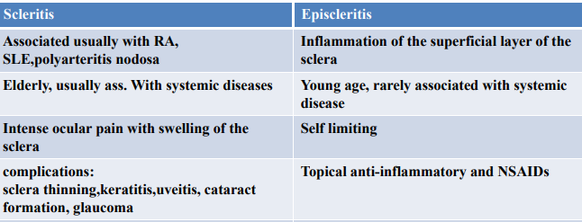
Scleritis vs episcleritis
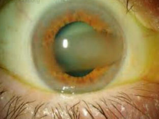
Classification of glaucomas
1-Primary glaucoma:
Chronic open angle.
Acute and chronic closed angle.
2-Congenital glaucoma:
Primary.
Secondary to maternal rubella infection.
Secondary to inherited ocular disorders (ex:aniridia-absence of the iris).
3-Secondary glaucoma(causes):
Trauma.
Ocular surgery.
Associated with other ocular dx, uveitis.
Raised episcleral venous pressure.
Steroidinduced steroid induced.
Chronic open angle glaucoma Tx:
1-Medical:
drugs ; prostaglandin , B-blocker , carbonic anhydrase inhibitor.
2-laser tx: (laser burns to the meshwork).
3-surgical drainage procedures: TRAB (Trabeculectomy).
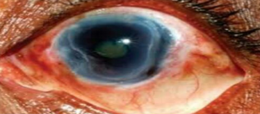
Acute closed angle glaucoma Tx
IV acetazolmide, topical pilocarpine , Bblocker , iridotomy by YAG laser or lensectomy