Week 9: Muscle Microanatomy and Histology
1/20
There's no tags or description
Looks like no tags are added yet.
Name | Mastery | Learn | Test | Matching | Spaced | Call with Kai |
|---|
No analytics yet
Send a link to your students to track their progress
21 Terms
skeletal muscle
striated
voluntary contractions from nervous system
acts on bone to produce motion
smooth muscle
non- striated
involuntary peristaltic
lines the gi tract, respiratory tract, blood vessels of the circulatory system, urinary tract, and reproductive organs
multinucleated
vascularized and innervated
cardiac muscle
striated
branching fibers connected by intercalated discs; heart beat
synchronized involuntary contraction
direct vs indirect attachment of smooth muscle
direct- straight attachment from muscle to bone via the periosteum or perichondrium
indirect - attachment via the tendon or apopneurosis (sheet of connective tissue)
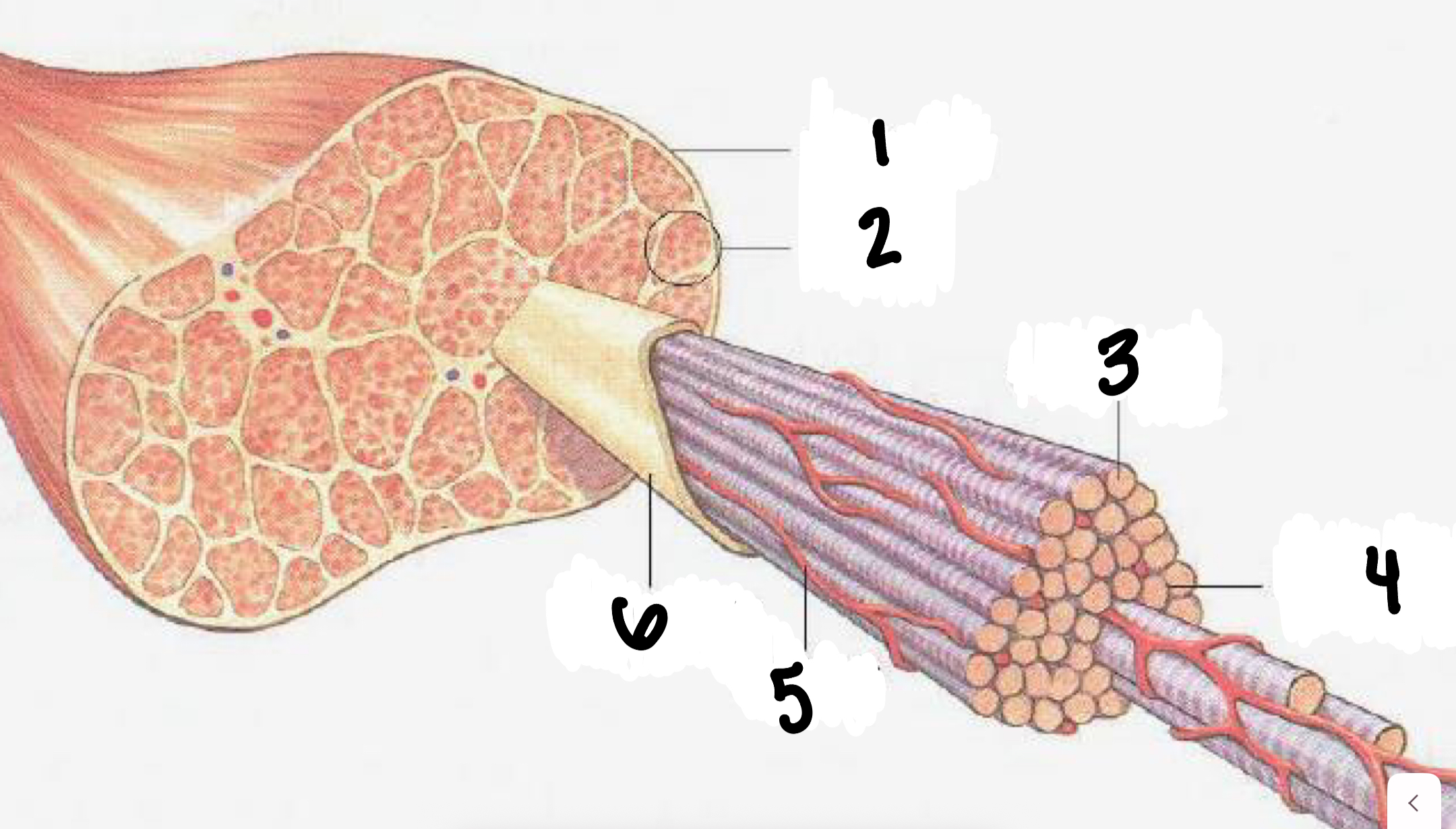
label 1-6 of the composition of skeletal muscle
epimysium, fasicle, muscle fibers, endomysium, capillary, perimysium
describe epimysium, perimysium, and endomysium
epimysium- outermost sheath that surrounds the entire muscle
perimysium- surrounds a bundle of muscle fibers called a fasicle
endomysium- smallest sheat; surrounds a singular muscle fiber
fasicle
bundle of muscle cells with motor units
motor unit
a motor neuron and all the muscle fibers it innervates
muscle fiber
made up of myofibrils
myofibrils
rod shaped organelles with sarcomeres
sarcomeres
smallest unit of muscle
contractile unit of muscle of myosin and actin
myosin and actin
contractile proteins (myofilaments) that slide past each other to create muscle contraction
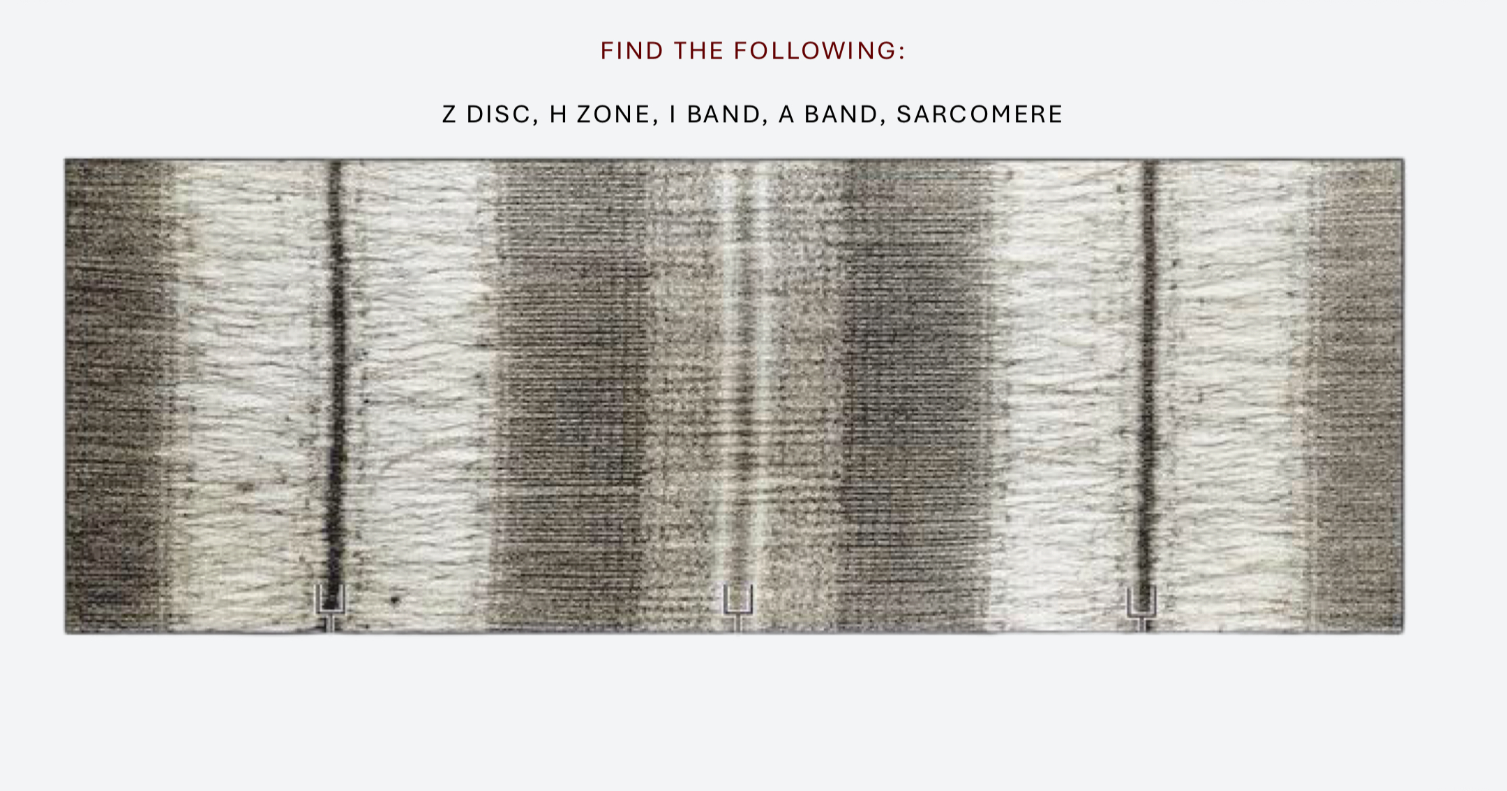
label and describe the Z disc, H zone, I band, A band, M line, and sarcomere
Z disc- represent the edges of the sarcomere
M line- represents the middle of the sarcomere
A band- represents the length of myosin **never changes in length
I band- represents the portion of actin that has no myosin overlap and contains only thin filaments ** will shorten when the muscle contracts
H Zone- represents the portion of myosin that has no actin overlap and contains only thick filaments ** will shorten/disappear when muscle contracts
sliding filament theory
actin and myosin slide past each other during muscular activity to shorten the muscle causing contraction
sarcolemma
plasma membrane of a muscle cell
terminal cisternae and sarcoplasmic reticulum
calcium storage, release and uptake
t- tubule
allows action potential into muscle fiber
triad
t-tubule between two terminal cisternae
describe EC coupling
Acetylcholine released from the axon terminal binds to receptor on the sarcolemma
an action potential is generated and travels down the t-tubule
calcium is released from the SR in response to the change in voltage
calcium binds troponin; cross bridges form between actin and myosin
acetylcholinesterase removes acetylcholine from the synaptic cleft
calcium is transported back into the SR
tropomyosin binds active sites on actin causing the cross- bridge to detach
powerstroke
movement of the myosin head, bound to actin, towards the M-line of the sarcomere which pulls actin towards the center of the sarcomere, shortening the I-band, H-zone, and the entire sarcomere
describe the cross-bridge cycle
binding of actin to myosin
power stroke
rigor (myosin in low-energy form)
unbinding of myosin and actin
cocking of the myosin head (myosin in high-energy form)