CVRS anatomy
1/64
There's no tags or description
Looks like no tags are added yet.
Name | Mastery | Learn | Test | Matching | Spaced | Call with Kai |
|---|
No analytics yet
Send a link to your students to track their progress
65 Terms
what heart is this?
dog
pig
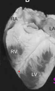
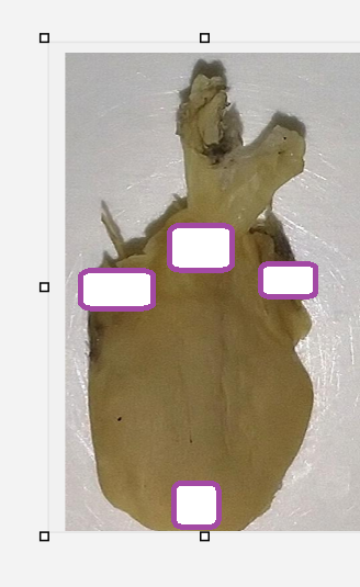
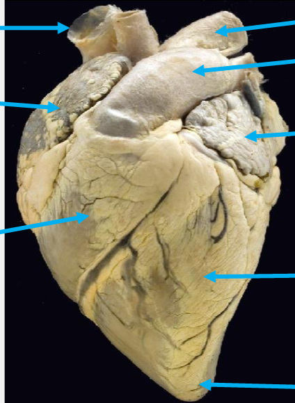
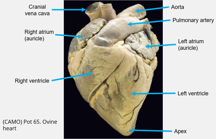
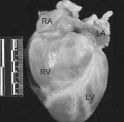
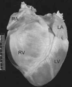
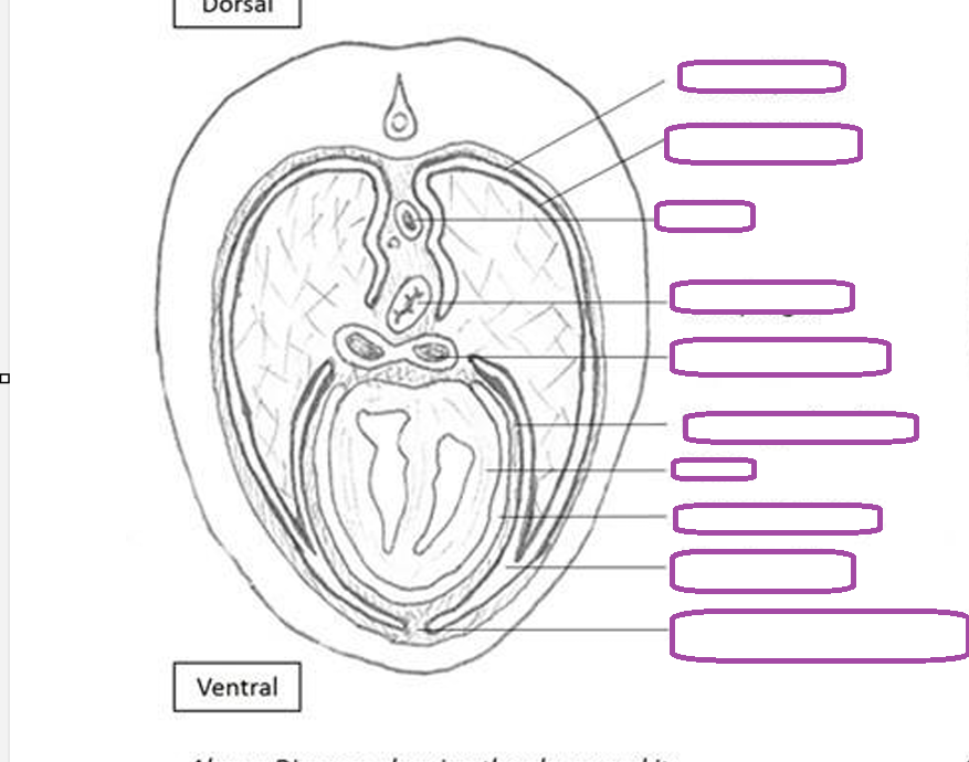
what heart is this
sheep heart
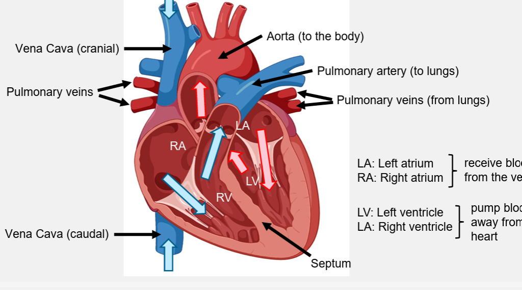
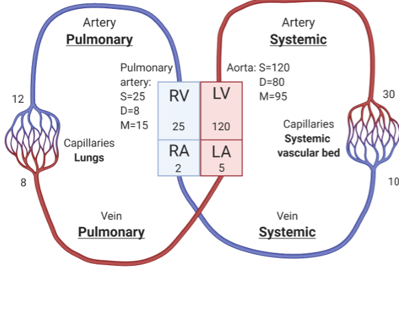
aorta
where does it pump blood
pressure
to rest of body- systemic circulation, 100mgHg
pulmonary artery
to the lungs, 12mmHg
pulmonary circulation
what enters the pulmonary vein
oxygenated blood
7mmHg
what enters the vena cava
deoxygenated blood
3mmHg
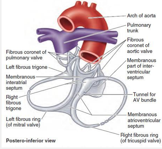
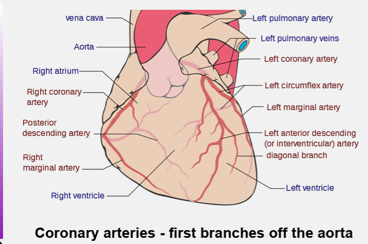
what are the coronary arteries
fibroblast
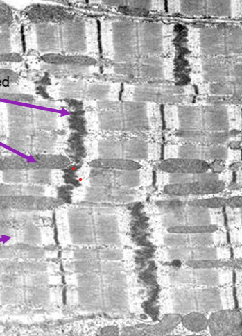
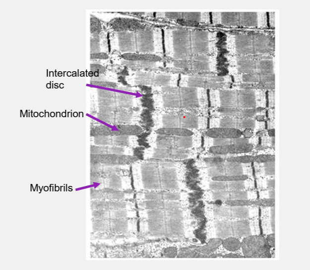
intercalated disks
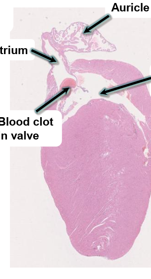
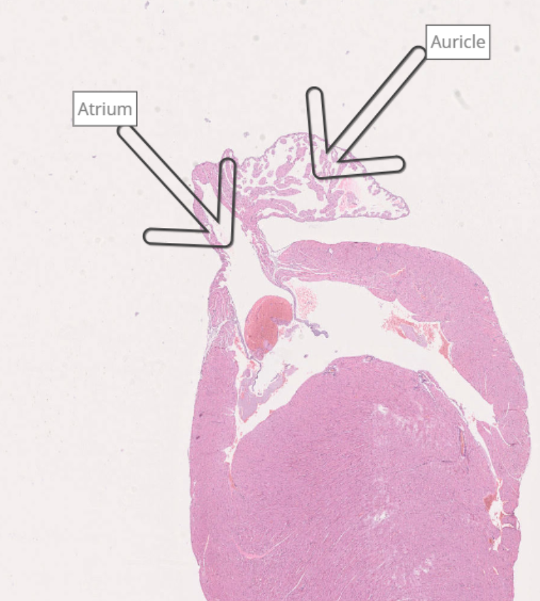
top of ventricle
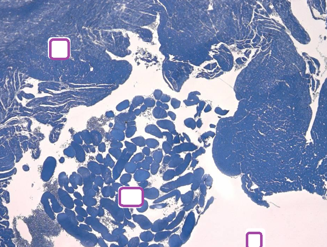
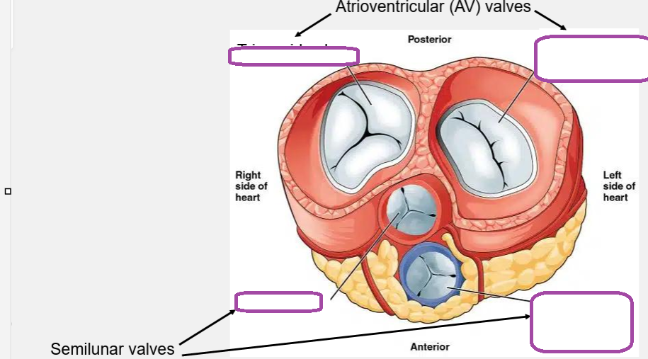
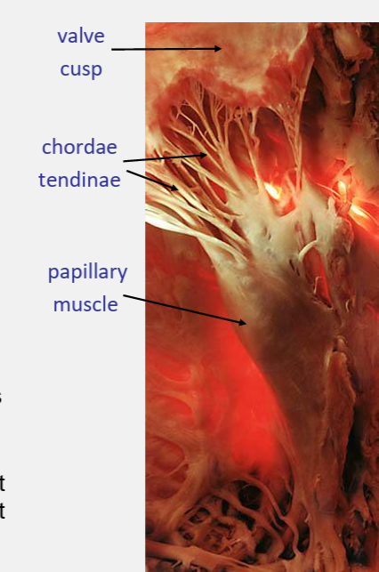
label the parts of the cardiac muscle
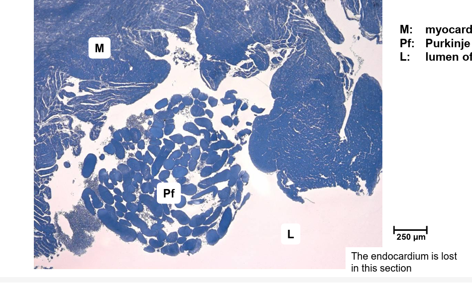
what type of cells make up purkinje fibres
what do they form and where
impulse conducting muscle cells
they forms the atrioventricular bundle and its branches in the walls of the ventricles
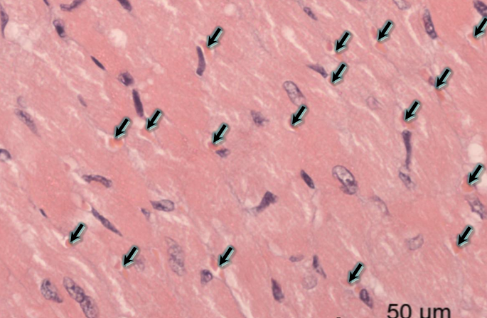
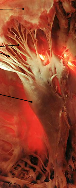
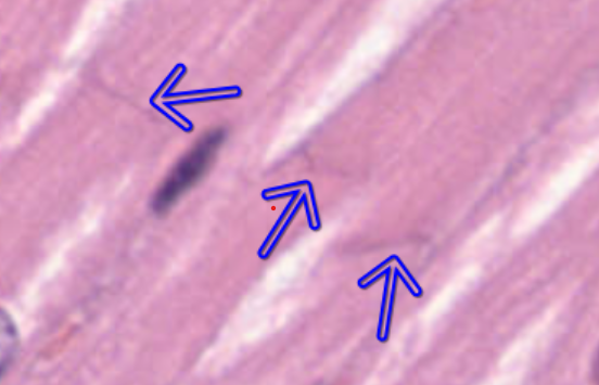
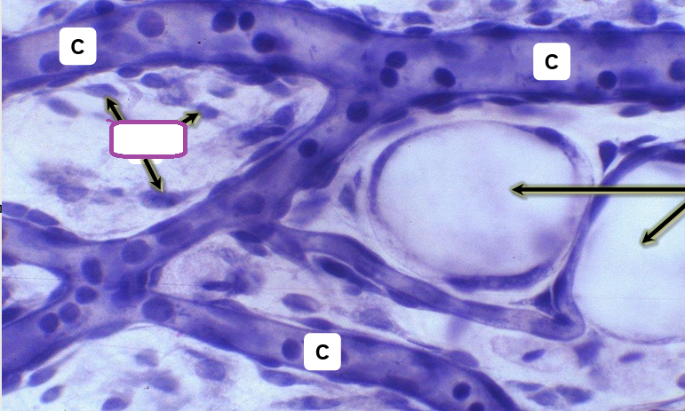
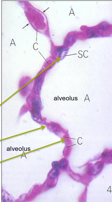
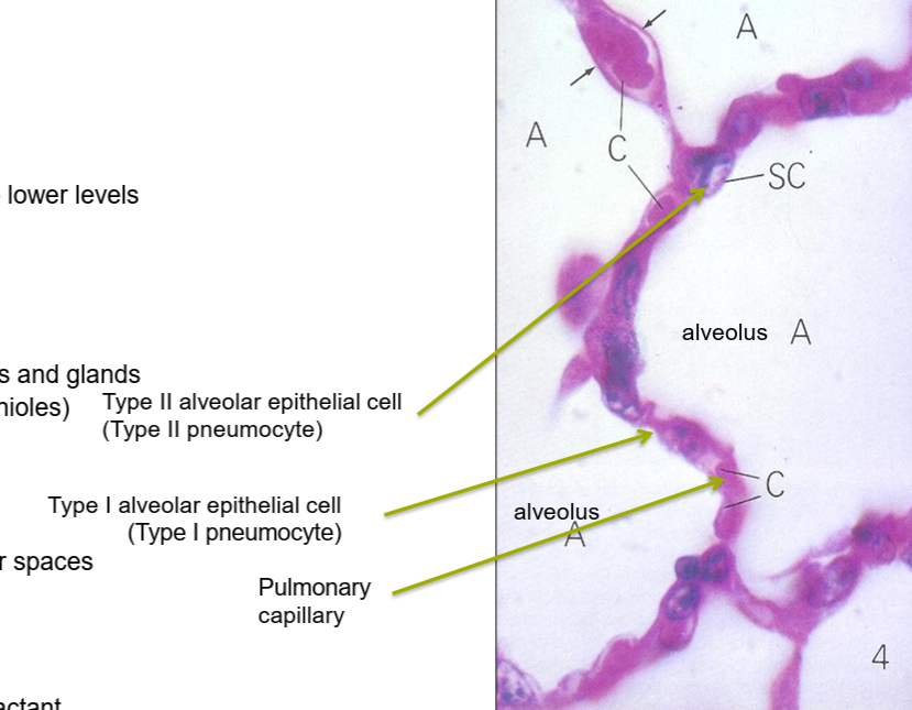
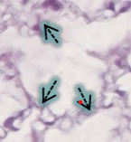
what are these arrows pointed to
capillaries
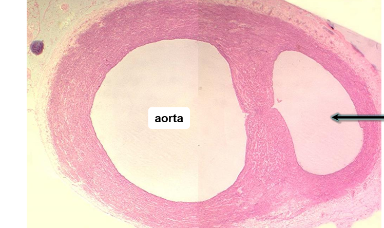
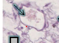
brachiocephalic trunk
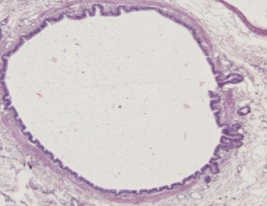
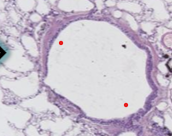
tunica intima
endothelial lining
sub endothelial connective tissue
internal elastic membrane
tunica media
circular smooth muscle layer
elastic and collagen fibres
external elastic membrane
tunica externa
outer covering
connective tissue sheath
what structures are present in the tunica adventitia
blood vessels- vaso vasorum
autonomic erves- nervi vasorum
arrangement of smooth muscle cells in the tunica media of arteries
near tight circular helical arrangement
muscular arteries control blood flow to various organs
what is the function of elastin in veins
provides elastic recoil to maintain vascular tone and allows the vessels to expand to store to the needs of the network as a whole
type of epithelium in capillary in loose connective tissue
simply squamous epothelium
active transport and movement of gases and metabolites
fibroblasts
apidocytes
state 3 differences between capillaries and sinusoids
1. Sinusoids usually larger and more varied shape than capillaries.
2. Sinusoids may be fenestrated, have gaps and pores in the endothelium.
3. They have a discontinuous basal lamina.
what structures connect individual endothelial cells
gap or tight junctions
how does the capillary network in the brain differ from that of other organs
tight junctions and pericytes in the brain contribute to the blood brain barrier
where are cardiac myocytes and capillary endothelium found
within the myocardium
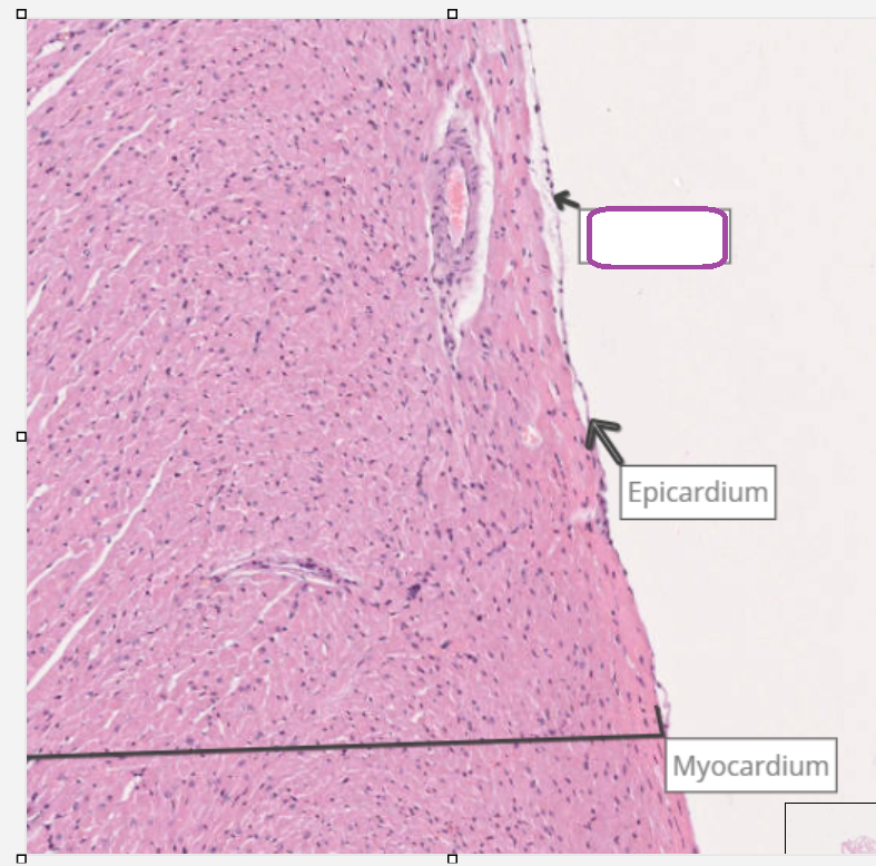
where are fibroblasts found
within the epicardium
which structures make up the intercalated disks
Fascia adherens – Connects the cells and provides a link to the myocyte’s actin network
Desmosomes – Provides a strong mechanical connection between the cells
Gap junctions – Allows passage of ions, allowing transmission of the wave of depolarisation
what are the structures that allow cardiac muscle to act as a functional syncytium
intercalated disks- gap junctions
branched fibres- lateral junctions
rich vasculature
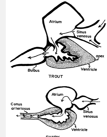
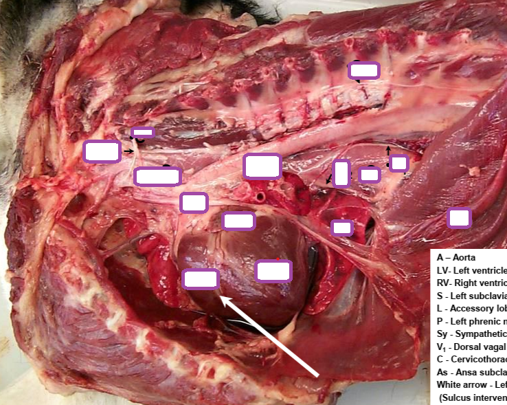
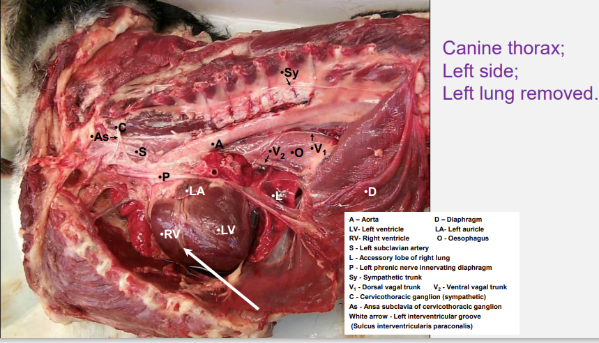
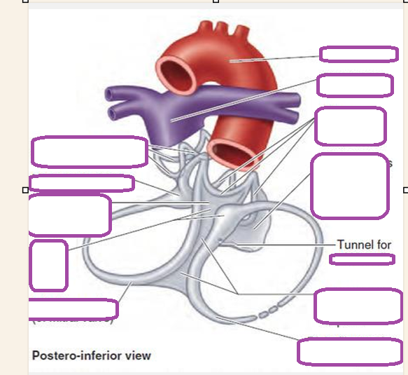
where do the coronary arteries originate and where do the coronary veins drain
aortic root
coronary sinus which drains into the right atrium
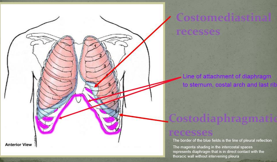
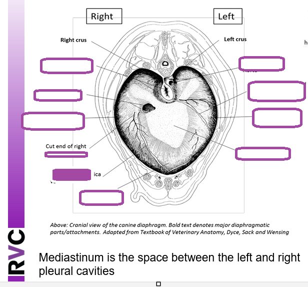
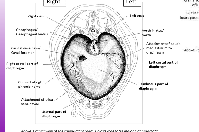
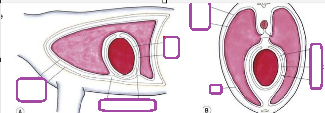
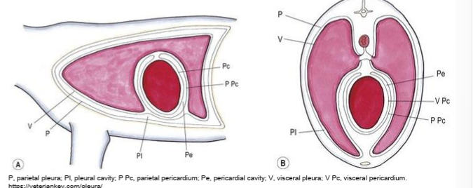
what is meant by the line of pleural reflection
the line where the pleura lining the inner surface of the ribs and intercostal muscle continues onto the surface of the diaphragm at the latters attachment to the body wall
caudoventral to the line of pleural reflection the diaphragm is in direct contact with the intercostal tissues and the ribs and conseqeuntly the more thoracic wall is not in contact with the pleural cavity
liver biopsy can be used to access the abdominal cavity without penetrating the pleural cavity
bronchus
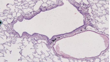
bronchiole
terminal bronchiole
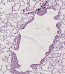
respiratory bronchiole
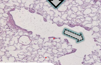
respiratory bronchiole to alveolar duct
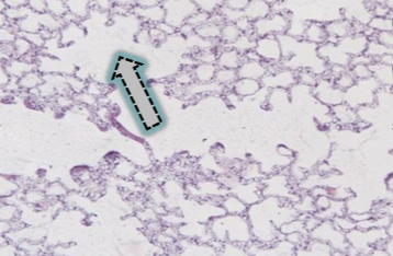
alveolar duct to alveolar sac
alveolus to capillary
capillary to venule
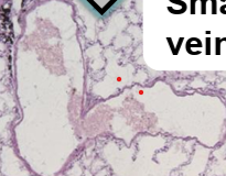
small vein
what anchors myosin heads to the z disk
titin
acts like a molecular spring
where is cap z found
on the z disk
heart beat

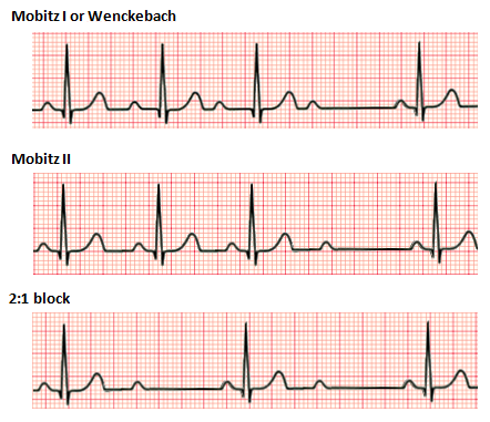
first degree av block
prolonged PR intervals
contraction delayed due to increased time for AVconduction
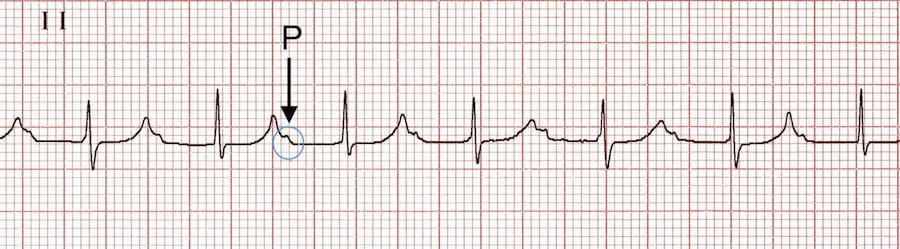
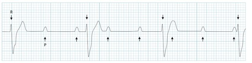
second degree AV block
AV node fails to trasmit all atrial impulses
more P waves than QRS complexes
atria beat more than once for each ventricular contraction
third degree AV block
transmission of impulse from atria to ventricles fails
atria and ventricles beat independently of each other
P and QRS complexes completely dissociated