bio 99 midterm 4
1/97
There's no tags or description
Looks like no tags are added yet.
Name | Mastery | Learn | Test | Matching | Spaced | Call with Kai |
|---|
No analytics yet
Send a link to your students to track their progress
98 Terms
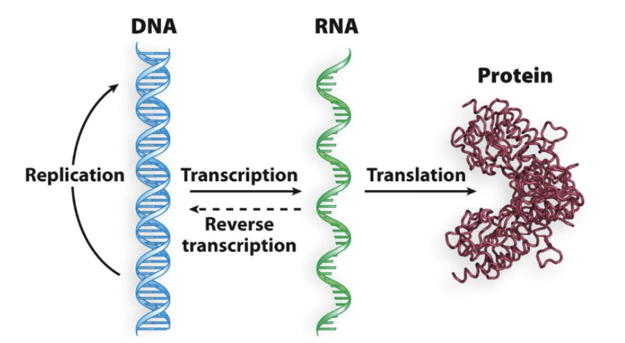
The central dogma of Molecular biology
DNA —(Transcription)—> RNA —(Translation)—> Protein
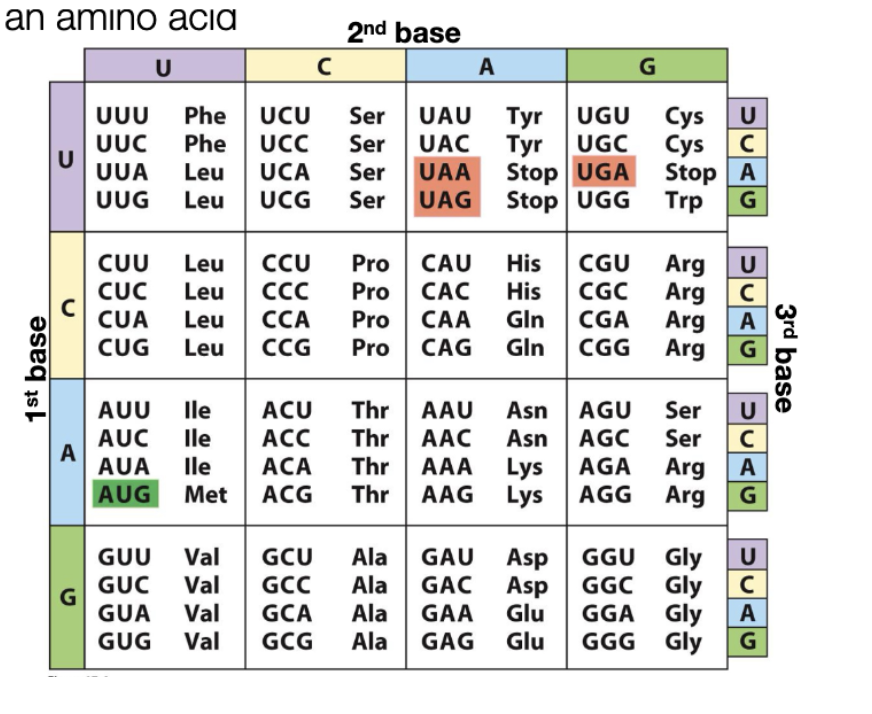
What is a genetic code? - Codon
trinucleotide sequence that codes for an amino acid (AUG)(UAA)(UAG)(UGA)
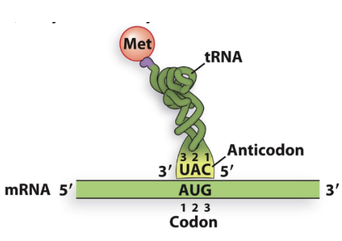
Anticodon
triplet nucleotode sequence on tRNA that base pairs with codon on mRNA
Degenerate
multiple codons can encode the same amino acid
consequence of there being 64 possible codons but only amino acids
Codon family
- when 4 codons specify the same amino acid
start codons
AUG —> encodes Met
Stop codons
UAA, UAG, UGA
there are no tRNA that recognize stop codons
Open reading frame
sequence that has a start codon, long stretch of codons, then a stop codon
find AUG to start the reading frame
Decoding the genetic code by tRNAS
codons with U or C in the 3rd position always code for the same animo acid

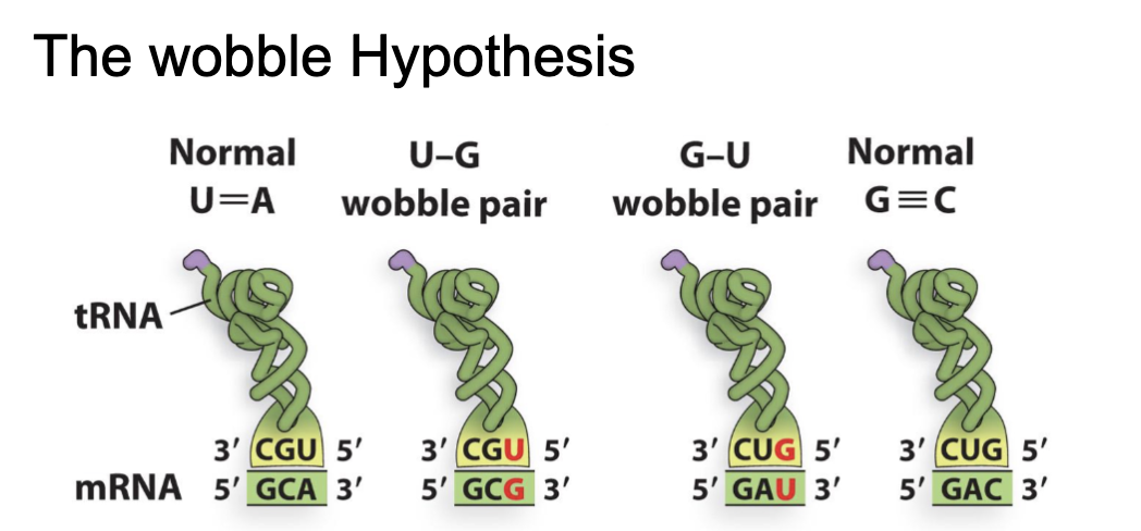
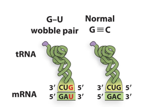
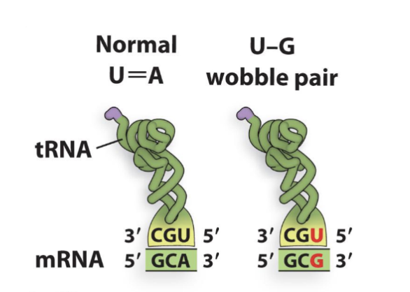
The wobble hypothesis
fewer than 45 transfer RNA (tRNA) molecules can decode all 61 messenger RNA (mRNA) codons****** redo this after wtaching a video
….
U-G Wobble Rule (Part 1)
Anticodon (3’to 5’) xxG recognizes xxC and xxU codons
U-G Wobble Rule (Part 2)
Anticodon (3’5’) xxU recognizes xxA and xxG codons
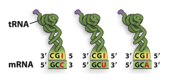
Inosine
Adenosine(A) in 1st anticodon position (5’) of tRNA is converted to inosine (I)
can only pair with C, U, A
“I” Wobble Rule
Anticodon(3’5’) xxI recognizes xxC, xxA, and xxU codons
Wobble rules
wobble base pairing only occurs between the last 3’ position of the codon and the first 5’ position of the anticodon
Types of mutations
note* mutations happen to DNA NOT RNA, but affect mRNA sequence
Single base substitutions
silent
missense
nonsense
SIngle base substitutions
change of a single base in the DNA sequence of a gene
Silent mutation
change in codon that does not change the amino acid sequence
GAA = Glu …. —> mutated to—> GAG = Glu
Missense mutation
change in codon that results in a different amino acid encoded
GAA(glu) —mutated—> GAC (Asp)
Nonsense mutation
change in codon that creates an early stop codon
GAA(glu) —mutated—> TAA (UAA) or (stop)
Frameshift mutation
insertion or deletion of nucleotides that alter the reading frame of the coding sequence
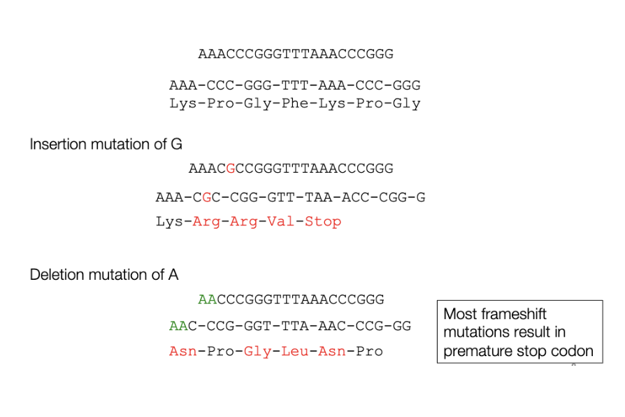
deletion mutation
deletion of one or more nucleotides
transition mutation
a purine is substituted for another purine. A → G or G → A. Most common type of mutation. Can lead to all 3 types of substitutions
in-frame insertion mutation
insertion or deletion of 3 nucleotides restores the reading frame (“in frame“)
Reversion mutations
where a mutation that is deleterious to the function of the encoded protein acquires a second mutation that allows the protein to function again
in order for a reversion mutation to be identified, protein function must be restored (full or partially)
Reversion mutation: Missense mutation
(Lys to Glu) in the active site causes the protein to be non-functional. A second mutation returns the amino acid to lysine
Same situation as above, but a second mutation changes the Gly to Arg, which is similar to lys and the protein is mostly functional.
Reversion mutation: Nonsense mutation
(Tyr to STOP) creates a truncated protein that is non-functional. A second mutation changes STOP back into Tyr.
Same situation as above, but the second mutation changes STOP to Ser, allowing a full length protein and resources function.
Reversion mutation: Frameshift mutation
caused by the insertion of a single nucleotide. A second mutation causes a deletion of a single nucleotide near the site of the insertion. Protein function is restored.
Minor mutation
found in other regions of protein(not active site), substituting similar amino acid, new stop codon near the back (C-terminus), & if a reversion mutation restores reading frame mutations are close together
Catastrophic mutation
found in active site of protein, substituting dis-similar amino acid, new stop codon near the front (N-terminus), if a reversion mutation restores reading frame mutations are far apart
How does molecular nature of proteins protect from mutations
deleting an entire DNA binding domain of a protein and the transactivation domain will still work, vice versa
Biochemical basis of muatation
single base substitution —> chemical carcinogens
frameshift mutation—> intercalating agents
ionizing radiation—> x rays & large deletion
Intercalating agents
these amino acids insert themselves into the double helix causing stretching of the DNA that leads to polymerase errors during DNA replication
acridine orange is a intercalating agent
ionizing radiation
type of radiation that causes the release of electrons from molecules, ionizing them can directly or indirectly damage DNA.
leads to double strand DNA BREAKS
during repair= leads to large deletion mutations
x-rays are common form of ionizing radiation used to create mutations
large deletion
deletion of 7 or more nucleotides
Cause of Mutation: singel base substitution
Silent, missense, nonsense mutations (carcinogens)
Cause of Mutation: frameshift mutations
insertion or deletion (intercalating agents)
Cause of Mutation: Deletion mutation
loss of a large chuck of sequence (ionizing radiation)
Standard Deviation (SD)
a way to measure the amount of spread of a set of data [e.g. mean +/- SD (1 SD (plus/minus) captures about 68% of the data points)]
![<p><span style="background-color: transparent;">a way to measure the amount of spread of a set of data [e.g. mean +/- SD (1 SD (plus/minus) captures about 68% of the data points)]</span></p><p></p>](https://assets.knowt.com/user-attachments/320fe7a0-d896-4aa3-a7c1-4a7af04c1ce9.png)
error bars
a graphical way to visualize the spread of data in bar graphs, without showing teh individual data points (usually based on SD but not always)
T-Test
a way to compare the means and SDs of two data sets to assess statistically if the difference between them is significant
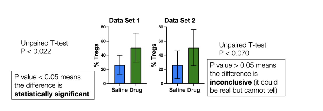
P-value
a p-value <0.05 means theres < a 5% chance (or 1 in 20 chance) that the difference is due to random chance (and 95% chance that its real)
P value >0.05 means the difference is inconclusive (it could be real but we cannot tell)
P-value < 0.05 means the differnece is statistically significnat
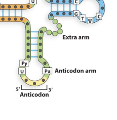
tRNA structure
has 4 -5 arms
anticodon arm
amino acid arm
others arms
tRNA structure: Anticodon arm
contains the anticodon, interacts with mRNA sequence
tRNA structure: amino acid arm
attaches to amino acid
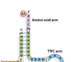
tRNA structure: other arms
structural, interact with ribosome, tRNA synthetase
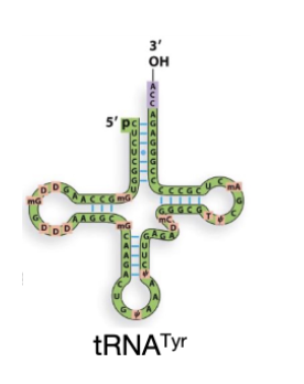
tRNA^Tyr
tRNA that recognizes a codon for Tyrosine, but does not necessarily have a Tyrosine amino acid attached to it.
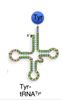
Tyr-tRNA^Tyr
tRNA that recognizes a codon for tyrosine, and is “charged” with a Tyrosine amino acid
Aminoacyl
tRNA with an amino acid attached to it
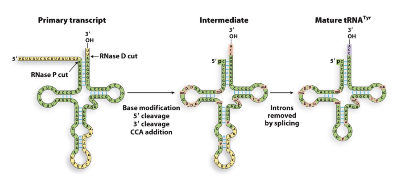
tRNA processing
tRNA is processed post-transcriptionally into its mature form.
5’ cleavage- pre-tRNA is transcribed with extra sequence at its 5' end(leader sequence). This is cleaved off by an endonuclease called RNase P to generate the mature 5' end
3’ cleavage- the extra sequence at its 3' end (the trailer) is trimmed by exonucleases (RNase D cut)
CCA addition- (Cytosine-Cytosine-Adenine) attached to 3’ end
Intron splicing- introns within anticodon loop(yellow) are excised by an endonuclease complex, then two mature exons of tRNA are ligated together to form a continuous anticodon loop
tRNA processing: Base modification
specific nucleotides are modified
tRNA processing: Cleavage
the end of transcription are removed
5’ cleavage
3’ cleavage
5’ cleavage & RNaseP
pre-tRNA is transcribed with extra sequence at its 5' end(leader sequence). This is cleaved off by an endonuclease called RNase P to generate the mature 5' end
3’ cleavage & RNase D cut
the extra sequence at its 3' end (the trailer) is trimmed by exonucleases (RNase D cut)
tRNA processing: CCA addition (amino acid)
a CCA is attached to the 3’ end of the transcript. This is what the amino acid attaches to.
tRNA processing: Intron splicing- introns removed (only in eukarotes)
introns within anticodon loop(yellow) are excised by an endonuclease complex, then two mature exons of tRNA are ligated together to form a continuous anticodon loop
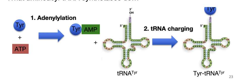
tRNA Activation or aminoacylation
Each tRNA must have its correct amino acid attached to it.
2 steps: 1) Adenylylation and 2) tRNA charging
both steps catalyzed by aminoacyl-tRNA synthetase (AATS)
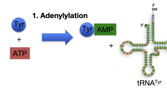
tRNA Activation: Adenylylation
amino acid + ATP —→ aminoacyl-AMP + PPi
AMP attaches to carboxyl group of amino acid
(Amino Acid Activation): The enzyme binds a specific amino acid(Tyr) and an ATP molecule. It transfers the AMP portion of the ATP to the amino acid, -creates—> aminoacyl-adenylate intermediate.
This reaction releases inorganic pyrophosphate (PPi) and stores vital energy for later use
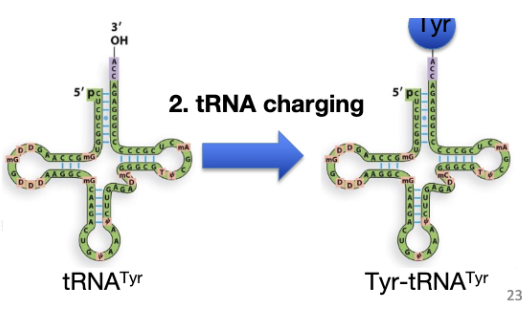
tRNA Activation: tRNA charging
aminoacyl transferred off of AMP onto → tRNA’s CCA arm
While bound to the enzyme's active site, the appropriate tRNA molecule attaches to the complex. The enzyme transfers the activated amino acid to the 3' hydroxyl end of the tRNA, releasing the AMP molecule in the process.
second genetic code
aminoacyl-tRNA synthetase interaction with the different tRNAs
Bacteria: Initiator Met tRNA
tRNA^fMet
Bacteria: Internal Met tRNA
tRNA^Met
Bacteria: fMet
Formyl group attaches to the N-terminus of fMet, preventing fMet from being able to attack to an Amino acid in front of it. fMet can only be the first Amino acid
cannot be added internally
tRNA^fMet is the only tRNA that is recognized by the ribosome initiation complex
Transformylase
converts Methionine to N-formylmethionine (fMet)
Eukaryotes: Initiator tRNA
- tRNAi^Met
the only tRNA that is recognised by the ribosome initiation complex
Eukaryotes: Internal tRNA
- tRNA^Met
Met is not modified
aminopeptidases
in both bacteria and eukaryotes, they often remove the N-terminus Met —> so many mature proteins DONT have Met as the first amino acid
Necessary(required)
something is necessary for a function when you need it to carry out that function
NOT necessary= when removed there is no effect
Sufficient(all you need)
something is sufficient for a function when you can get function with only that thing
NOT sufficient- if you add and there is no effect
Bacteria: Small subunit- 30s
rRNA- 16s
Protein- 21 total subunits
Function- mRNA , tRNA assembly
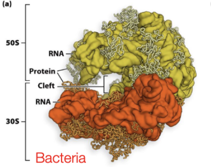
Bacteria: Large subunit
RNA- 5s and 23s
Protein- 36 total subunits (L1 to L36)
Function- catalyze peptide bond formation
Eukaryotes
similar to bacteria (2 subunits) but slightly more complex, more proteins
40s small and 60s large
Svedberg units(S)
roughly proportional to size, but not linear relationships, based on sedimentation rates during ultracentrifugation
rRNA Processing (only in eukaryotes)
rRNA transcribed as one long transcript (30s), then later proceeded into 16s, 23s, and 5s mature RNA (note that these do not add up to 30)
Harry Noller
Ribosome catalytic activity- what is necessary and/or sufficient for peptide bond formation? rRNA, protein, or both?
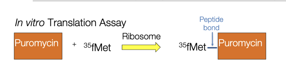
Puromychin
mimics tRNA, but binds directly to the large subunits, ribosomes will attach it to a growing polypeptide chain
New amino acids cannot be attached to puromycin, so it terminates translation and is therefore a powerful antibiotic
In this experiment, puromycin is just the substrate for forming peptide bonds
Ribozyme
RNA with enzymatic activity
Closest protein is 18A away from the active site of large subunit.
How manhy tRNA binding sites does the ribosome have
3! A, P, & E site

Ribosome: A site
Acceptor site, where new tRNAs enter ribosome
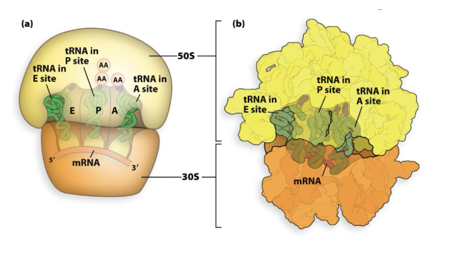
Ribosome: P site
Polypeptide site, where growing polypeptide chain is held

Ribosome: E site
Exit site, where tRNA are expelled after amino acid removal
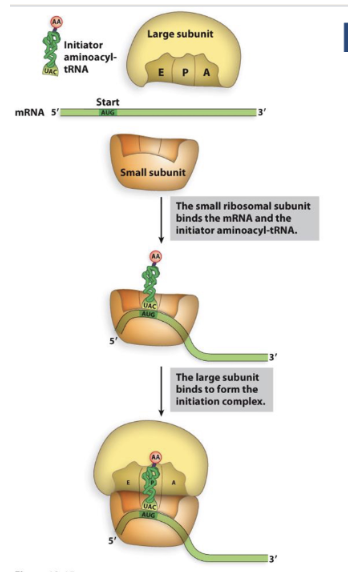
Ribosome: Initiation
Small subunits binds mRNA
Small subunits bind initiator tRNA
Large subunits binds to the rest
Final product has= small subunits, large subunit, mRNA(start codon at p site), initiator tRNA (paired with start codon)
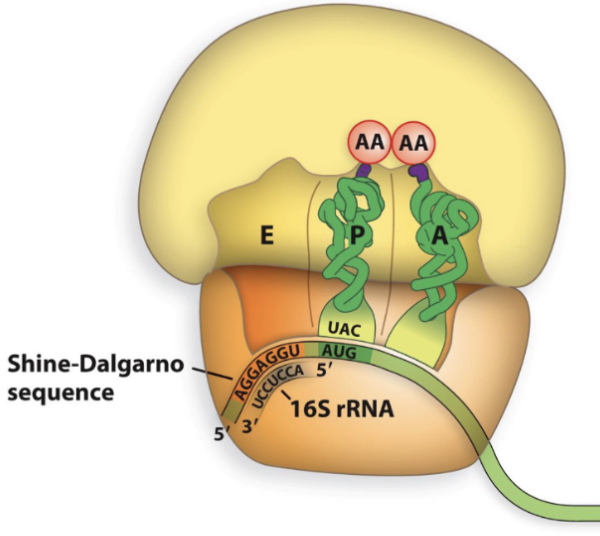
Shine-Dalgarno sequence
consensus sequence in front of start codon
AGGAGGU
Recruits mRNA to small subunit
Directs mRNA start site to correct position on ribosome
Base pairs with 16S rRNA on small subunits
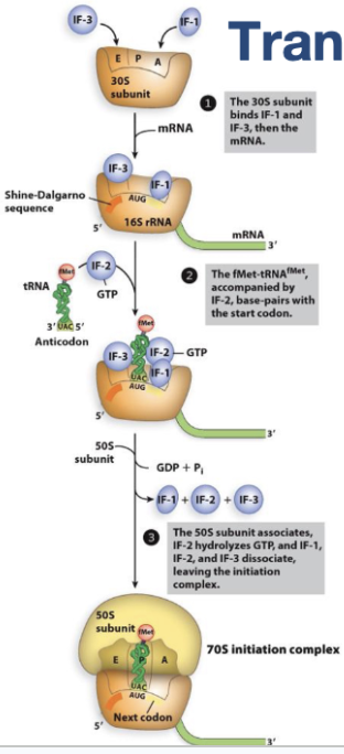
Translation Initiation in Bacteria
Bacterial proteins involved in initiation
IF-1 - fills A site to prevent tRNAs from binding
IF-2 - escorts initiator tRNA
IF-3 - prevents large subunit from binding
GTP hydrolysis powers formation of the initiation complex
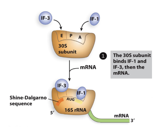
Translation Initiation in Bacteria: step 1
1a- blocking site on a small subunit
IF-1 and IF-3 bind to 30S small subunit
IF-1- blocks A site, prevents tRNA binding
IF-3 blocks large subunit form binding
1b- loading mRNA
mRNA attached to 30S small subunit
Uses shine-dalgarno sequence to position start codon right at P site
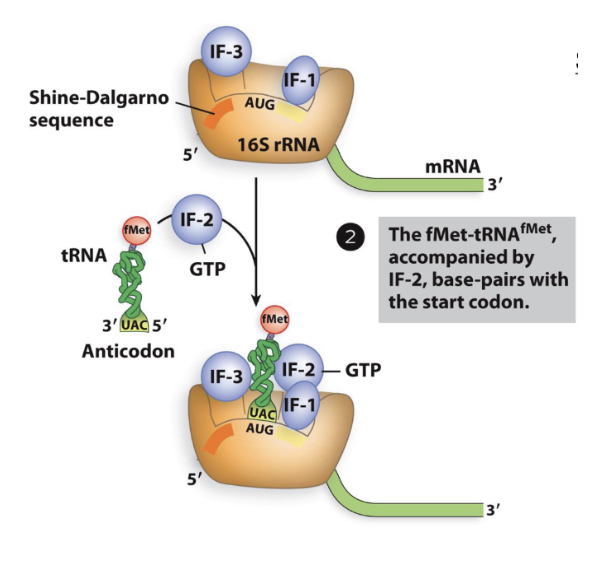
Translation Initiation in Bacteria: step 2
Step 2- recruitment of initiator tRNA
IF-2 binds to initiator tRNA
IF-2 is bound to GTP
Has GTP-hydrolase activity
Initiator tRNA (fMet-tRNA^fMet)
binds to start codon
rRNA binds to unique sequence on initiator tRNA (reason why internal tRNA^Met does not bind)
tRNA^fMet can only bind to the P site
No other tRNAs can bind to P site
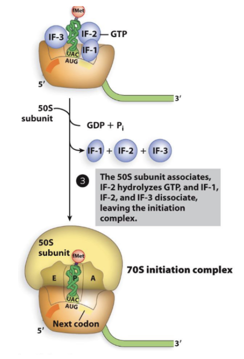
Translation Initiation in Bacteria: step 3
Step 3- recruitment of large subunit
30S changes conformation to kick out IF-3, allowing 50S large subunit to bind
Hydrolysis of GTP to GDP causes IF-1 and IF-2 to leave
Initiation complex completed
Both large and small subunits bound
mRNA with start codon lined up in P site
Initiator tRNA bound to start codon in P site of ribosome
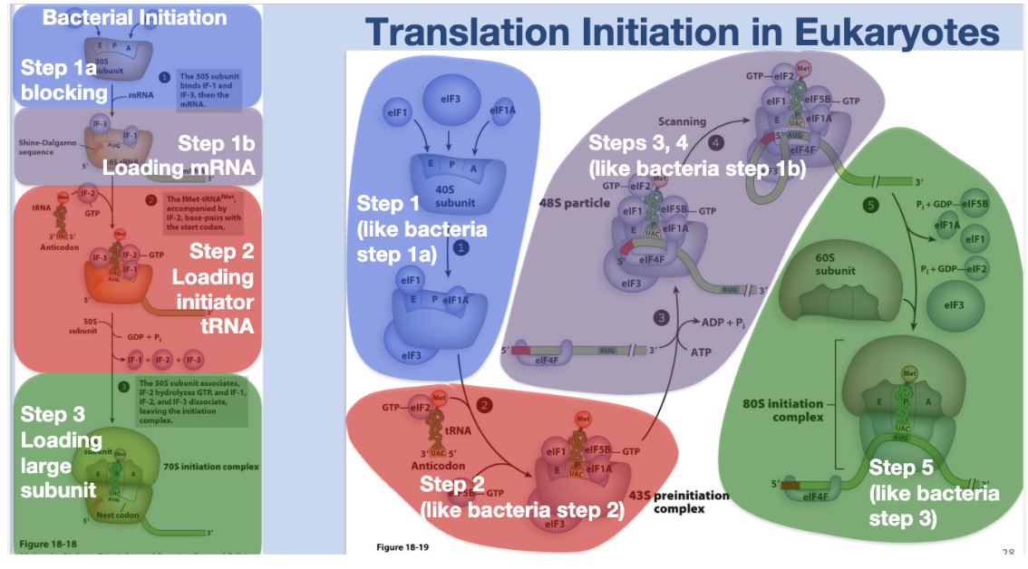
Translation initiation in Eukaryotes
the process where ribosomes assemble on mRNA with initiator tRNA to locate the start codon (AUG)
Initiation in Eukaryotes: Step 1
Initiation in Eukaryotes: Step 2
Initiation in Eukaryotes: Step 3
Initiation in Eukaryotes: Step 4
Initiation in Eukaryotes: Step 5
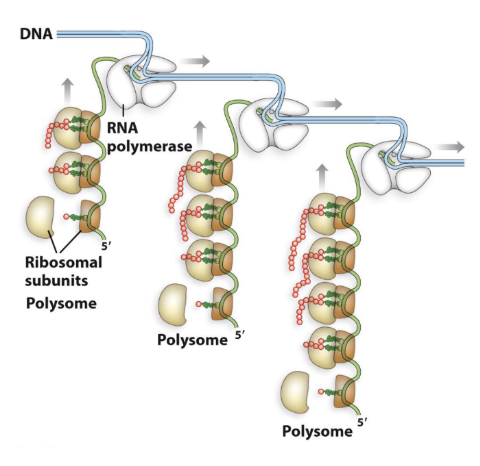
Bacterial Polysome
Multiple ribosomes can translate the same mRNA simultaneously
In bacteria, this can even happen while the mRNA is still being transcribed
Multiple RNA polymerases can also be transcribing the same DNA at once
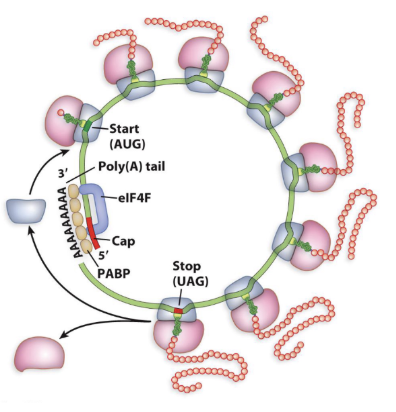
Eukaryotic Polysome
elF4G (part of elF4F complex) can bind to poly-A binding protein (PABP)
Connects 5’cap to poly-A tail, forming a circle
Facilitates translation regulation
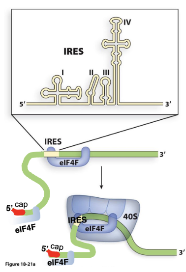
IRES/ Internal Ribosomal Entry Site
a specialized RNA sequence that allows ribosomes to directly bind and initiate protein translation in the middle of a messenger RNA (mRNA) molecule, independently of the 5' cap structure typically required
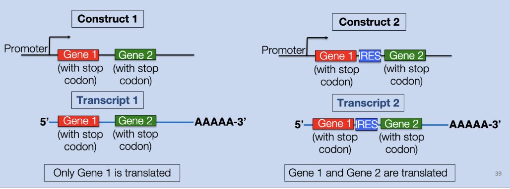
polycistronic transcripts
a single messenger RNA (mRNA) molecule that encodes multiple different proteins.
IRES is also used by molecular biologist to express two genes off the same transcript
IRES makes eukaryotic polycistronic transcripts