HC 4: Linking identity and function
1/8
There's no tags or description
Looks like no tags are added yet.
Name | Mastery | Learn | Test | Matching | Spaced | Call with Kai |
|---|
No analytics yet
Send a link to your students to track their progress
9 Terms
Functional genes as markers
Instead of looking only at broad identification genes (like 16S rRNA), researchers can target functional genes that encode specific traits or processes.
nifH → nitrogen fixation
amoA → ammonia oxidation (nitrification)
antibiotic resistance genes → resistance traits
Functional gene markers help answer: “What can this microbial community do?” rather than only: “Who is there?”
SIP
PLFA + labeled substrate (13C)
analyses df=ifferent microbial groups as peaks
The idea is simple: you feed a microbial community a labelled compound, and only the microbes that actively metabolise it will incorporate the isotope into their cells. You then detect the “labelled” microbes using different molecular or biochemical approaches.
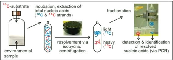
SIP: DNA/RNA vs PLFA
Heavy DNA/RNA = active users of the substrate
Labelled PLFAs indicate active microbial biomass using the substrate (Only living, active cells can incorporate the isotope into membrane lipids)
Single-cell approaches
MAR-FISH
NanoSIMS
RAMAN Micro/Spectroscopy
MAR-FISH (/STAR-FISH)
You have two different steps:
1.fluorescent in situ hybridization (FISH);
you have a probe that’s specific for specific taxa; sticks to the taxa where the probe has affinity for.
2.looking at autoradiogram/ at radioactive substrate
you add radioactive glucose
every place where you see a spot, is where the substrate has been taken up.
Then you overlay the FISH and the radioactive results:
you can see which organisms are the taxa of interest AND are being active in taking up the substate.
With different fluorescent labels you cat target different taxa, and you can determine which ones are taking up substrate and which ones aren’t.
NanoSIMS
You zap a cell, all the chemicals get scattered and land on detectors, so heavy and light atoms land on different places
NanoSIMS tells you where specific elements or isotopes are located at a very small scale, allowing researchers to link microbial identity with actual activity
useful for studying microbial interactions, nutrient cycling, and host–microbe relationships.
SIP + NanoSIMS
a method that tracks isotope uptake to directly see which individual microbial cells are actively metabolising a substrate, at extremely high spatial resolution.
Raman spectroscopy
shining a laser on a single cell and measuring how the light is scattered.
Most light bounces off unchanged, but a small part interacts with the molecules inside the cell (like proteins, lipids, and DNA) and changes energy
»» creates a unique spectral pattern (biochemical fingerprint)this is compared to a reference database of known cells
» Based on how similar the patterns are, the cell can be classified as most likely belonging to a certain species/functional group
Raman-SIP
combines Raman spectroscopy with Stable Isotope Probing (SIP) to link microbial identity with activity at the single-cell level
If they actively metabolise it, the isotope is incorporated into their biomass, which causes a detectable shift in their Raman spectrum —> identify which individual cells are actively using a specific substrate (without cultivation or DNA extraction)
“shows who is active and what they are consuming at single-cell resolution”