1.3: development of the nervous system
 Call Kai
 Learn
Learn Practice Test
 Spaced Repetition
 Match
Match Flashcards
 Knowt Play
1/47

There's no tags or description
Looks like no tags are added yet.
Name | Mastery | Learn | Test | Matching | Spaced | Call with Kai |
|---|
No analytics yet
Send a link to your students to track their progress
48 Terms
everything starts as a
flat sheet of cells that turn into our nervous system
starts with a tube
stages of neural tube development
pre-embryonic stage
embryonic stage
fetal stage
pre-embryonic stage
conception to second week
after sperm and egg join and egg is fertilized
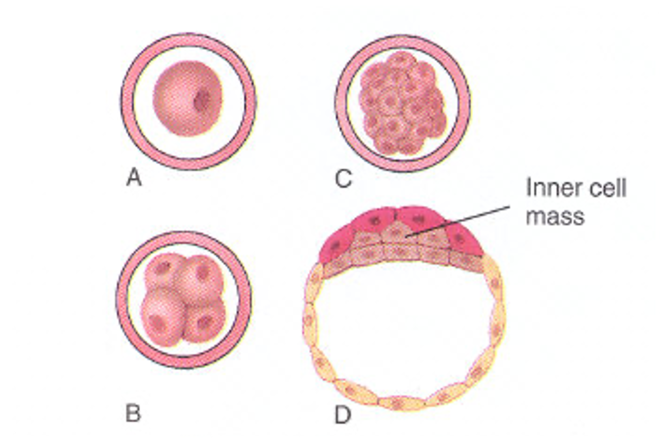
embryonic stage
2nd week → 8th week
disc of cells (musculoskeletal, bone, and visceral) suspended in a giant water balloon
fetal stage
8 weeks → birth
differentiation occurs during this stage
when neural tube starts to differentiate (to bones, muscles, everything)
develops into recognizable parts of the nervous system
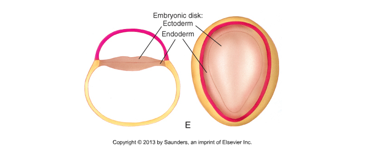
embryonic disk
turns into the neural tube
endoderm (inferior - forms digestive tract and respirayory system)
ectoderm (superior - will form NS and skin)
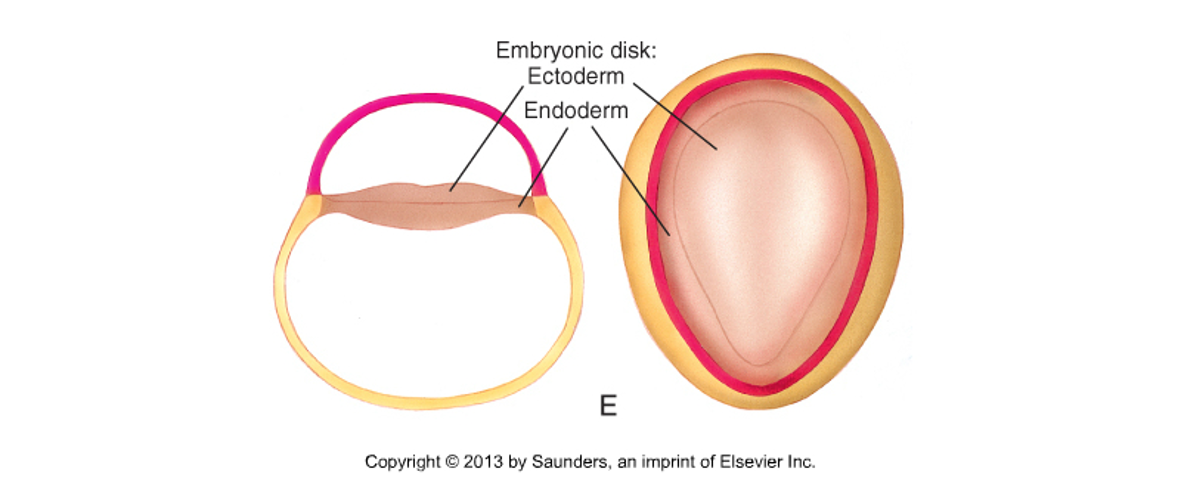
embryonic disk structure
neural plate: most dorsal part of the disk
made up by nerve cells
nerve cells start posterior (dorsal) and grow forward (ventral)
endoderm: most ventral part of the disk
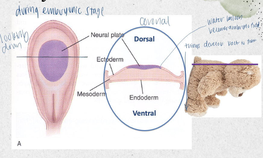
embryonic disk (dorsal to ventral)
neural plate
ectoderm
mesoderm
endoderm
neural plate
most dorsal part of embryonic disk
made up of nerve cells
nerve cells start dorsal and grow forward
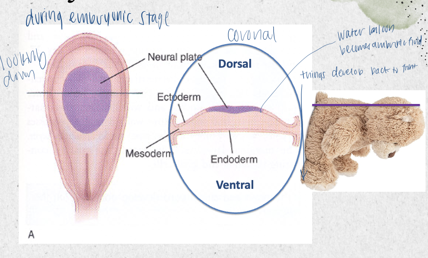
ectoderm
includes neural plate
most dorsal part of embryonic disc
becomes sensory organs, epidermis, and nervous system
what does the ectoderm become
sensory organs
epidermis
nervous system
mesoderm
somites live here
becomes dermis, muscles, skeleton, and circulatory
(support system)
where do somites live
mesoderm
endoderm
gut, liver, pancreas, and respiratory system
neural tube formation
the neural plate buckles in the middle to form the neural groove
the sides of the groove (the folds) come up and bend toward each other; eventually they fuse at midline
somites appear and develop along the neural tube
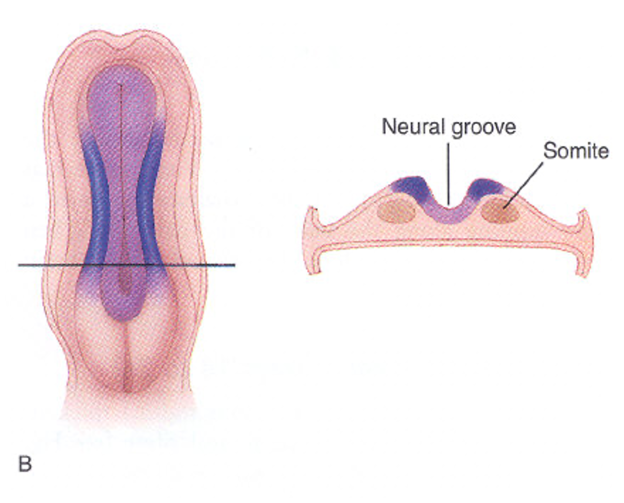
initially, the neural tube is __
open at both ends like a cannoli so amniotic fluid can go in and out
eventually it will close at both ends
the somites (bumps) that are lined up on either side
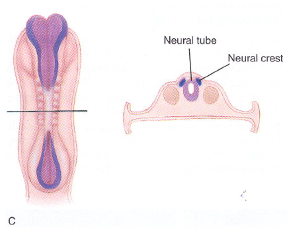
somites in the neural tube
lined up on either side
turn into the part of us that isn’t neural
they can turn into dermatomes/myotomes to help with movement and sensation
why is the neural tube initially open at both ends
so amniotic fluid can go in and out
neuropores
the temporary openings at the top and bottom of the neural tube
should close around end of week 4
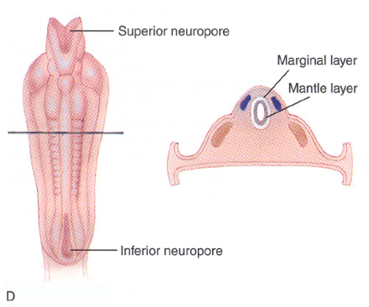
superior vs inferior neuropore
superior affects brain (higher) - anencephaly
inferior affects spinal cord (lower) - spina bifida
layers of inside of neural tube
marginal layer (outside) - becomes white matter
mantle layer (inside) - becomes gray matter of CNS
eventually flips, as white matter tends to be deep to gray
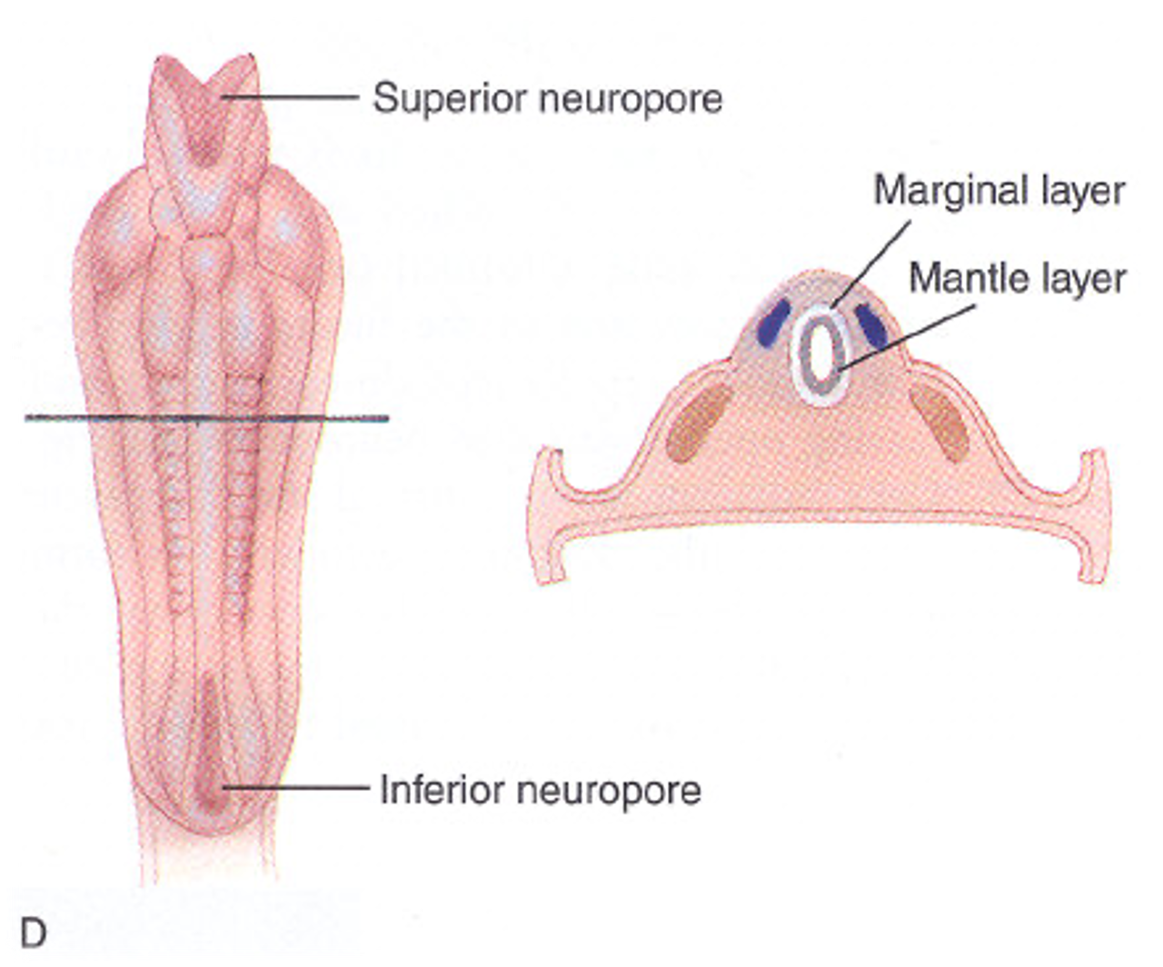
mantle layer
middle, thick cellular layer that develops into the gray matter of the CNS
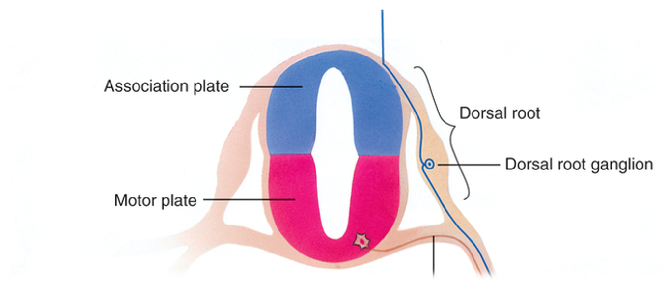
further differentiates into motor and association (sensory) plates
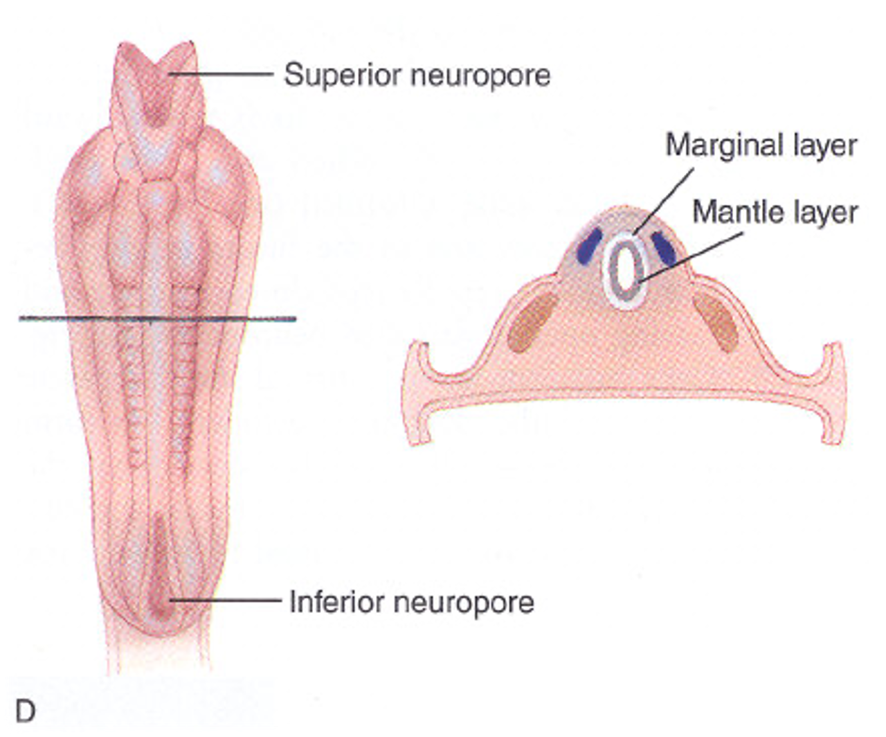
mantle layer splits to
(inner layer)
2 plates:
association plate
houses sensory cells (not al sensory cells are housed here, but theya re sensory in nature)
dorsal
motor plate
ventral
somites differentiate to
3 different parts:
dermatome - part of somite that becomes the skin
myotome - part of somite that becomes muscle
sclerotome - part of somite that becomes mucle
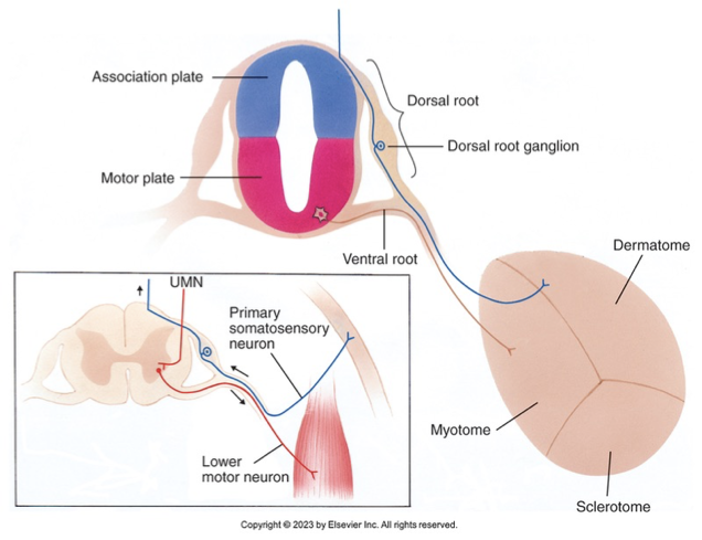
developing vs mature somite parts
developing (in utero) dermatomes, myotomes, and sclerotomes are the skin, muscle, and bone.
mature ““ have to do with innervation
somite vs neural tube
each somite is a neighbor to a SEGMENT of the neural tube
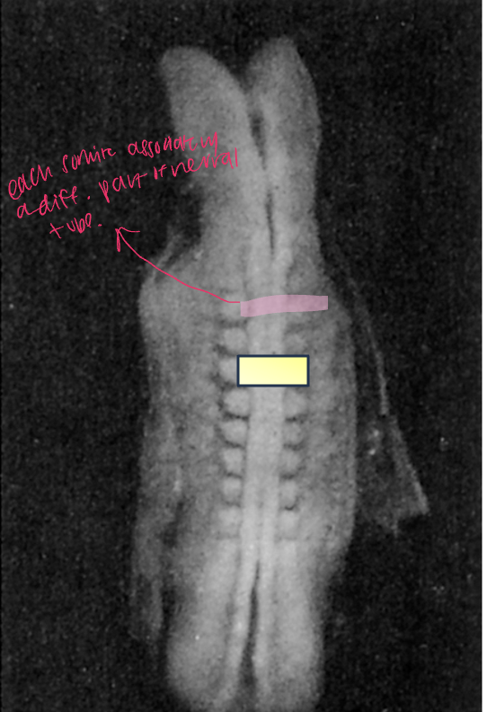
somite segments
each level has a somite on each side
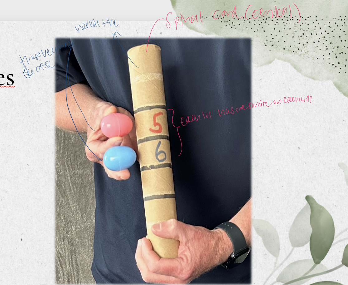
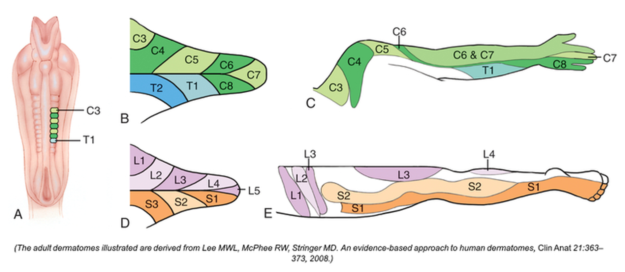
dermatomes in development
a part of the somite that becomes the skin
dermatomes once developed
refers to a part of the skin that is innervated by a singular spinal nerve
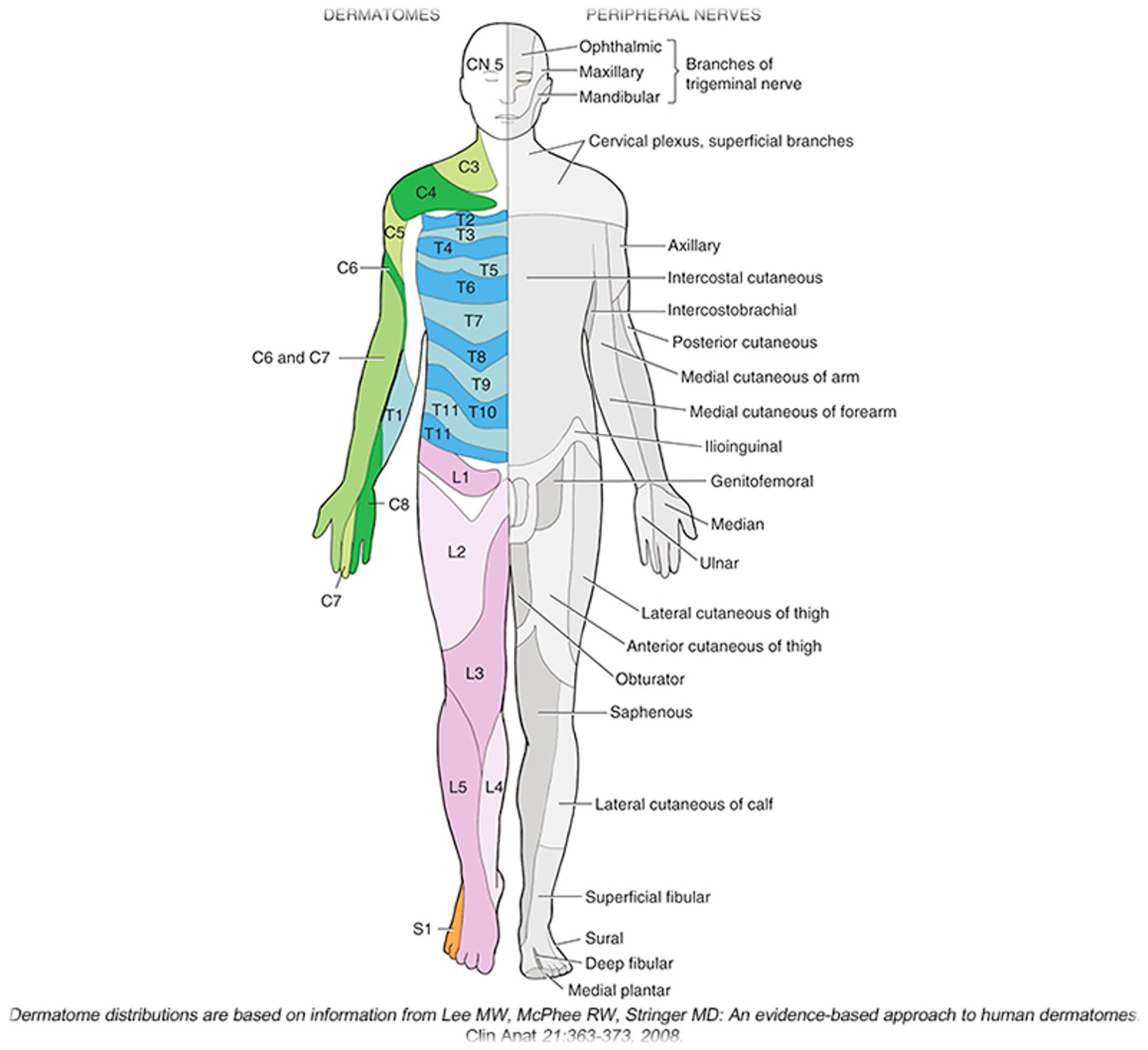
neural tube & dermatomes
arms and legs but out of neural tube
start in a stack, but as it turns into fetus, the dermatomes pull each other out (why arms and legs have uneven/warped dermatomes
myotomes in development
part of the somite that becomes muscle
myotomes once developed
represents all muscles innervated by a singular spinal nerve
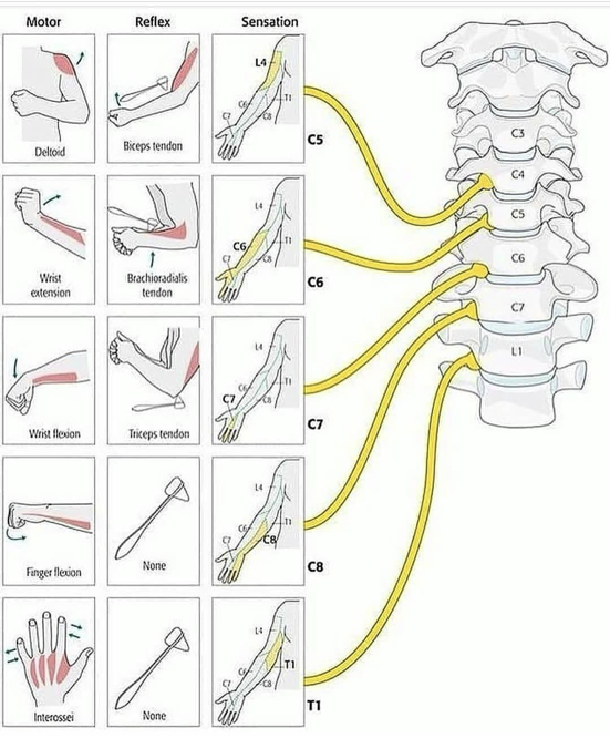
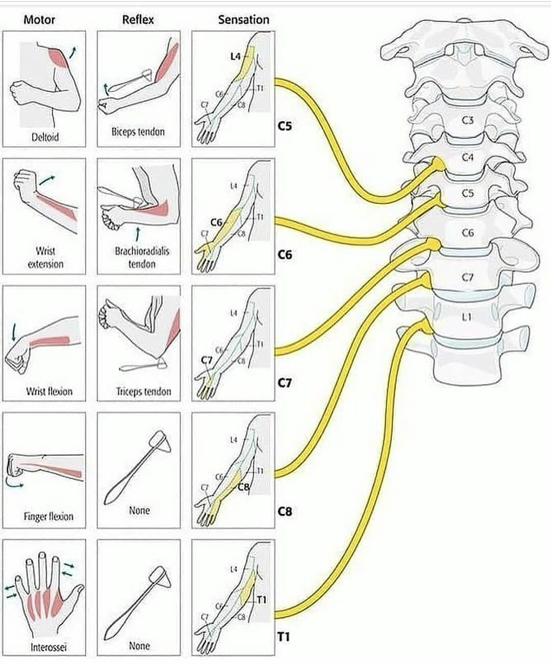
spinal nerves vs muscle innervation
a single spinal nerve can innervate multiple muscles
multiple spinal nerves can innervate the same muscle
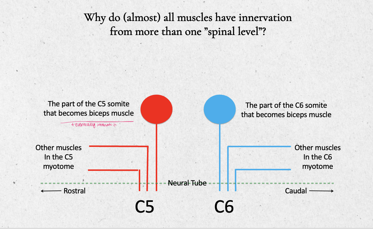
why do almost all muscles have innervation from more than one “spinal level”
ex. C5 and C6 somites both become the biceps muscle (and eventually innervates it)
C5 and C6 still become other muscles in the myotome as well
the “bicep” cells from C5 and C6 somites start migrating away from the neural tube toward their final destination on the anterior humerus
if one of the spinal nerves for biceps is completely cut, then the biceps has only lost part of its connection to the neural tube (nervous system) - it will be weak
spinal vs peripheral nerves
spinal nerves live close to the spinal cord
as they move away they come together to form a peripheral nerve
if the peripheral nerve is cut/gone, then the muscle WON’T work because the peripheral nerve is both, therefore there is no innervation to that muscle
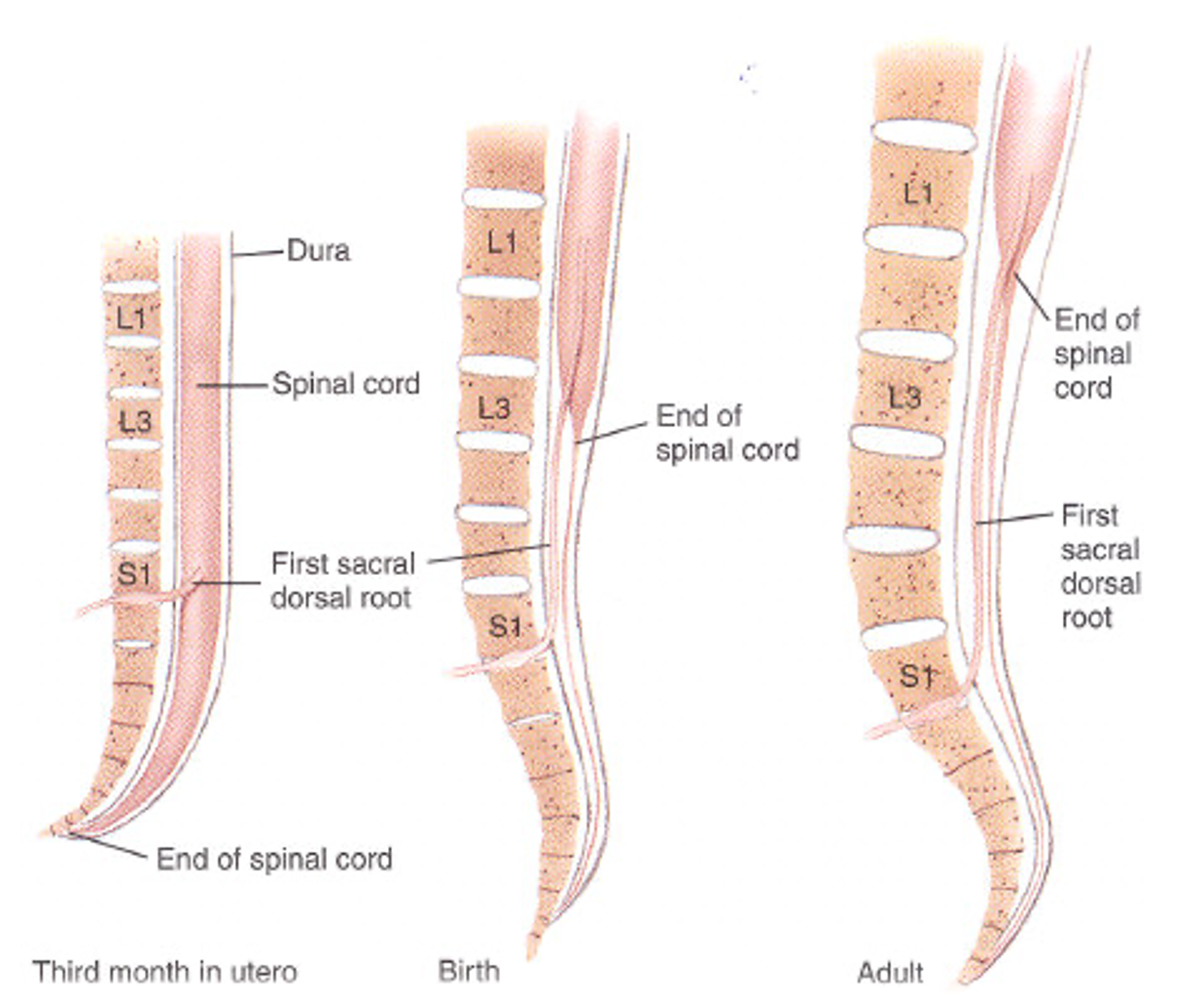
spinal cord in 3rd month of utero
spinal cord lines up with end of vertebrae
as we get older, the spinal cord ends around L1 but the bone continues to grrow
leaving a part of the spinal cord you can draw CSF fluid from (spinal tap/lumbar puncture)
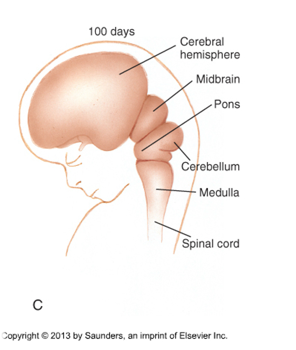
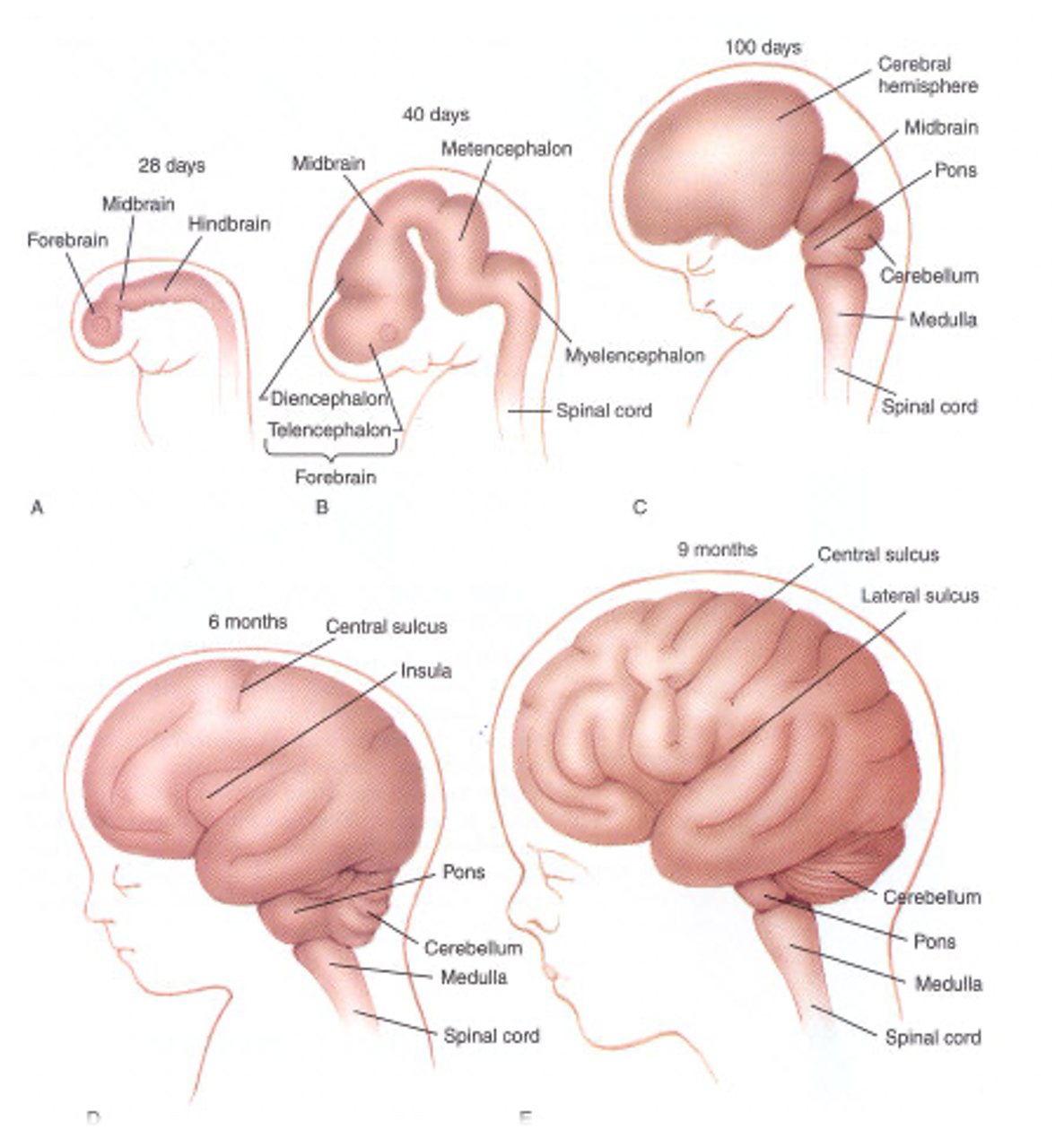
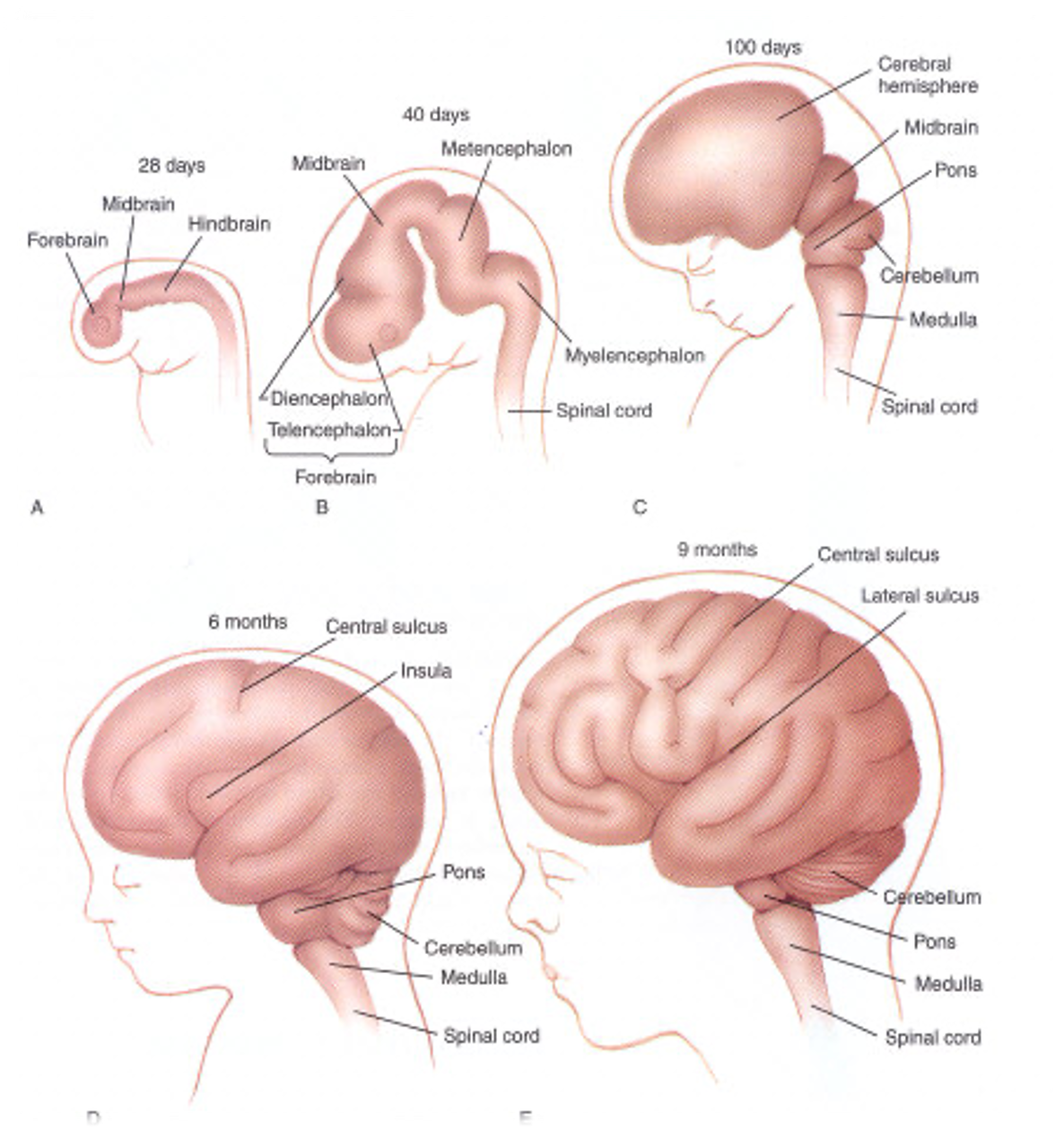
the brain starts as
a simple forebrain, midbrain, and hindbrain
then those develop into parts of the brain that we know
hindbrain becomes
pons
medulla
cerebellum
4th ventricle
PONS =
coffee machine - sends messages
belly
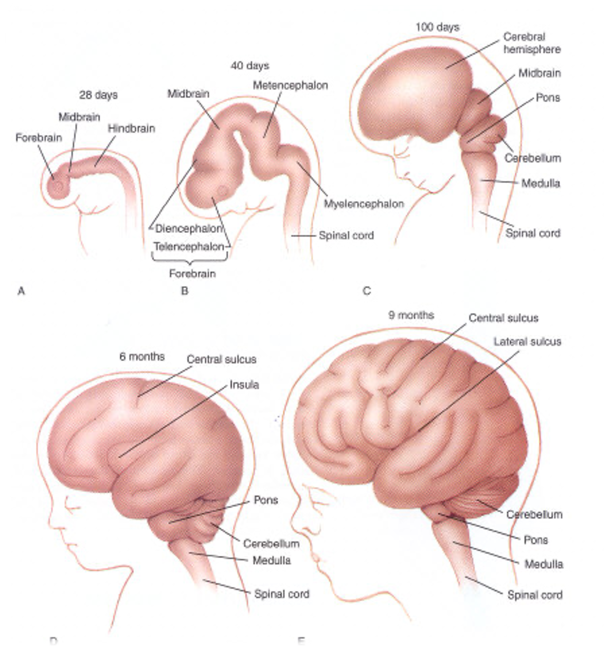
forebrain becomes
diencephalon (thalamus, hypothalamus, 3rd ventricle)
telencephalon (cerebral hemispheres, basal ganglia, cerebral cortex, and lateral ventricles)
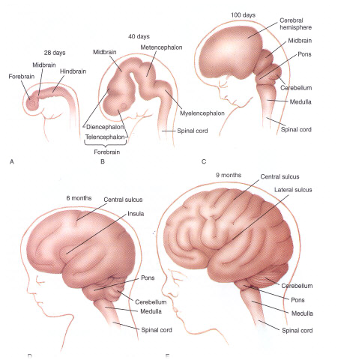
telencephalon
cerebral hemispheres
basal ganglia
cerebral cortex
lateral ventricles
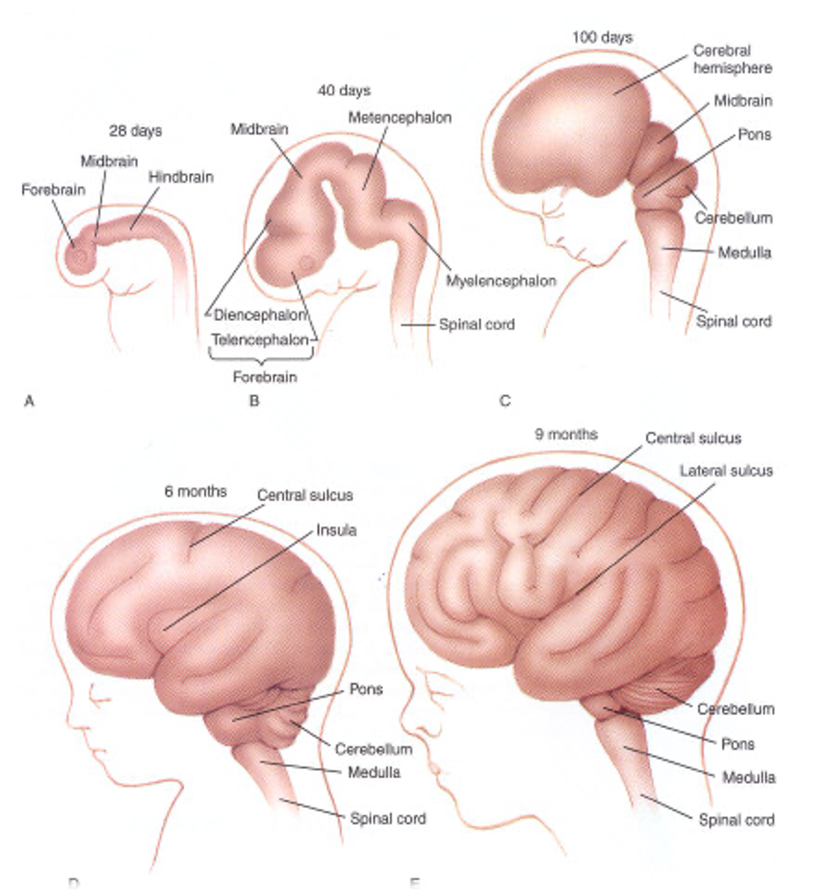
midbrain
brainstem
stays the same
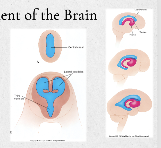
ventricles of the brain develop from
the central cavity of the embryonic neural tube
these later form the choroid process to crease CSF
this cavity expands to form the ventricles, eventually lining them with chorid plexi to produce CSF
spina bifida
incomplete closure of the posterior (caudal/inferior ) neuropore
can happen anywhere on the spine
ranges from mild to severe(spina bifida occulta, meningocele, or myelomeningocele)
when the neural tube does not close all of the way, the backbone is uable to fully develop to protect the spinal cord
this can cause damage to the spinal cord and nerves
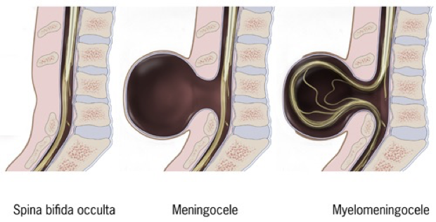
spina bifida myelomeningocele
a neural tube defect where the backbone and spinal canal fail to close before birth, causing a fluid-filled sac containing parts of the spinal cord and nerves to protrude through the baby's back
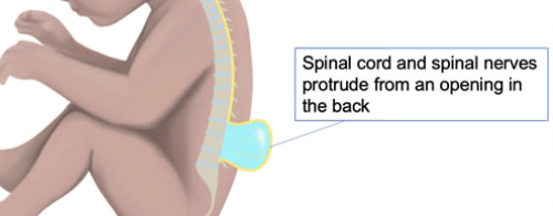
3 types of spina bifida
occulta: small gap in spine - spinal cord and nerves remain in normal place and not exposed
meningocele: rarest type -
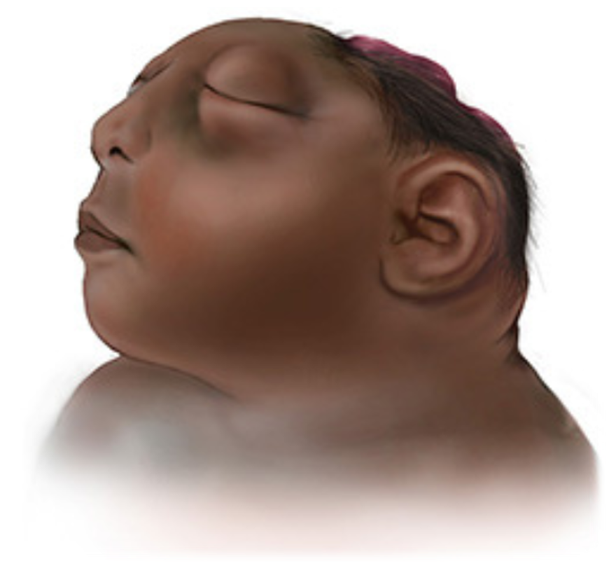
anencephaly
incomplete closure of the superior (rostral) neuropore during development
results in lack of skull and parts of brain
unfortunately, there is no cure, fatal
cerebral palsy
permanent, non-progressive damage to developing brain
in utero or very shortly after birth
similar to stroke
can affect movement, posture, and coordination
brain injury doesn’t worsen over time