Lecture 18 Digestive System
1/20
There's no tags or description
Looks like no tags are added yet.
Name | Mastery | Learn | Test | Matching | Spaced | Call with Kai |
|---|
No analytics yet
Send a link to your students to track their progress
21 Terms
peritoneum
serous membrane surrounding (some of) the abdominal viscera
two types:
parietal peritoneum is the outer layer. inside of the whole abdominal wall
visceral peritoneum: the inner layer. directly wraps around and covers some of the abdominal organs
mesentery: directly connects the parietal peritoneum to the visceral peritoneum
intraperitoneal organs
surrounded by visceral peritoneum
suspended by a layer of mesentery
mobile
retroperitoneal
BEHIND; pinned to the posterior abdominal wall by the parietal peritoneum
not mobile
these organs only touch the parietal peritoneum, do NOT touch visceral
the organs are behind (posterior to) the parietal peritoneum
Retroperitoneal organs
SAD PUCKER
Suprarenal glands
Aorta (and Inferior Vena Cava)
Duodenum
Pancreas
Ureters
Colon (ascending and descending only)
Kidneys
Esophagus
R*ctum
mesentery
double-layered ligaments of Peritoneum
Greater Omentum
greater curvature, apron of fat
Lesser Omentum
lesser curvature
Mesentery (capital M)
small intestines
Transverse mesocolon
transverse colon
Sigmoid mesocolon
sigmoid colon
alimentary canal
other name for GI tract
the continuous, muscular tube that runs from the mouth to the anus
what are the layers of the alimentary canal (superficial to deep)?
visceral peritoneum
serosa
muscularis externa
submucosa
mucosa (epithelium and lamina propria)
lamina propria
deep to epithelium of the mucosa
filling
stomach histological identifiers
gastric pits (not that deep)
lots of cells inferior to the gastric pits
duodenum
first section of small intestine
directly distal to stomach (right after)
where Bile and Pancreatic enzymes enter
retroperitoneal (in saD pucker) so stuck behind the parietal peritoneum
ileum
most distal part of small intestine
proximal to Large Intestine (cecum)
jejunum and ileum are intraperitoneal
suspended by Mesentery
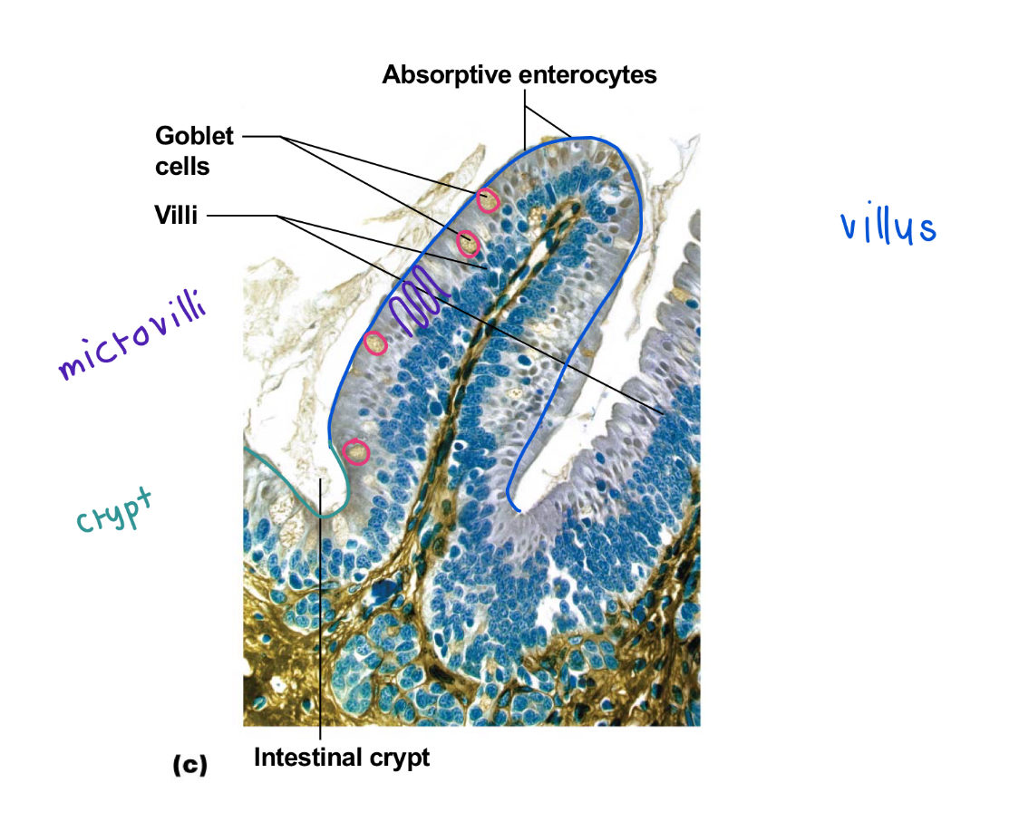
small intestine histology identifiers
villi
made of columnar epithelium, have microvilli
crypts (holes)
few goblet cells show up fully WHITE
scattered around
large intestine histology identifiers
a LOT of goblet cells, white dots
liver
2 surface lobes, big right and small left
separated by the falciform ligament
ligament hanging off is ligamentum teres
4 anatomical lobes
right lobe
left lobe
caudate lobe
quadrate lobe
posterior liver
inferior vena cava has the left and right hepatic veins draining into it
caudate and quadrate lobes
caudate is on top (C is first in alphabet)
microscopic anatomy of liver
hepatocytes: functional cells of the Liver
unit
hepatocyte units make up:
Lobule: functional unit of liver, formed by many Hepatocytes
central vein —> hepatic veins —> IVC —> right atrium
what is the function of hepatocytes
detoxify the blood and make bile
liver histology identifiers
HEXAGONS
pancreas
sits in the C of the duodenum
head, body, and tail
makes digestive enzymes (exocrine) and hormones (endocrine)
pancreas histology
acini (with little pizza cuts)
Islet of Langerhans
lighter colored clump surrounded by acini
no space around the islet of langerhans
duct lumens of the acini are very small (compared to Nephrons)
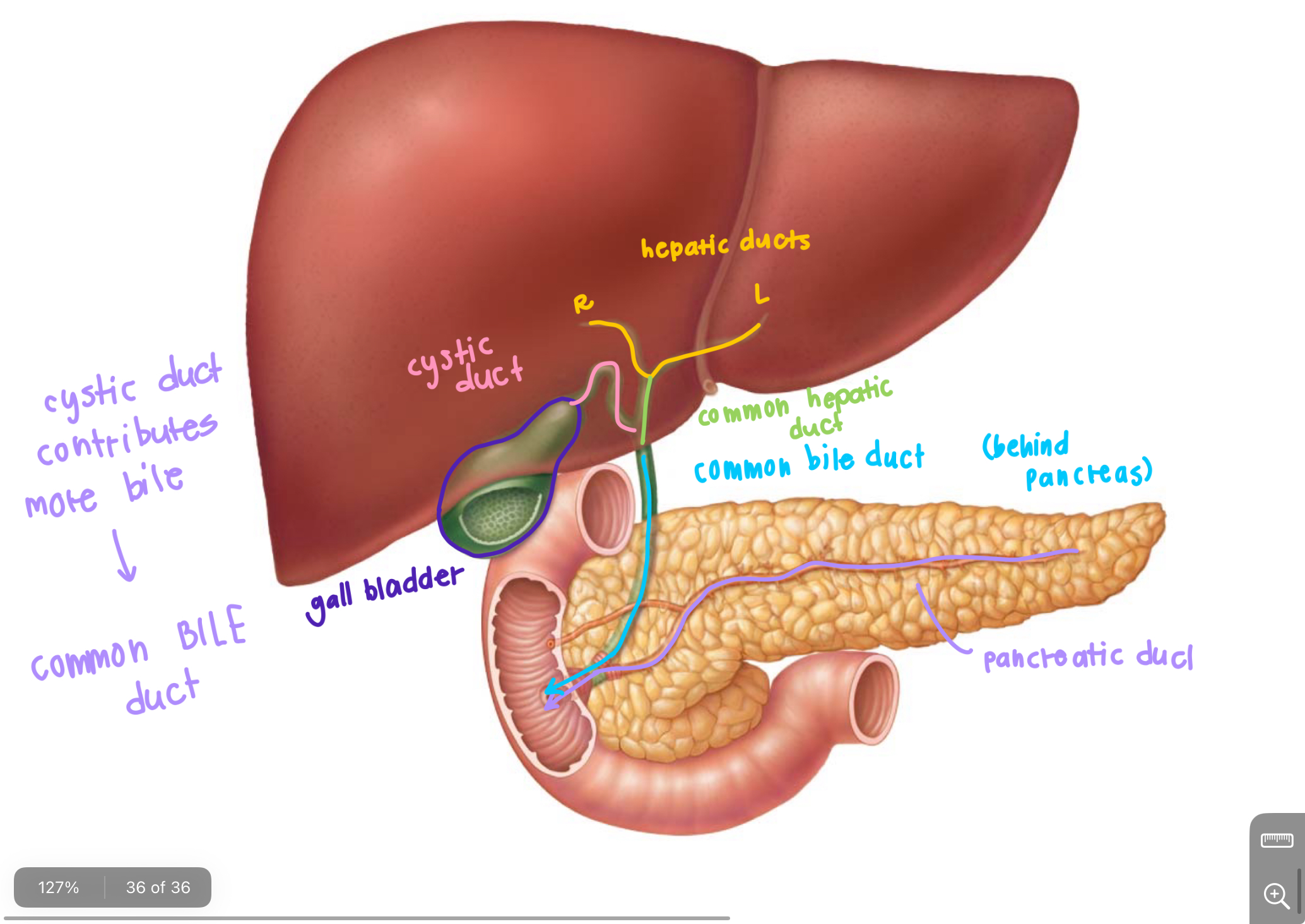
Duct System
carries bile and pancreatic enzymes
left and right hepatic ducts —> Form common hepatic duct
add on cystic duct from gall bladder → becomes common bile duct
pancreatic duct joins to drain into the duodenum together