Cytology
1/49
There's no tags or description
Looks like no tags are added yet.
Name | Mastery | Learn | Test | Matching | Spaced | Call with Kai |
|---|
No analytics yet
Send a link to your students to track their progress
50 Terms
Light Microscope
uses visible light and lenses to magnify images of samples that produces simple images with the inability to discern veryTra small details in tissue and cell morphology
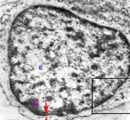
What kind of electron microscope produces this image?
Transmission Electron Microscope (TEM)
What kind of microscope produces this image?
Scanning Electron Microscope (SEM)
What can cause variations in the results of the staining process? (3)
user error
pH differences
poor fixation
Hematoxylin and Eosin (H&E Stain)
Hematoxylin stains acidic tissue components (RNA/DNA) purple, and eosin stains basic tissue components pink-orange
Basophillic
acidic tissues that are stained purple due to attracting the basic stain
Acidophillic
basic tissues that are stained pink-orange due to attracting the acidic stain
Silver Stain
uses silver salt to make reticular fibers (basement membrane) turn black
Masson’s Trichrome
three color staining protocol in which connective tissue is blue, nulcei and muscle are dark red/purple, and the cytoplasm is stained red/pink
Periodic Acid-Schiff Stain (PAS)
staining protocol that stains the basement membrane, glycogen, and carbohydrates magenta and the nuclei blue
Wright’s Stain
a common stain used for blood samples that is essentially a modified H&E
What is the most common staining technique used in veterinary medicine?
H&E
Phases of Mitosis (5)
Interphase
Prophase
Metaphase
Anaphase
Telophase
Interphase
genetic material inside the nucleus is dispersed equally to facilitate DNA transciption
Prophase
centrosomes duplicate and begin moving toward opposite ends
“ball of yarn nucleus”
Metaphase
chromosomes are at the middle of the cell and microtubules pull the centromeres to separate the sister chromatids
Anaphase
Sister chromatids are dragged toward the poles (hand like projections)
Telophase
nuclear envelope starts to reform between the two new cells
Nucleolus
site of RNA transcription that assembles around clusters of rRNA gene repeats
How does the golgi apparatus appear under the microscope?
clear cytoplasm as it has a neutral pH
ONLY visible in cells releasing a lot of material extracellularly
Cillia
coordinate movements to move material along the surface of the cell
Microvilli
NEVER mobile; used to increase surface area of the cell surface for absorption
Cell Inclusions
nonliving components of the cell that do not possess essential metabolic activity
What are the most common inclusions?
Glycogen, lipid droplets, crystals, and pigments
What is the moston biological pigment? comm
Melanin
Hemosiderian
Staining Artifact
staining variations in tissue due to handling; edges may be lighter than the center
Parenchyma
living, functional portion of any tissue or organ
Stroma
non-living, supportive portion of any tissue or organ
Folding Artifact
tissue has folded over onto itself
Tearing Artifact
tissue was torn during process
Lobule
grouping of parenchyma in some organs and tissue
Septa
stroma that separate lobules
When do shrinkage artifacts occur?
when water is removed from the tissue it can cause an artificial space to appear that is NOT present in the living animal
Basal Side
side of a cell that is the closest to the basement membrane (aka Basal Lamina)
Apical Side
side of the cell farthest away from the basement membrane or closest to the lumen or surface
Acini / Acinus
describes a cellular arrangement in a raspberry formation
Closed-Faced Nucleus
little variation between euchromatin (light) and heterochromatin (dark) and means the cell is metabolically inactive
Open-Faced Nucleus
variation between euchromatin (light) and heterochromatin (dark) and means the cell is metabolically active
Zymogen Granules
eosinophilically stained granules considered cell inclusions
True or False: all neuron nuclei are open-faced.
TRUE
What color does the Golgi apparatus stain?
neutral
Cilia
Specializations of the cell surface that can help move material along the surface of the cell
Microvilli
Specialization of the cell surface that increase the surface area of the cell for absorption
Cell Inclusion
Nonliving components of the cell that do not possess essential metabolic activity - OR - anything inside a cell that the cell does not need to live
Glycogen
Cell inclusion that is used as a storage material when glucose supplies are high; common in Liver and Skeletal muscle (PAS stain = magenta)
Lipids
Cell inclusion that is stored as droplets for energy source; appears clear as they are lost during processing
Melanin
Most common biological pigment that protects cells from UV radiation
Hemosiderin
Cell inclusion in macrophages that is the residue of blood cell destruction
Lipofuscin
Celll inclusion that appears yellow-brown granules that are the sign of incomplete lysosomal digestion; aging cells