Lecture 2 -- Nematodes of Sheep
1/30
There's no tags or description
Looks like no tags are added yet.
Name | Mastery | Learn | Test | Matching | Spaced | Call with Kai |
|---|
No analytics yet
Send a link to your students to track their progress
31 Terms
Which endoparasites are found in the abomasum of ruminants?
Teladorsagia circumcincta
Trichostrongylus axei
Haemonchus contortus
Which endoparasites are found in the small intestine of ruminants?
Bunostomum spp.
Nematodirus spp.
Cooperia spp.
Trichostrongylus spp.
Which endoparasites are found in the large intestine of ruminants?
Oesophagostomum spp.
Chabertia ovina
Trichuris ovis
Which endoparasites have a similar trichostrongyle-type life cycle?
Trichostrongylus spp
Teladorsagia circumcincta
Cooperia spp
Describe the life cycle of typical trichostrongyle life cycle.
Step 1: Adult male and female parasites live in the GI tract of the sheep (abomasum or small intestine) and mate.
Step 2: Only the fertilised female produces and lays eggs → Eggs pass through intestinal lumen and are excreted in faeces
Step 3: Eggs develop in faecal pat → L1 hatches 孵出 and feeds on bacteria in faecal pat → Grows to L2 → Moults to L3, which is ensheathed
Step 4: L3 released from the faecal pat by rain splash (L3 is the infective stage, found on grass)
Step 5: When the sheep is grazing the pasture, it picks up the L3 larvae and ingests the infected larvae
Step 6: Depending on what species that parasite is, it either goes to the abomasum or small intestine → It will most to L4 inside the host and matures as L5
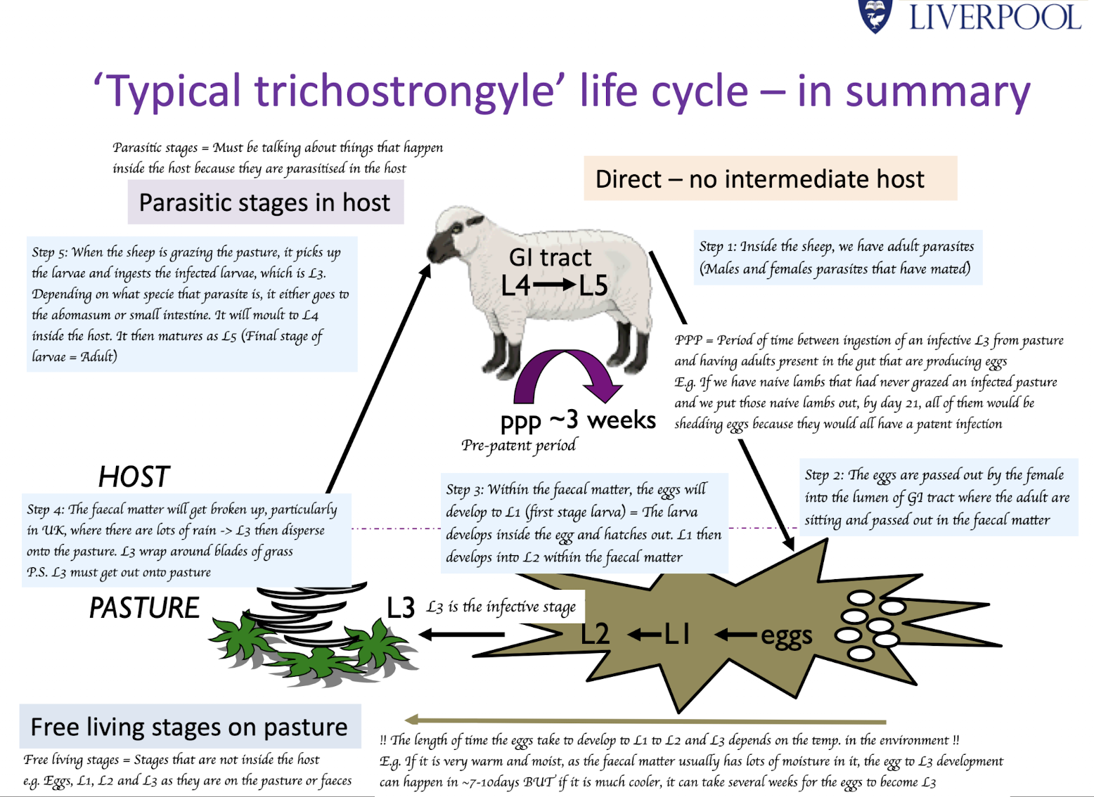
What is the pre-patent period (PPP)?
Period of time from ingestion of infective L3 from the pasture to the detection of eggs in the faeces
E.g. If we have naive lambs that had never grazed an infected pasture and we put those naive lambs out, by day 21, all of them would be shedding eggs because they would all have a patent infection
What is a patent infection?
An infection where adult parasites are producing eggs and one be detected (e.g. by faecal egg count)
How long is the pre-patent period (PPP) for PGE nematodes?
~ 3 weeks
What is hypobiosis?
Arrested development of early L4 larvae within the host
Those infective L3 larvae are ingested and penetrate the abomasal mucosa, where they develop into early L4 larvae
BUT they then pause their development and remain dormant for several month
When spring arrives and the temp. increases, the arrested larvae resume development, mature into adult females and males, and mate
What triggers hypobiosis?
Decreasing ambient temperature in autumn
P.S. If the winter is not cold enough, hypobiosis might not occur
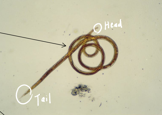
Which parasites are shown in this picture?
Teladorsagia circumcinta
What are the key morphological features of adult Teladorsagia circumcincta?
1cm in length
Both sex:
Fine cervical papillae at the head
Males:
Symmetrical bursa and spicules at the tail end
Spicules can retract/ extend through bursa
Describe the life cycle of Teladorsagia circumcincta in the sheep abomasum.
Step 1: Ingestion of L3
Sheep eat infective L3 larvae while grazing.
Step 2: Exsheathment and penetration
L3 Swallowed and reaches the abomasum → After L3 is swallowed, L3 lose their protective sheath and penetrate the abomasal mucosa and burrows into gastric glands
Step 3: Larval development:
Within the gastric glands, L3 moult to L4 and then L5.
Step 4: Emergence
L5 (immature adults) emerge from the gastric glands into the abomasal lumen
Step 5: Maturation
L5 develop into adults, mate, and fertilised females lay eggs in the lumen, which are passed out in the faeces.
How many Teladorsagia circumcincta larvae are needed to cause parasitic gastroenteritis in sheep?
At least 40,000 larvae.
How do Teladorsagia circumcincta larvae cause disease in sheep?
L4 and L5 develop in gastric glands → Damage glands → Parietal cells replaced by undifferentiated epithelial cells → Consequences:
↓ Acid production = ↑ Abomasal pH → Loss of bacteriostatic effect
No conversion of pepsinogen to pepsin
↑ Mucosal permeability = Lose the tight junction between cell → “leaky” abomasum → Diarrhoea
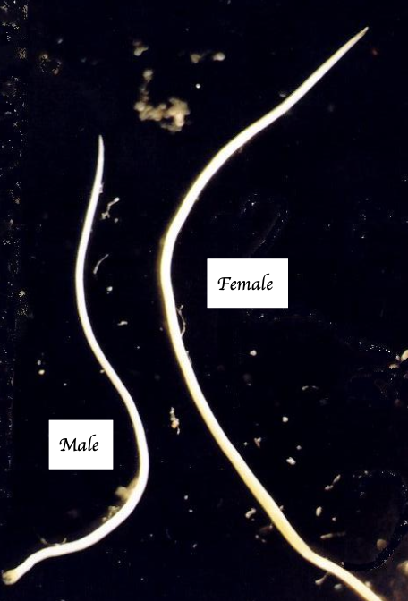
What are the key morphological features of adult Trichostrongylus?
Very small, hair like worms (4-7mm)
Smallest gastrointestinal nematodes in sheep
Excretory pore at head end of the worm
Female:
NO vulval flap
<12 eggs uterus can be seen in the uterus
Male:
Bursa present
Short, unequal and dissimilar spicules
How do Trichostrongylus spp. larvae cause disease?
L4 and L5 develop deep in sub-mucosa and mucosa → Form sub-epithelial tunnels → Cause villous atrophy, haemorrhage, oedema because of protein loss, and diarrhoea (black scour)
What are the clinical signs of Trichostrongylus infection in lambs?
Black scour
Weight loss/ poor weight gain
Poor skeletal growth
When does Trichostrongylus disease typically occur?
Autumn
Which animals are commonly infected by Cooperia spp.?
Cattle and sheep
What are the key morphological features of adult Cooperia spp.?
Presence of cephalic vesicle = Cuticular inflation on the head end
Striation on the head
Body coiled in the middle of its body = “Watch-spring”appearance
Male:
Short, stumpy spicules
How pathogenic is Cooperia spp.?
Usually mild, but heavy infections can cause disease.
What are the pathological effects of heavy Cooperia spp. infections?
Reduced weight gain
Catarrhal enteritis
Villous atrophy
Oedema of the intestinal mucosa
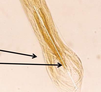
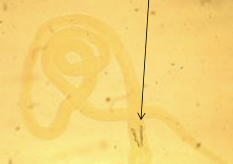
Which parasites are shown in this picture, and which morphological feature supports this conclusion?
Teladorsagia circumcincta (Male)
Symmetrical bursa and spicules
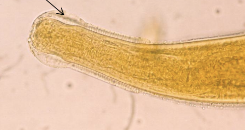
Which parasites are shown in this picture, and which morphological feature supports this conclusion?
Teladorsagia circumcincta (Both sex)
Cervical papillae at head end
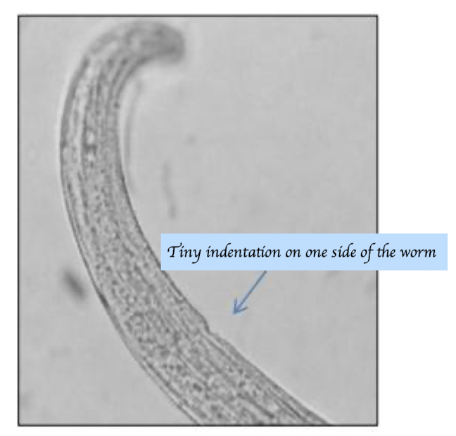
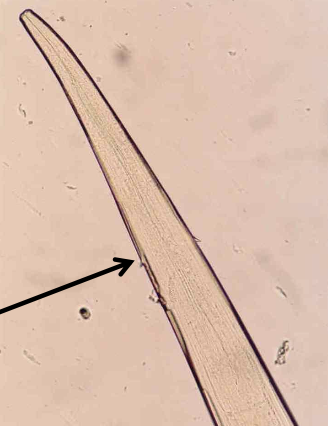
Which parasites are shown in this picture, and which morphological feature supports this conclusion?
Trichostronglyus spp.
Excretory notch
Which parasites are shown in this picture, and which morphological feature supports this conclusion?
Cooperia spp.
Presence of cephalic vesicle and cervical vesicle
Cervical vesicle (Inflated but not bubble like)
Striation on the head
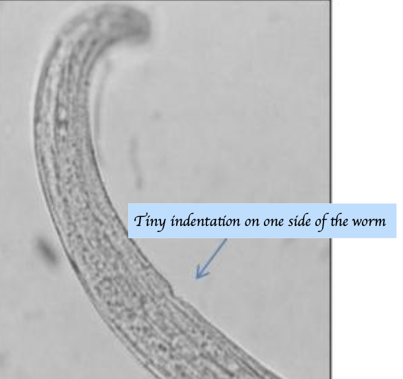
Which parasites are shown in this picture, and which morphological feature supports this conclusion?
Cooperia spp.
Coiled body (= “Watch spring” worm)
Which parasites are shown in this picture, and which morphological feature supports this conclusion?
Coopria spp. (Male)
Short, stumpy spicules
What are the differences between type 1 and type 2 PGE?
Type 1 PGE:
First season grazing lambs = Lambs that have never been grazed
Happen in the mid summer
Type 2 PGE:
Caused by hypobiosed larvae → Simultaneous emergence of L5
Happen in late winter/ early spring because of the increased temp
What are the clinical sings of PGE (Both type 1 and type 2)?
Profuse watery diarrhoea
Weight loss
Inappetence
Dehydration
Poor weight gain
Reduced appetite
Reduced feed intake
Loss of plasma proteins into gastro-intestinal tract
Death