Urinary System: Last Study
1/34
There's no tags or description
Looks like no tags are added yet.
Name | Mastery | Learn | Test | Matching | Spaced | Call with Kai |
|---|
No analytics yet
Send a link to your students to track their progress
35 Terms
The Kidneys
Filtration of metabolic wastes and toxins
Hormone production
Homeostasis of body fluids
Receives 20-25% of resting cardiac output
What hormones are produced by kidneys?
Calicitriol: Works with PTH to increase plasma [Ca+]
Erythropoeitin: Increases RBC production
Renin (not a hormone) → First step to angiotensin II
Detoxification in detail
When you want to metabolize those amino acids to turn them into glucose or burn them off for ATP, we can deal with the carboxyl group, BUT we need to get rid of the amide group (NH2) because it forms ammonia.
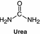
We convert ammonia to UREA
Nitrogenous wastes
Urea
amino acids → NH2 removed → forms ammonia → processed by liver and converted to urea
Uric Acid
nucleic acid catabolism
Creatinine
creatine phosphate catabolism
Renal failure
Azotemia: Increased nitrogenous wastes in blood (blood urea nitrogen)
Cortex
Medulla
Filtrate vs Urine
Filtrate can be modified— the ability to gain and lose ions and salts and water. Concentration can be increased or decreased. However as it leaves the renal papilla, draining into the minor calyx/renal pelvis, it becomes URINE.
Blood Flow into the Kidney (Entering cortical nephron)
Renal artery
Segmental artery
Lobar arteries that travel up the pyramid
Interlobar arteries bringing them up toward the cortex between lobes
Arcuate artery “archs” at the top of the pryamid
Cortical radiate arteries radiate from the arcuate
Radiate arteries branch off into the glomerulus → AFFERENT ENTERS the tuft EFFERENT leaves
Efferent arterioles then wrap around the peritubular capillaries (tubular network aka “collecting ducts”)
Cortical radiate veins, arcuate veins, interlobar veins, and finally the exit at the renal vein
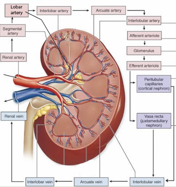
Collecting ducts
They form that peritubular network that the efferent arteriole wraps around
They drain into the renal papilla into the minor calyx
Nephron route
Begins at the renal corpuscle (glomerulus and capsule)
PCT
Loop of Henle → Goes into the medulla
DCT
Juxtamedullary vs Cortical
Cortical:
Make up 85% of our nephrons
next to the cortex
filter and modifies filtrate
Juxtamedullary:
Make up 15% of our nephrons
filter and modifies filtrate
creates a concentration gradient in medulla in order to CONCENTRATE our urine → makes the environment to make concentrated urine
Renal corpuscle
Bowman’s capsule + glomerulus
Where filtration occurs (where filtrate is separated into the PCT)
Proximal Convuluted Tubule (PCT)
Reabsorbing 2/3 of filtrate
Heavily packed with microvilli for absorption
Decending Loop of Henle
Allows for water to pass through it— nothing else
Ascending thin loop of Henle
Only allows salts to move through them— no water
Ascending thick loop of Henle
Distal Convuluted Tubule
Principal ducts
Collecting Ducts
Principal and intercalated cells
Vasa recta
protects the gradient that juxtamedullary nephrons create
Urine Formation Steps
1. Filtration
occurs in the renal corpuscle; forms filtrate
water, ions, small proteins, amino acids, hormones, ect.
2. Reabsorption
most occurs in the PCT, reclaiming all the good stuff
glucose, lipids, amino acids
3. Secretion
unwanted substances are added to filtrate; occurs throughout the nephron
H+, urea, creatinine, uric acid
Renal Corpuscle
Double layered
Parietal layer (outer):
Visceral layer (inner): podocytes around the squamous endothelial cells of the glomerulus → these can contract and close pores or relax and open pores
Juxtaglomerular apparatus (JGA)
the grey cells (mesengial) near vascular pole and orange cells on the walls of the afferent arteriole (where afferent or efferent arterioles come in)
mesengial cells are smooth muscle cells that relax to create more surface area and increase amount of filtration
if reducing filtration, we contract to reduce SA
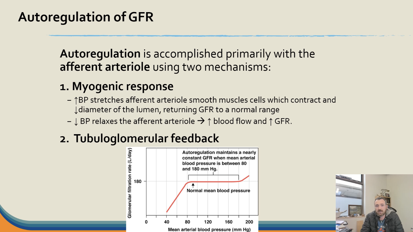
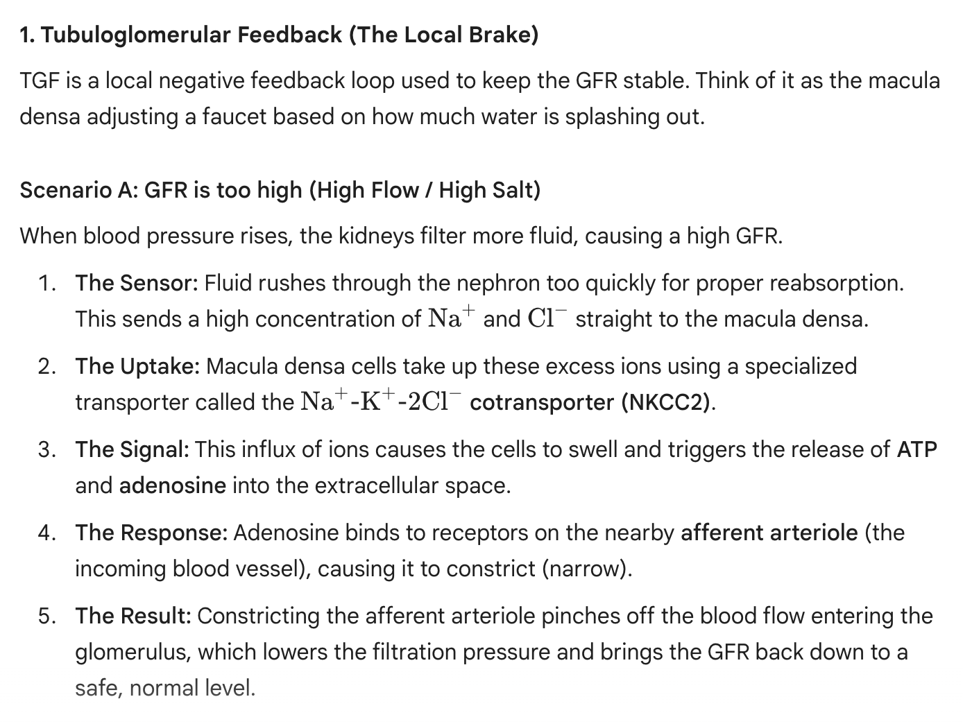
How is this regulated?
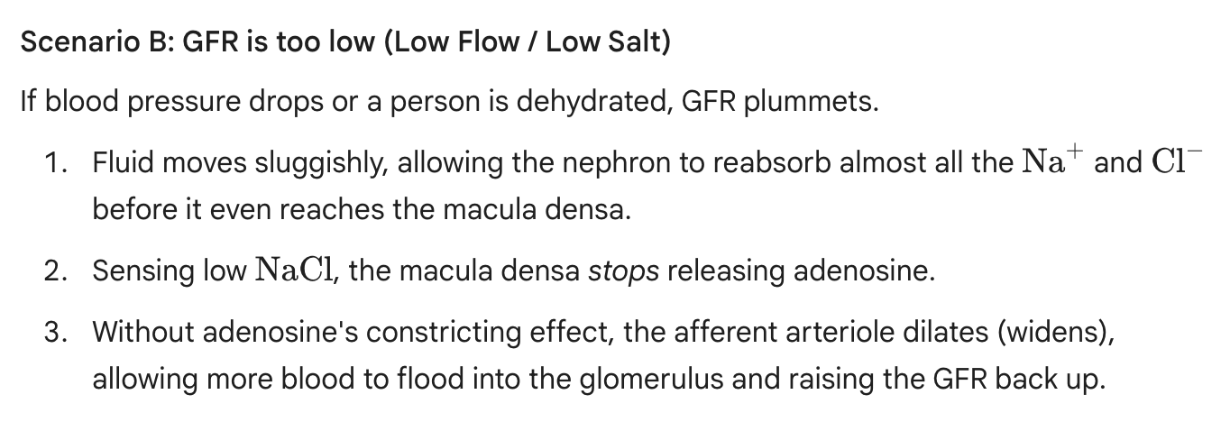
The macula densa cells: Located in the wall of the distal convoluted tubule (where it loops back and touches its own glomerulus), it monitors fluid flow and salt concentration.
Macula densa cells sees if things are moving too slow or too fast— it will them take steps to modify the glomerular filtration rate (GFR) by telling these cells to constrict or relax
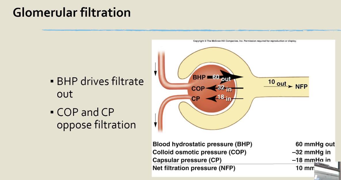
Glomerular filtration
driven by hydrostatic pressure inside the glomerular capillaries
approximately 20% of plasma becomes filtrate
180L/day filtered, 0.5-2L is excreted as urine
3 layers:
Innermost → Fenestration of endothelial cell to prevent cells and platelets
Middle → Basal lamina of glomerulus to prevent large proteins
Outer (closest to podocyte) → medium proteins get blocked
small inorganic and organics molecules can pass
Glomerular filtration rate (GFR)
Volume of filtrate produced by both kidneys per minute
eg. 125mL/min (filtrate) x 60 mins