lec 11 - axis formation, neural induction, and neural tube patterning
1/48
There's no tags or description
Looks like no tags are added yet.
Name | Mastery | Learn | Test | Matching | Spaced | Call with Kai |
|---|
No analytics yet
Send a link to your students to track their progress
49 Terms
the spemann-mangold organizer is ____ located at ____
a group of mesendodermal progenitor cells
dorsal blastopore lip
during gastrulation, the spemann-mangold organizer migrates
anteriorly
the spemann-mangold organizer gives rise to three distinct tissues
anterior endoderm
prechordal mesoderm
notochord
anterior endoderm contributes to ___ and is from the ____ organizer
head structures
early
prechordal mesoderm contributes to ____ and is from the ____ organizer
anterior structures
early
notochord contributes to _____ and is from the _____ organizer
patterning the overlying spinal cord
mid/late
who discovered the spemann-mangold organizer
hilde mangold and hans spemann
how did mangold and spemann discover the organizer
took cells from the dorsal blastopore lip of one xenopus embryo
transplant to ventral side of second embryo
second complete body axis forms, including second brain and spinal cord
those dorsal cells only formed a bit of the notochord, a transietnt structure
conclusions of the mangold-spemann experiments
the dorsal lip cells organize the patterning of the rest of the embryo - ‘organizer’
the organizer does its job via cell-cell signaling
organizer grafts - early vs late phenotypes + why
early: full split, complete second head and trunk
late: partial split, only second posterior parts
why?
organizer induces anterior structures first as it migrates
signaling molecules that organizer produces change over time
the organizer moves ______ly during gastrulation, _____ as it moves
anteriorly
patterning the AP axis
as the organizer migrates
it induces overlying ectoderm to form the neural plate
organizers inducing properties are achieved by secreting molecules that
inhibit Wnt and BMP signaling pathway activities
early organizer - BMP, Wnt, or both are blocked (and by what)
both are blocked
cerberus is most important - blocks both Wnt and BMP but BMP most
mid organizer - BMP, Wnt, or both are blocked (and by what)
Wnt a little, BMP a lot
cerberus is gone - BMP is blocked by BMP inhibitors chordin and noggin
mid organizer - BMP, Wnt, or both are blocked (and by what)
BMP is heavily blocked - added follistatin into the mix
Wnt is free
t/f - BMP expression is blocked everywhere
false - not blocked ventrally
what is the double gradient model
the organizer patterns the blastula through two perpendicular morphogen gradients
AP gradient of Wnt activity, from low Wnt anteriorly to high Wnt posteriorly
DV gradient of BMP activity, from low BMP dorsally to high BMP ventrally
each cell’s position can be read off a map like a cartesian plane
principle BMP is ____ which is expressed ____
BMP4
throughout early gastrula
principle Wnt is ____ which is expressed _____
Wnt8
marginal zone/wheere mesoderm is induced
what is the default state of ectoderm
neural tissue
_____ promotes brain formation in the anterior neural plate
______ promotes spinal cord formation
dual inhibition of Wnt and BMP signaling
BMP inhibition without Wnt inhibition
how is neural fate induced
epidermal fate is suppressed → neural fate induced in ectoderm
BMP antagonists chordin, noggin, and cerberus expressed in the organizer
diffuse from organizer and act on overlying ectoderm to suppress epidermal fate
neural induction
making neural plate from ectoderm
head induction requires inhibition of
Wnt and BMP
cerberus unique characteristics
blocks BMP and Wnt signaling
cerberus experiement
hypothesis: cereberus inhibits Wnt8 and BP4 → anteriorization and dorsalization of neural plate → head induction
ectopic cerberus mRNA injection → induced a second head
cerberus experiment conclusions
ectopic cerberus mRNA injection is sufficient for head induction
induction vs intrinsic differentiation
induction: extrinsic, a cell produces a signal that acts on another cell to change its fate
intrinsic: cells divide asymmetrically based on the specific cellular components within each cell
who proposed the activation-transformation model
hans spemann
the activation-transformation model explains
how the neural plate/tube acquires detailed AP patterning
explain the activation/transformation model
activation
early organizer secretes Wnt and BMP antagonists
neuroectoderm adopts anterior forebrain fate by default
transformation
mid → late organizer - Wnt secretion is attenuated (slowly increases)
posterior neural plate is exposed to multiple gradients of posteriorizing factors (Wnts, RA, FGFs)
forebrain fate is suppressed; over time, increasingly posteriorized neural fates are induced
neural plate elongates and transitions into the neural tube
what are the posteriorizing factors
Wnts
Retinoic Acid (RA)
FGFs
what is retinoic acid (RA)
diffusable small molecule
defivative of vitamin A
RA acts as a ligand for ____ which activate ____ in gene promoters
retinoic acid receptors
TFs that bind to Retinoic Acid Response Elements (RAREs)
FGFs activate ___ via ___
ETS TFs in the nucleus
cascade of serine-threonine kinases
activation-transformation involves
simultaneous integration of multiple signaling pathways (Wnt, RA, FGFs)
net result of the activation-transformation model
two opposing gradients
anteriorizing factors - BMP and Wnt antagonists (cerberus, chordin, noggin, dickkopf)
promote forebrain fate
posteriorizing factors - Wnts, RA, FGFs
suppress forebrain fate
promote increasingly posterior cell fates
hox genes
encode homeodomain TFs that interpret positional information from signaling molecules
once broad AP gradients are set up, TF in the neural plate:
interpret the signal levels
activate specific Hox genes at different positions
Hox genes determine what structure forms where
RA and Hox gene expression
RA establishes a posterior → anterior gradient
induces specific patterns of Hox gene transcription along different positions in the axis
different combos of Hox gene expression in the neural tube specify distinct domains of the spinal cord
hox genes are expressed in _____ order. explain this order
chromosomal
3' genes expressed first → more anterior
5' genes expressed later → more posterior
describe the stages of neural tube formation
flat neural plate
formation of neural furrow - by folding of the neural plate along the midline
elevation of neural folds - forms the neural groove
neural folds bend inward
neural folds fuse - closed neural tube + detachment of epidermis
neural crest cells migrate away
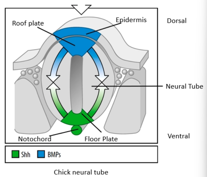
DV patterning of the neural tube - two opposing signaling systems
dorsal - BMPs secreted by the dorsal roof plate, induce dorsal neuronal fates
ventral - Shh secreted by the ventral Floor plate (and notochord), induces ventral neuronal fates
neural tube - BMPs are secreted by
dorsal Roof Plate
neural tube - Shh is secreted by
ventral Floor Plate + notochord
DV neural tube patterning - sequence
epidermal BMP signaling specifies roof plate; notochord Shh signaling specifies floor plate
BMPs from roof plate diffuse ventrally and pattern the dorsal half of the neural tube
Shh from floor plate diffuse dorsally and pattern the ventral half of the neural tube
in the dorsal neural tube, how are neuronal fates patterned
different members of the BMP family are expressed in different domains, and induce different dorsal neuronal fates
stepwise process
in the ventral neural tube, how are neuronal fates patterned
different concentrations of Shh induce different ventral neuronal fates