Kaplan Biology Chapter 2: Cell Cycle and Replication
1/144
There's no tags or description
Looks like no tags are added yet.
Name | Mastery | Learn | Test | Matching | Spaced | Call with Kai |
|---|
No analytics yet
Send a link to your students to track their progress
145 Terms
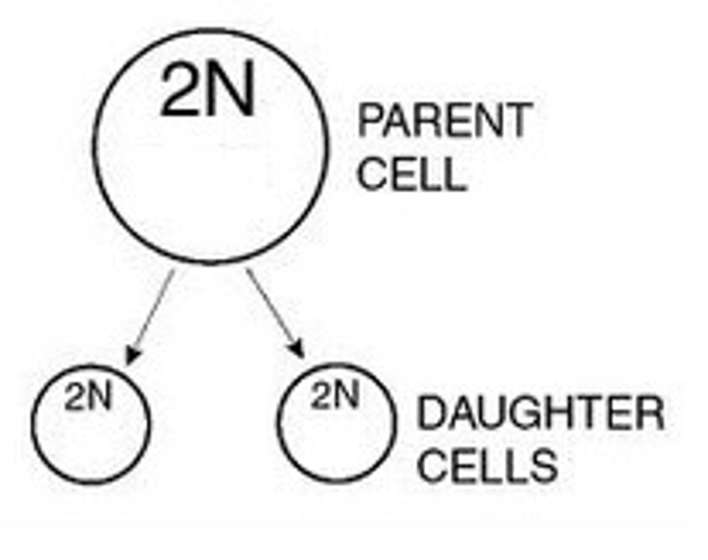
Diploid (2n)
This is having two copies of a chromosome and characteristic of autosomal cells
The number is 46 in humans
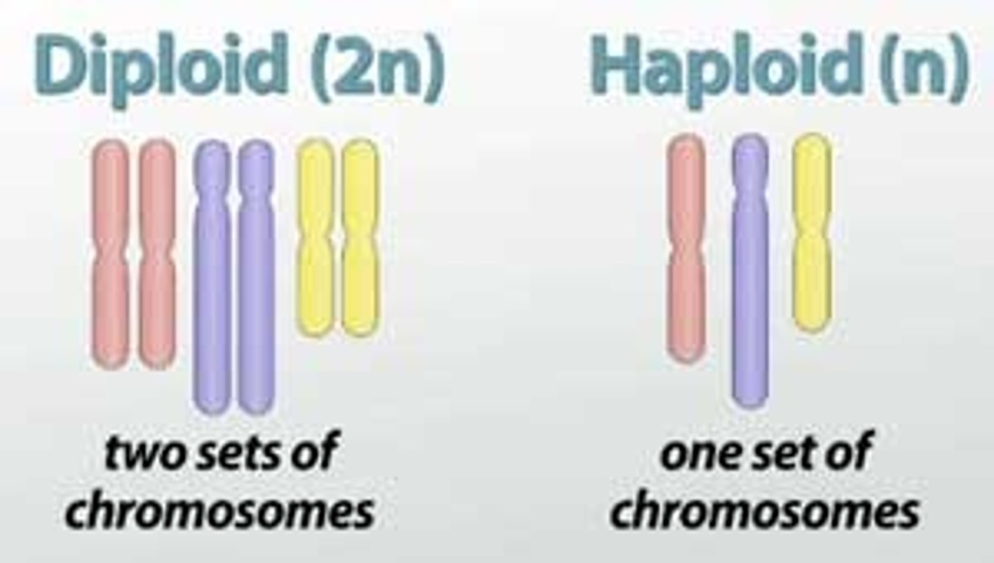
Haploid (n)
This is having only one copy of a chromosome and characteristic of germ cells
The number is 23

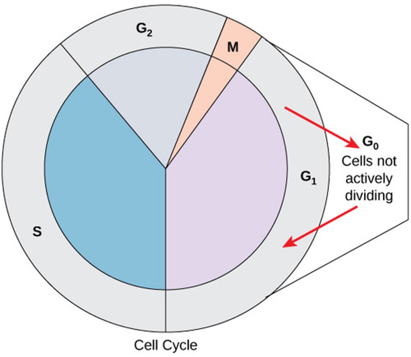
Cell cycle
This is the specific series of phases during which a cell can grow, synthesize its DNA, and divide
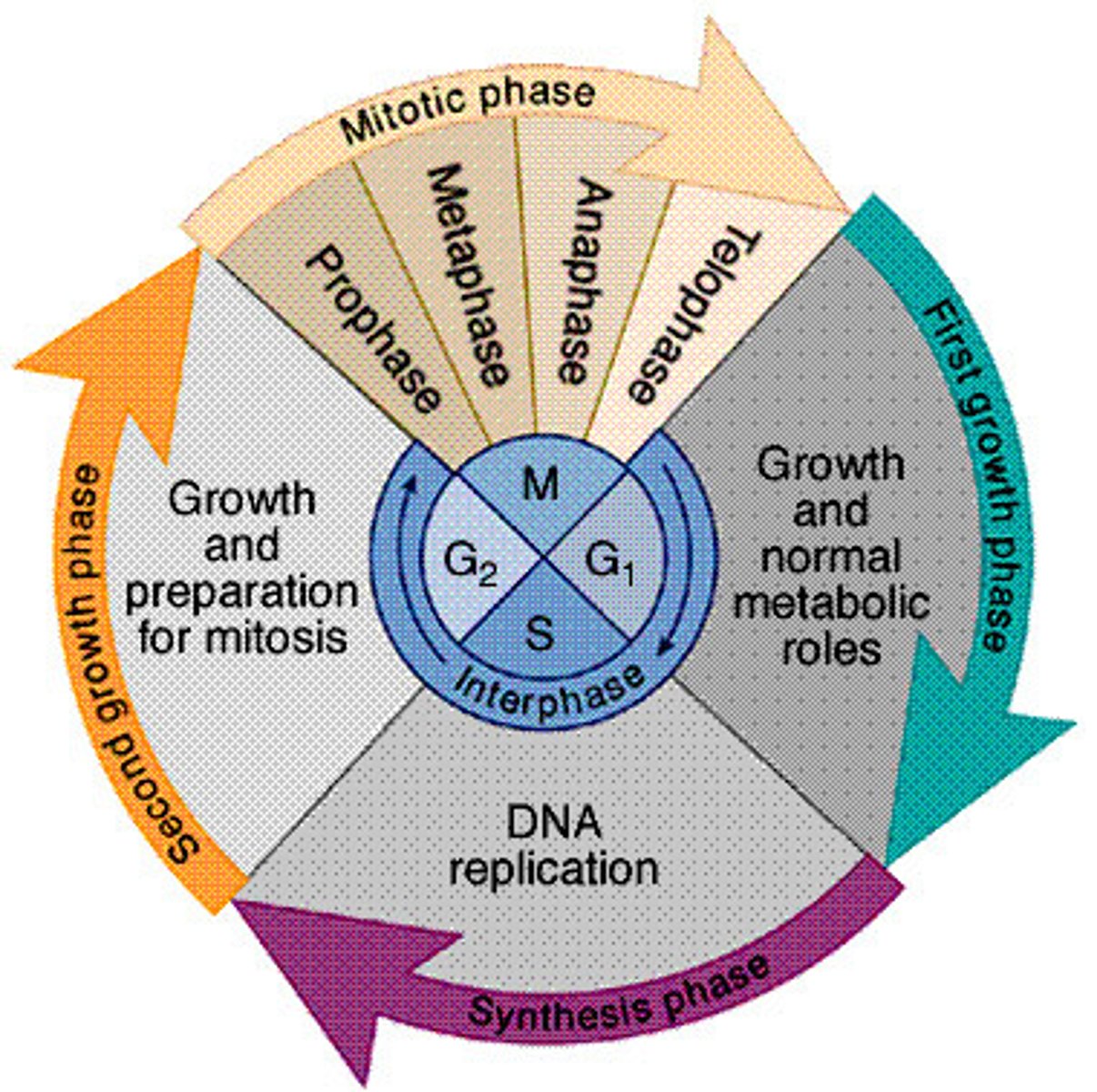
Four stages of the cell cycle
1. G1
2. S
3. G2
4. M
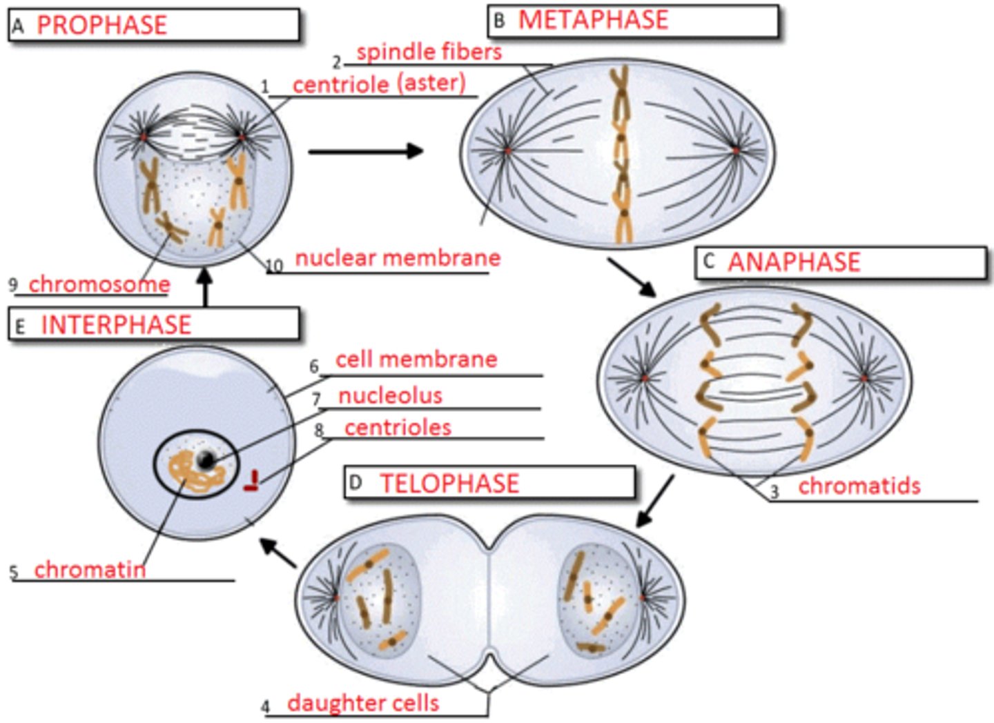
Interphase
This consists of G1, S, and G2
This is the longest part of the cell cycle, even actively dividing cells spend about 90% of their life in interphase

G0 phase
This is a phase that is an offshoot if the G1 phase and this is where cells that are simply living and serving their function without dividing go to just live without any preparation for cell division
What are the chromosomes like in interphase
They are not very condensed, they are the loose chromatin version of chromosomes during interphase in order for the DNA to be available for RNA polymerase so that the genes can be transcribed and replicated
What are the chromosomes like in mitosis
They are tightly coiled chromosomes in order to avoid losing any of their genetic material during their cell division
G1 stage: Presynthetic Gap
During this stage the cell creates organelles for energy and protein production (mitochondria, ribosomes, and endoplasmic reticulum) and they also will increase their size
G1 Restriction Point
In order for the cell to go from G1 into the S phase it must meet certain criteria such as containing the proper complement DNA in order to pass into the synthesis stage
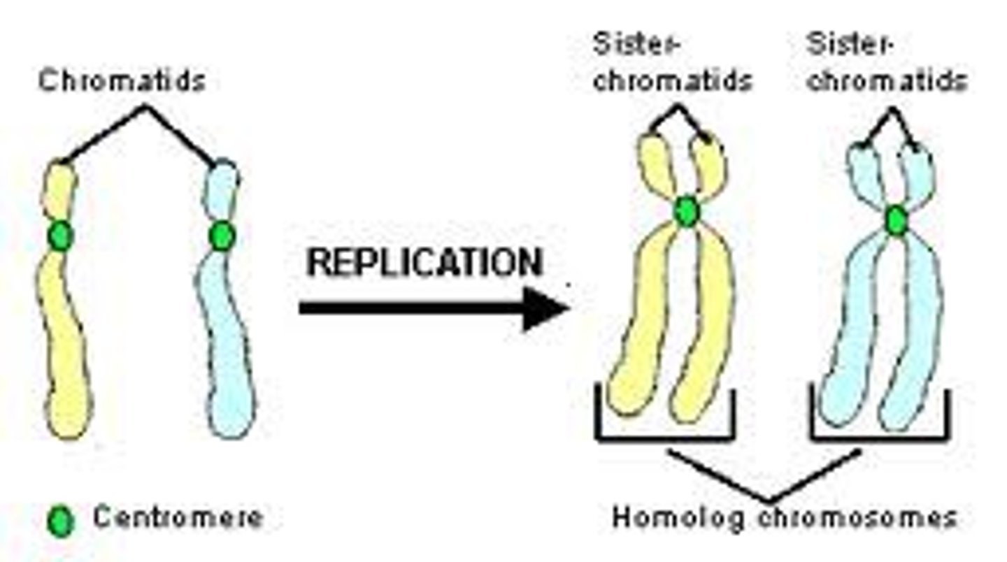
The S Stage: Synthesis Stage
This is the synthesis stage and this is where the cell replicates its genetic material so that each daughter cell will have identical copies

What happens to DNA in the synthesis stage
The chromosomes will replicate and afterwards they will consist of 2 sister chromatids that are bound by a centromere--the ploidy of the cell will not change
G2 Stage: Postsynthetic Gap
- the cell passes through another quality control checkpoint
- the cell will check to make sure that there are enough organelles and cytoplasm to divide between the two daughter cells
- checks that DNA replication proceeded correctly

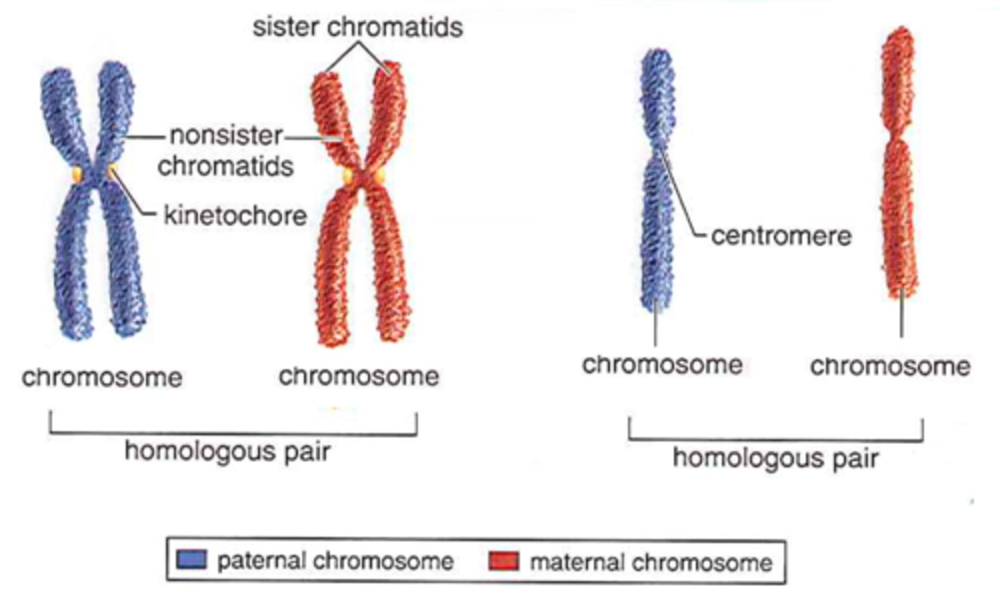
Chromatid
They are composed of a complete double stranded molecule of DNA and the sister chromatids are identical copies of each other
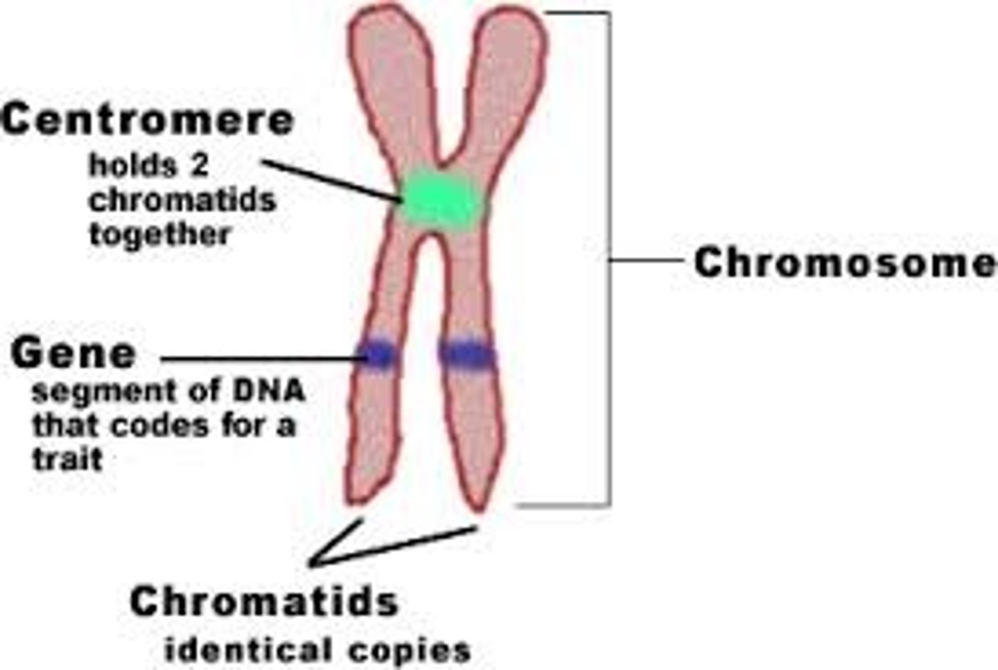
Chromosome
This can refer to either a single chromatid before the S phase or the pair of chromatids that are attached to the centromere after the S phase
Key idea about the daughter cells
1. In autosomal cells the division results in 2 genetically identical daughter cells
2. In germ line cells, division results in cells that are not equivalent
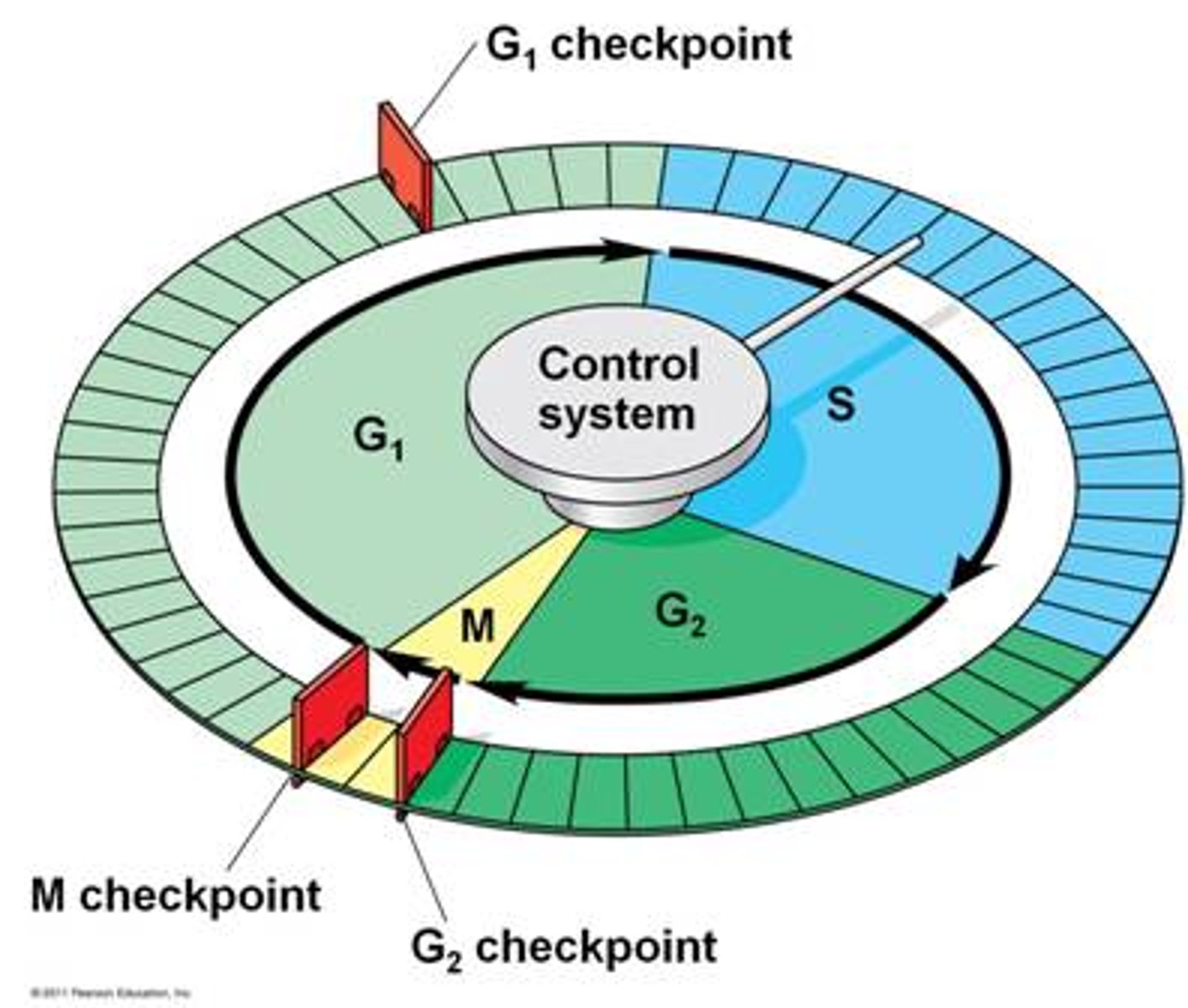
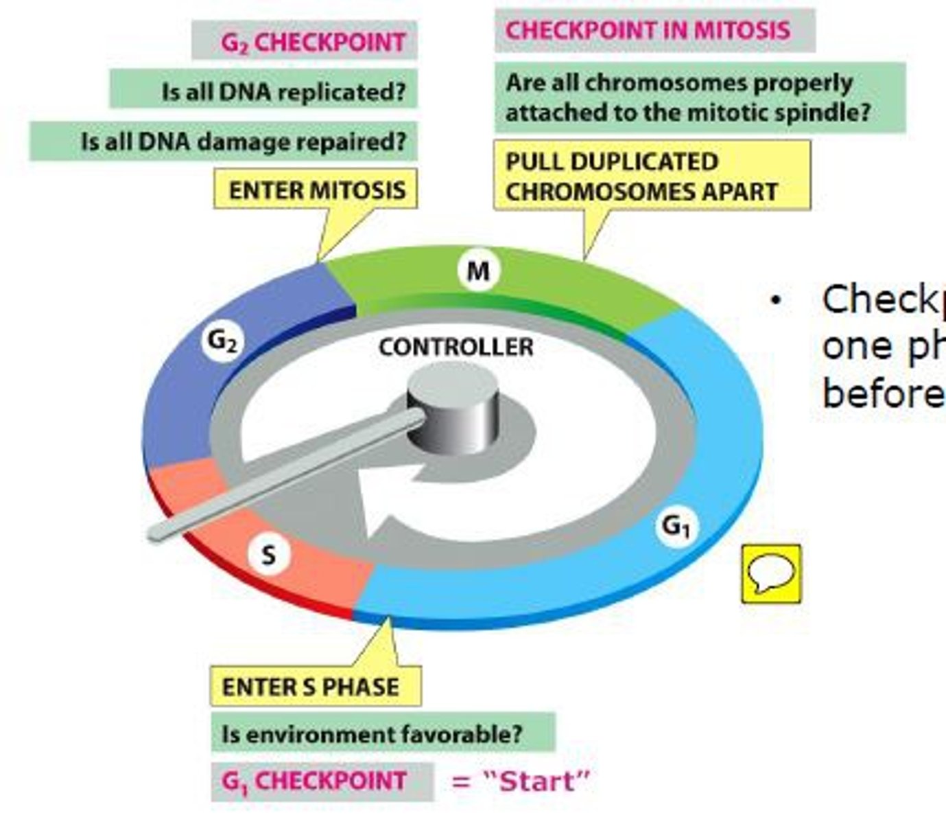
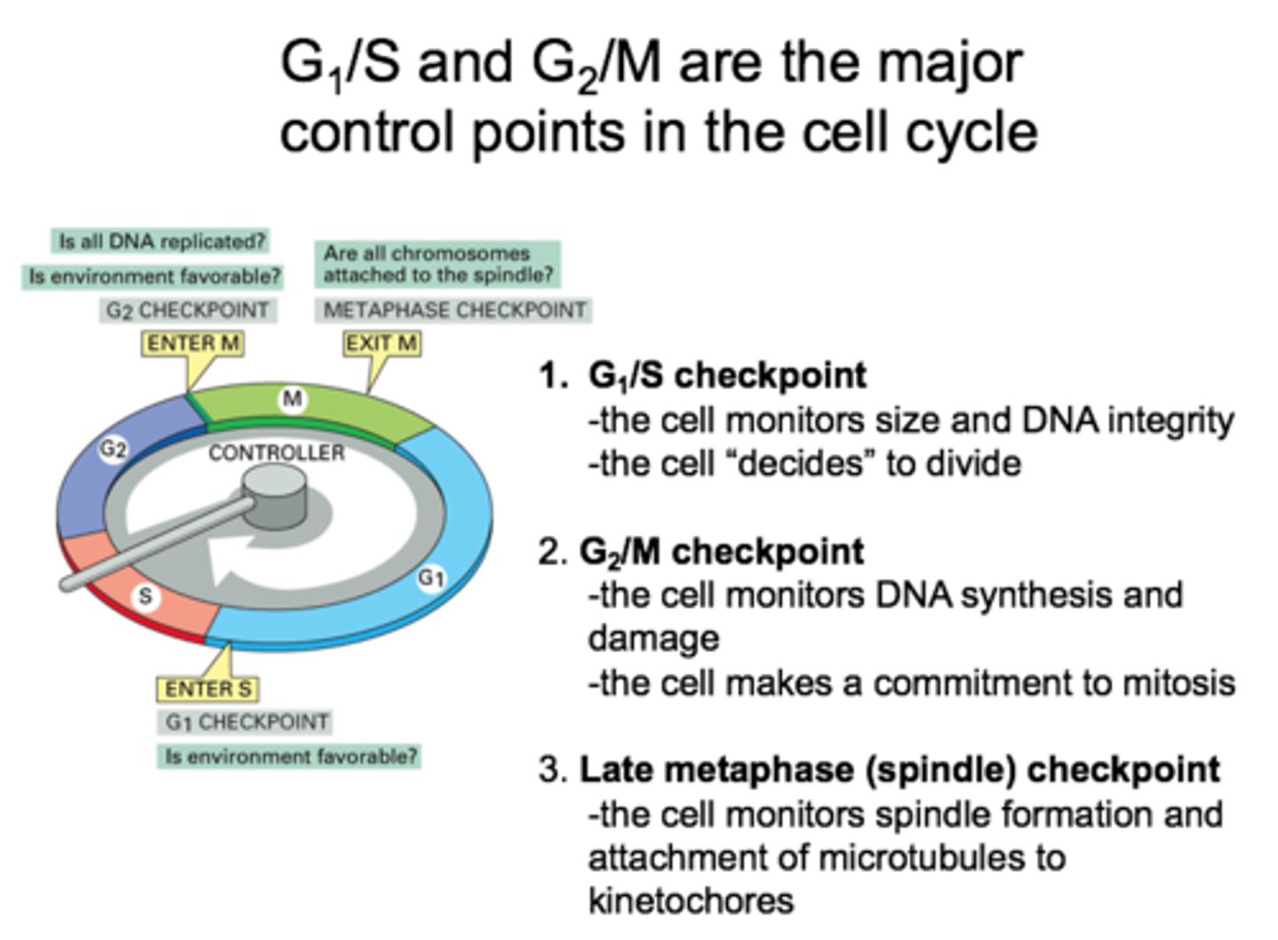
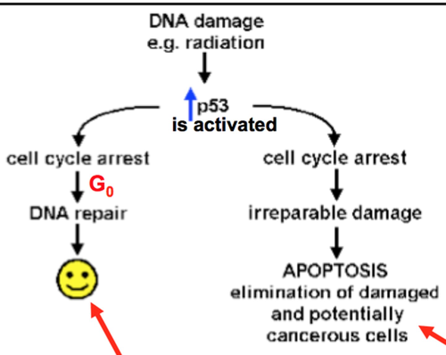
G1/S cell cycle checkpoint
- also known as the restriction point
- it will check to see if any of the DNA has been damaged and if it is ok for it to continue and synthesize more of this DNA
- the cell cycle goes into arrest if there is DNA damage and it will remain arrested here until the DNA is repaired
- p53 is main protein in control of this checkpoint
p53 protein
This is the protein that is responsible for having the cell cycle go into arrest until it can be repaired if there is some kind of damage - G1/S Checkpoint
- Also plays a role in G2/M checkpoint

G2/M checkpoint
This is the checkpoint that is mainly concerned with whether or not the cell has achieved the adequate size and the organelles have been properly replicated in order to support two daughter cells
p53 also plays a role in this
Cyclins
- one molecule responsible for cell cycle
- concentration fluctuate during cell cycle
- bind to cyclin dependent kinases creating activated CDK-cyclin complex
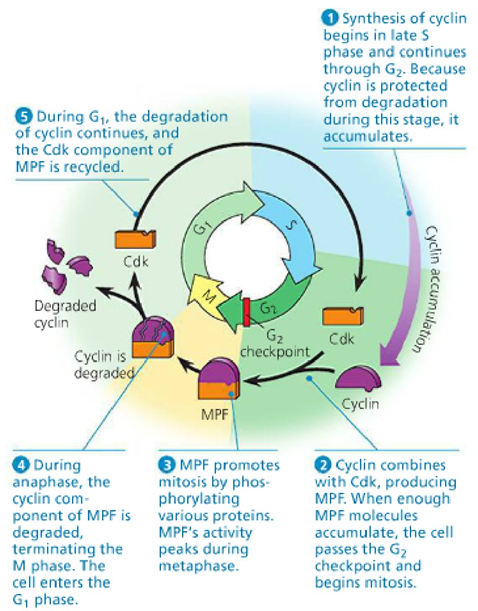
Cyclin Dependent Kinases (CDK)
- molecule responsible for cell cycle
- Activated by cyclins
- Once activated and CDK-cyclin complex is form, complex can phosphorylate transcription factors, which promote transcription of genes required for next stage of the cell cycle
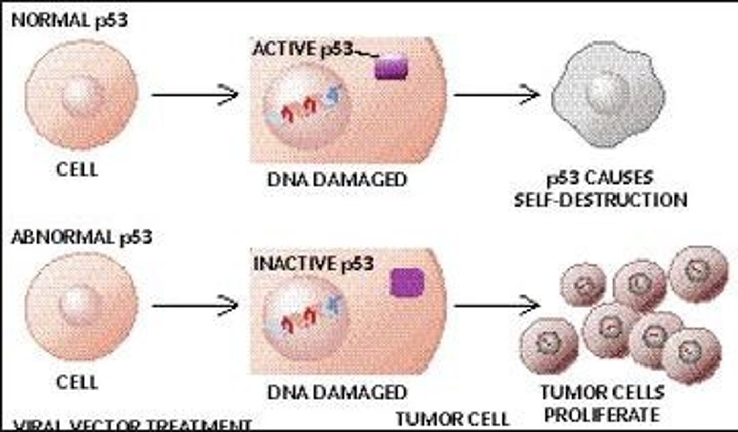
Cancer
This can result when there is inadequate cell cycle control
TP53
This is one of the most common mutations that will lead to cancer formation
- a mutation of the gene that produces p53
When this gene is mutated the cell cycle will not stop to repair the damaged or mutated DNA and this will allow for mutations to accumulate and then will allow the cancerous cell to continue to divide continuously without the regard for the quantity or quality of cells that are produced
Tumors
These are the results of rapid cell division

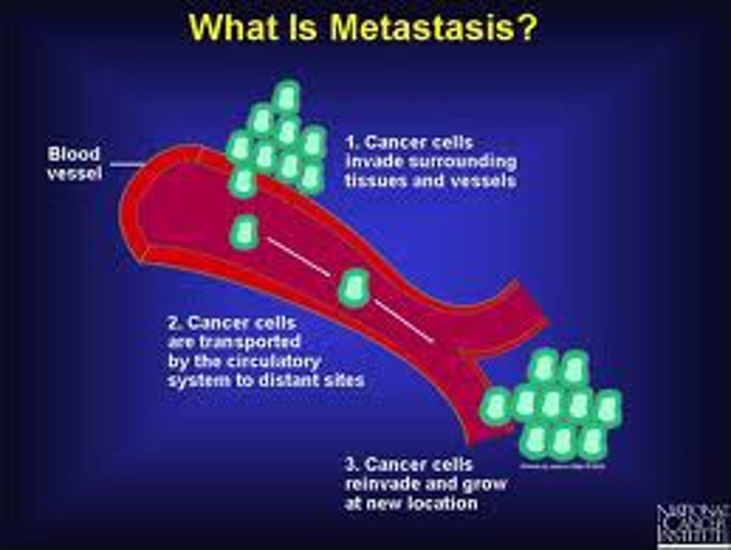
Metastasis
This is the distant spread of cancerous cells through the bloodstream or lymphatic system
- often due to the cancerous cells being able to make the right factors such as proteases to digest basement membranes and factors that will encourage blood vessel formation then the damaged cells can reach out to other tissues
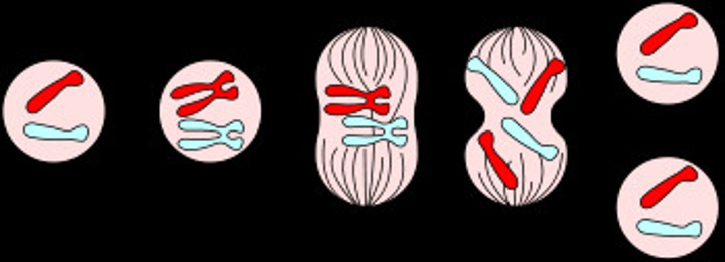
Mitosis
This is the process by which two identical daughter cells are produced from a single cell
This occurs in somatic cells which are cells that are not involved in sexual reproduction--these are cells that are just present in the body
Remember PMAT-these are the stages and order of the cell cycle

Tumor suppressor genes
These are genes that when they are mutated they will lose their ability to regulate or pause the cell cycle
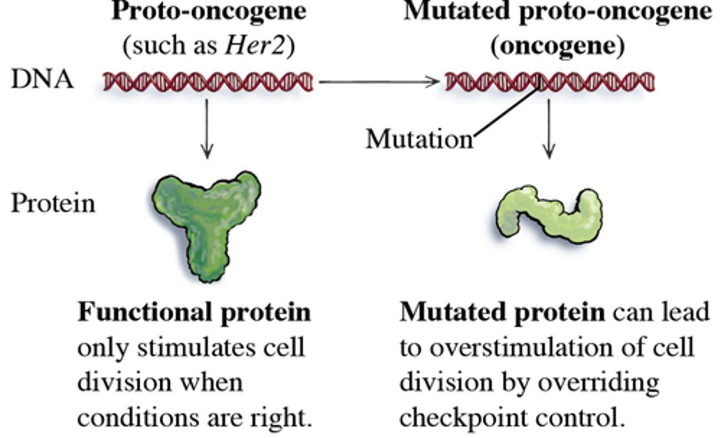
Oncogenes
These are genes that when they are mutated they will actively promote cell division
Mitosis Stages
The M stage consists of mitosis and cytokinesis
There are 4 stages of this:
1. Prophase
2. Metaphase
3. Anaphase
4. Telophase
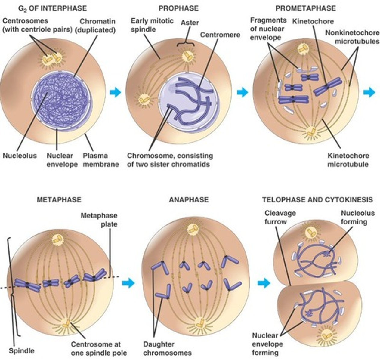
Prophase
1.CONDENSATION OF CHROMATIN INTO CHROMOSOMES (they become more condensed so that they are more easily able to be manipulated properly)
2. The centriole pairs will separate and move towards the opposite poles of the cell
3. Spindle fibers are formed from the centrioles
4. The nuclear membrane will also dissolve which will allow the spindle fibers to come into contact with the chromosomes
5. Nucleoli become less distinct or will disappear completely
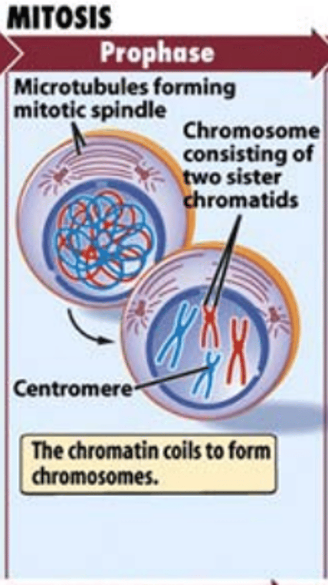
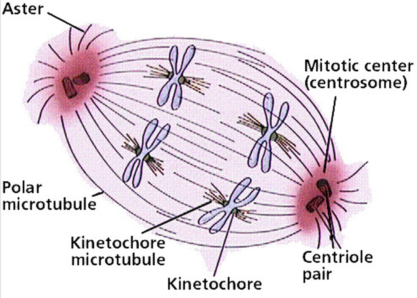
Spindle fiber formation during prophase
The fibers will radiate outwards of the centrioles
Some will form asters that anchor the centrioles with the cell membrane and others will radiate to the middle of the cell

Microtubule organizing center
This will create microtubules and microtubules like structures
The centrosome is included as one of these centers
The basal body of the flagellum or cilia are also a microtubules organizing center in the body
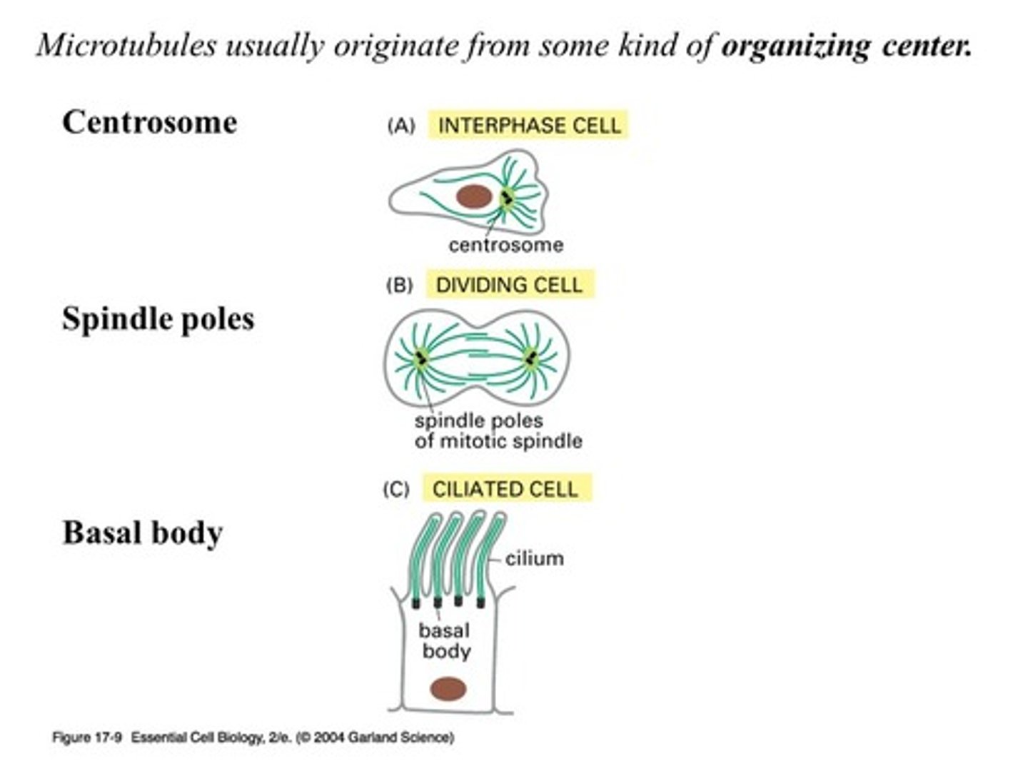
Centrosome
This is where the centriole pairs are stored and this is outside of the nucleus and this is responsible for the correct division of DNA
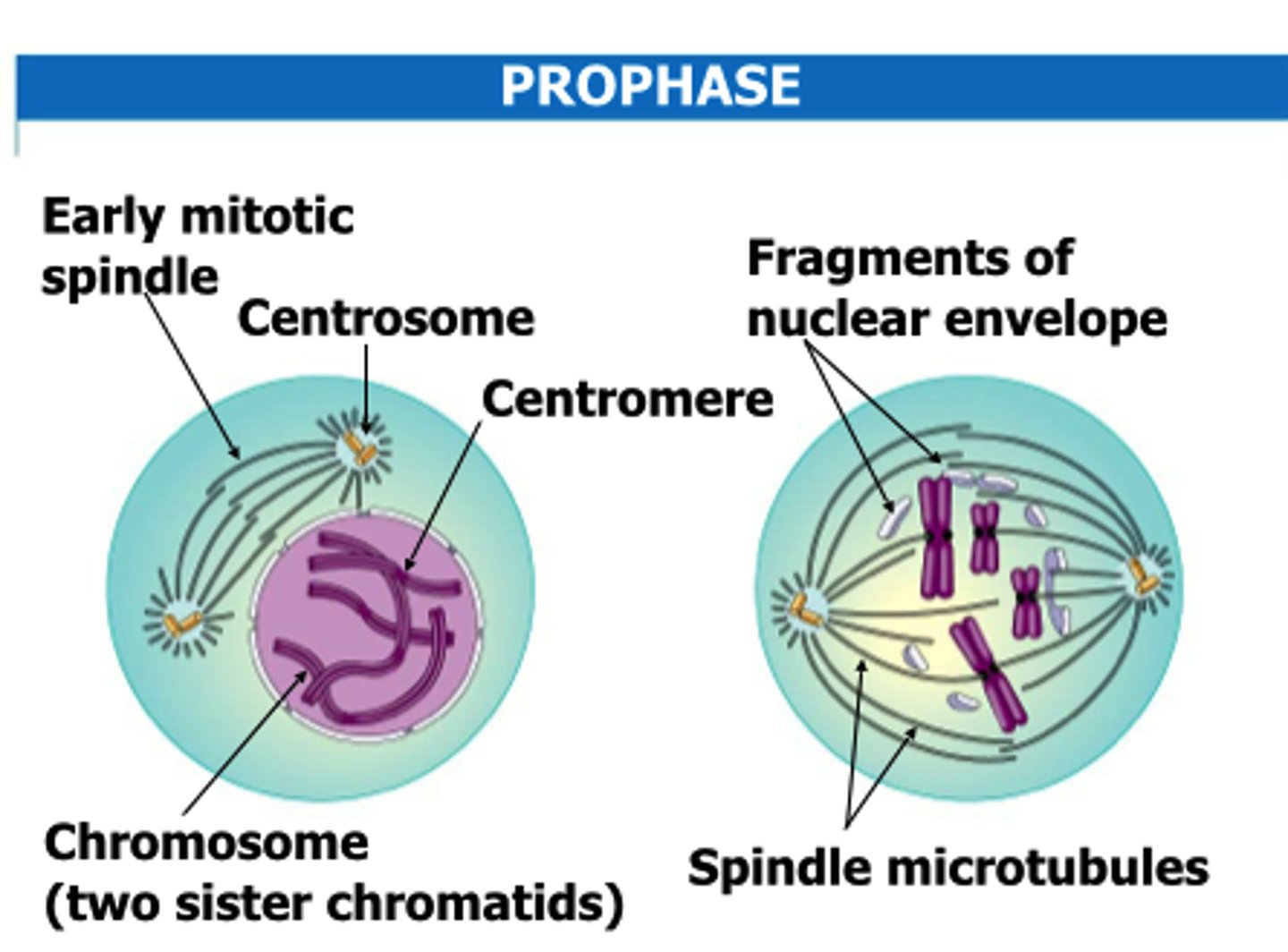
Kinetechores
These appear at the centromere of the chromosomes so that the spindle fibers can attach to them in the spindle apparatus
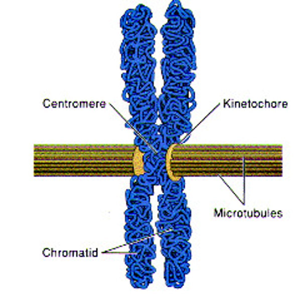
Kinetechore fibers
These are fibers that will attach to the Kinetechores that are located on the centromere of the chromosomes further making up the spindle apparatus
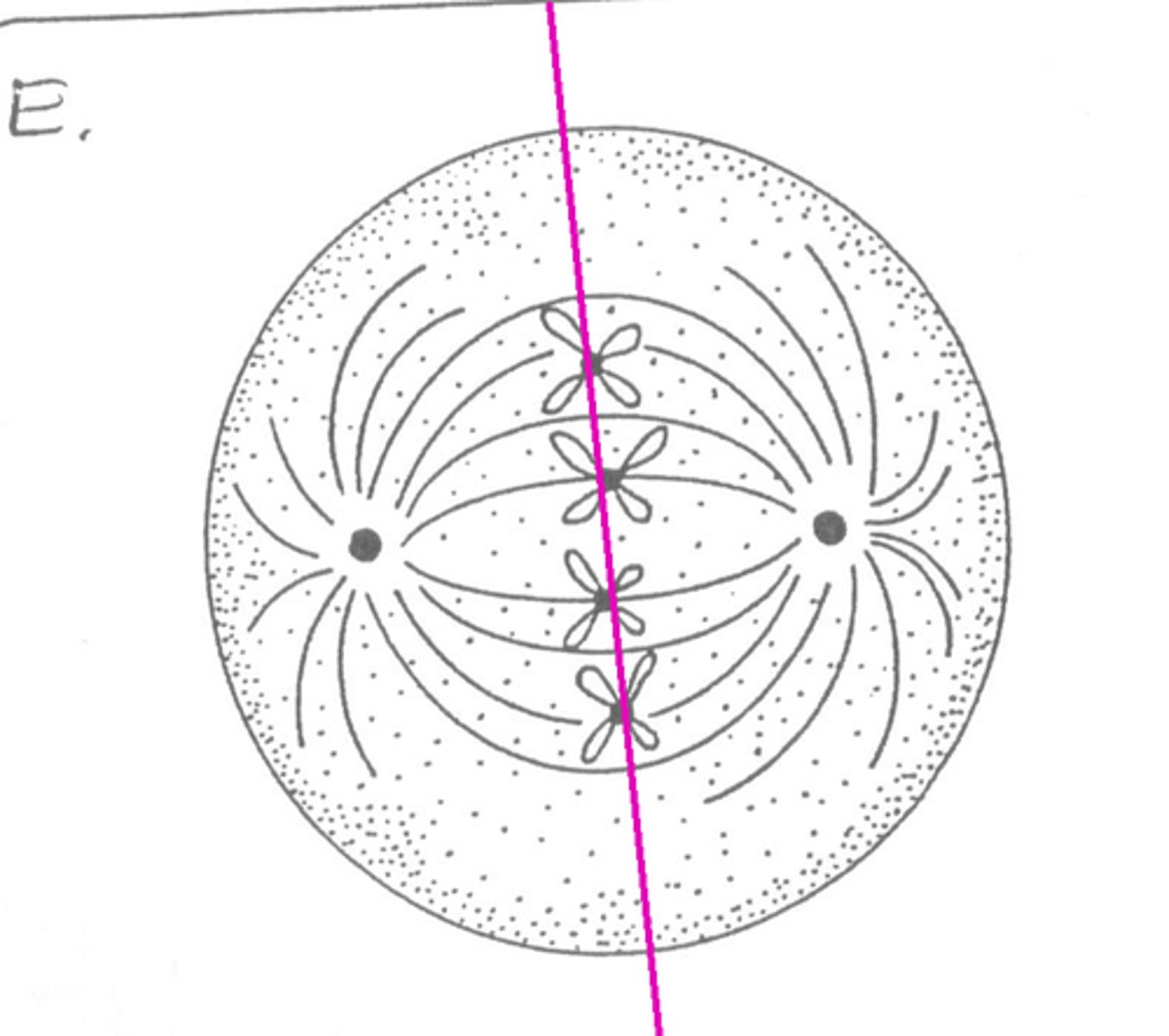
Metaphase
This is where the centriole pairs are on the opposite ends of the cell and the Kinetechores fibers will interact with the fibers of the spindle apparatus in order to align the chromosomes at the metaphase plate (equatorial plate)
- they will essentially be aligning itself in the middle of the cell on the fibers - equidistant from the two poles of the cell
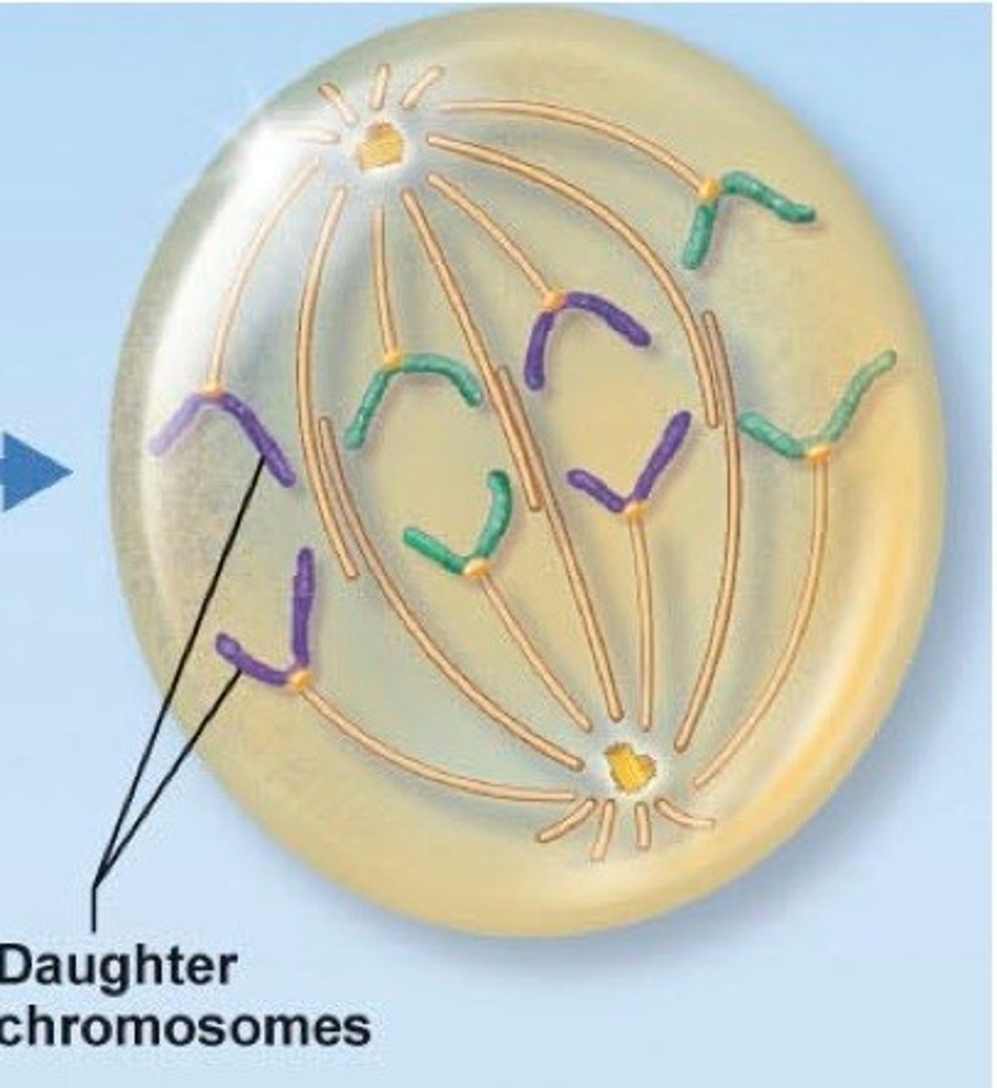
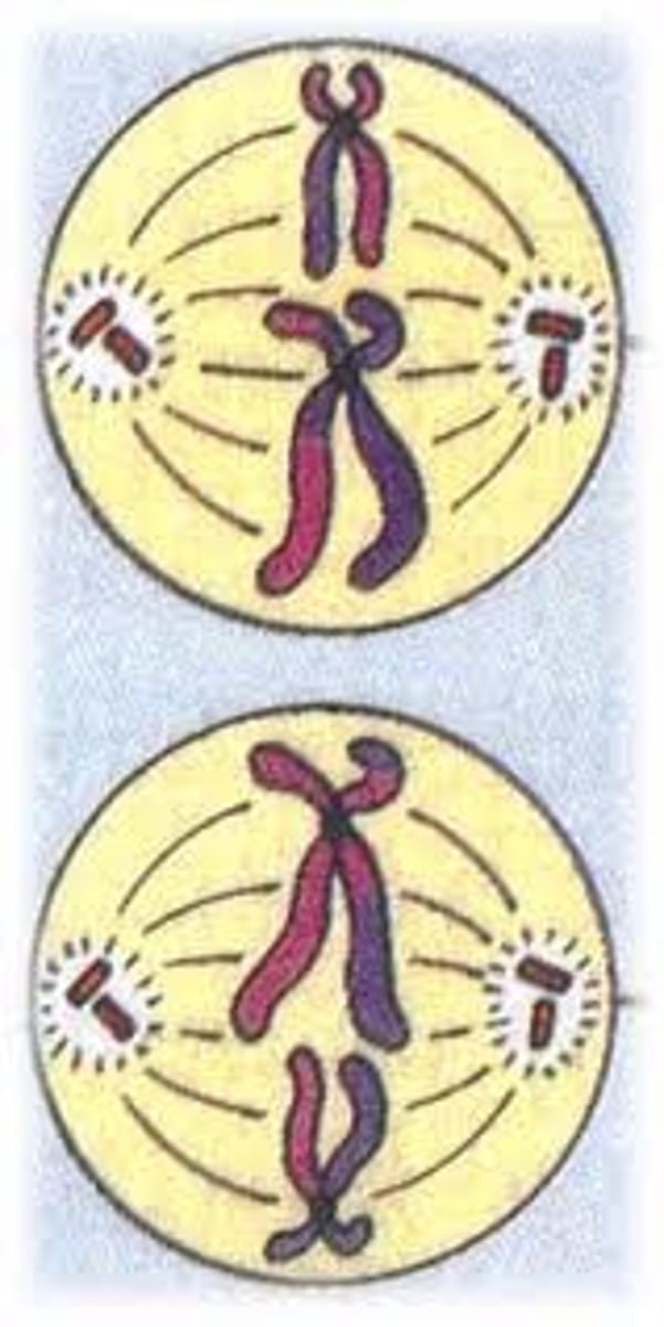
Anaphase
The centromeres are split so that each chromatid will now have its own distinct centromere and this allows the sister chromatids to separate
The sister chromatids are pulled towards the opposite poles of the cell by shortening of the Kinetechores fibers--the Kinetechore fibers will get shorter and allow for the splitting of sister chromatids

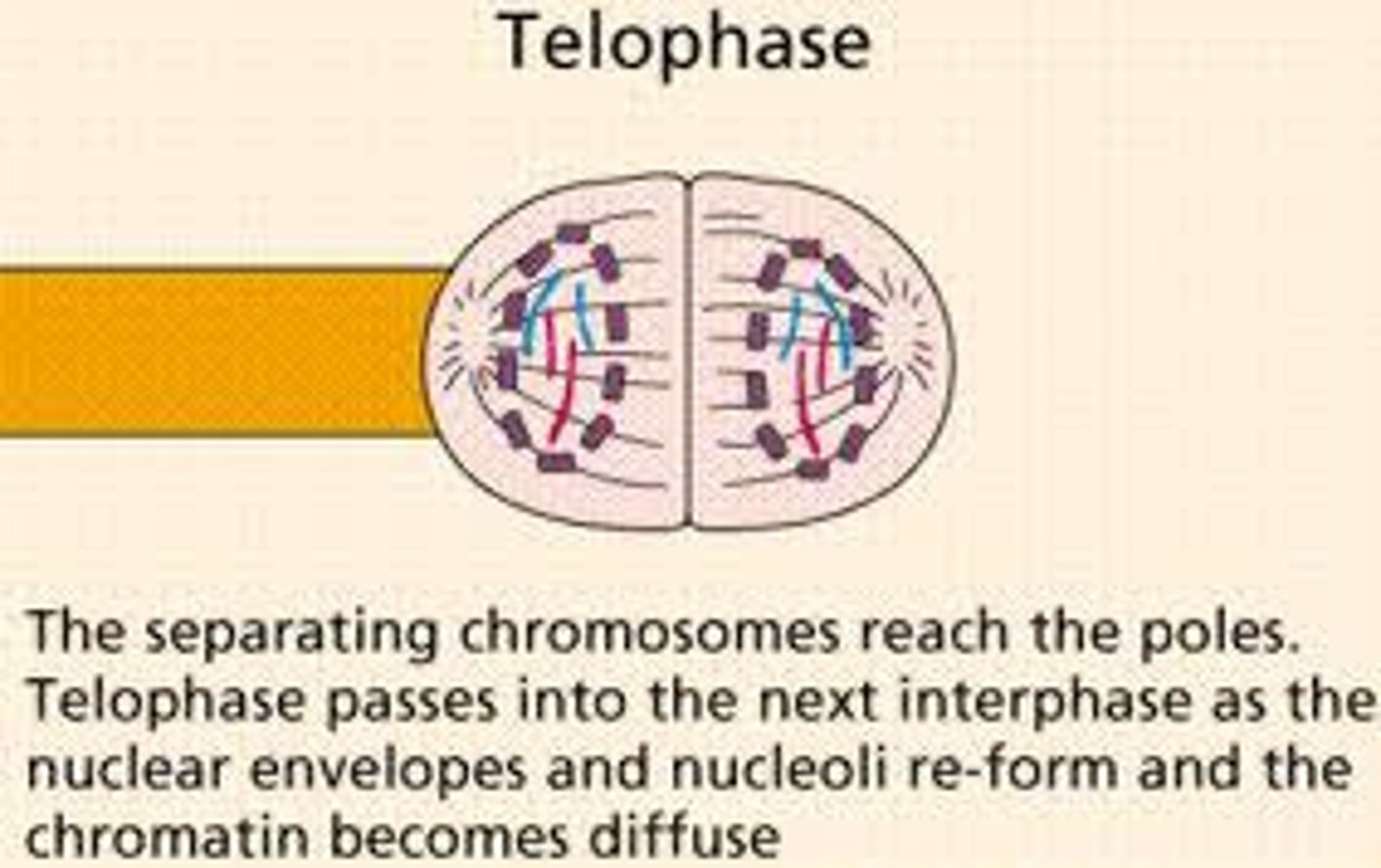
Telophase
This is essentially the reverse process of prophase-
1. The spindle apparatus will disappear
2. The nuclear membrane will reform around each set of the chromosomes
3. the nucleoli will reappear
4. Chromosomes will uncoil and return to their interphase form
There are now 2 new nuclei that have received a complete copy of the genome that is identical to the original genome and to each other
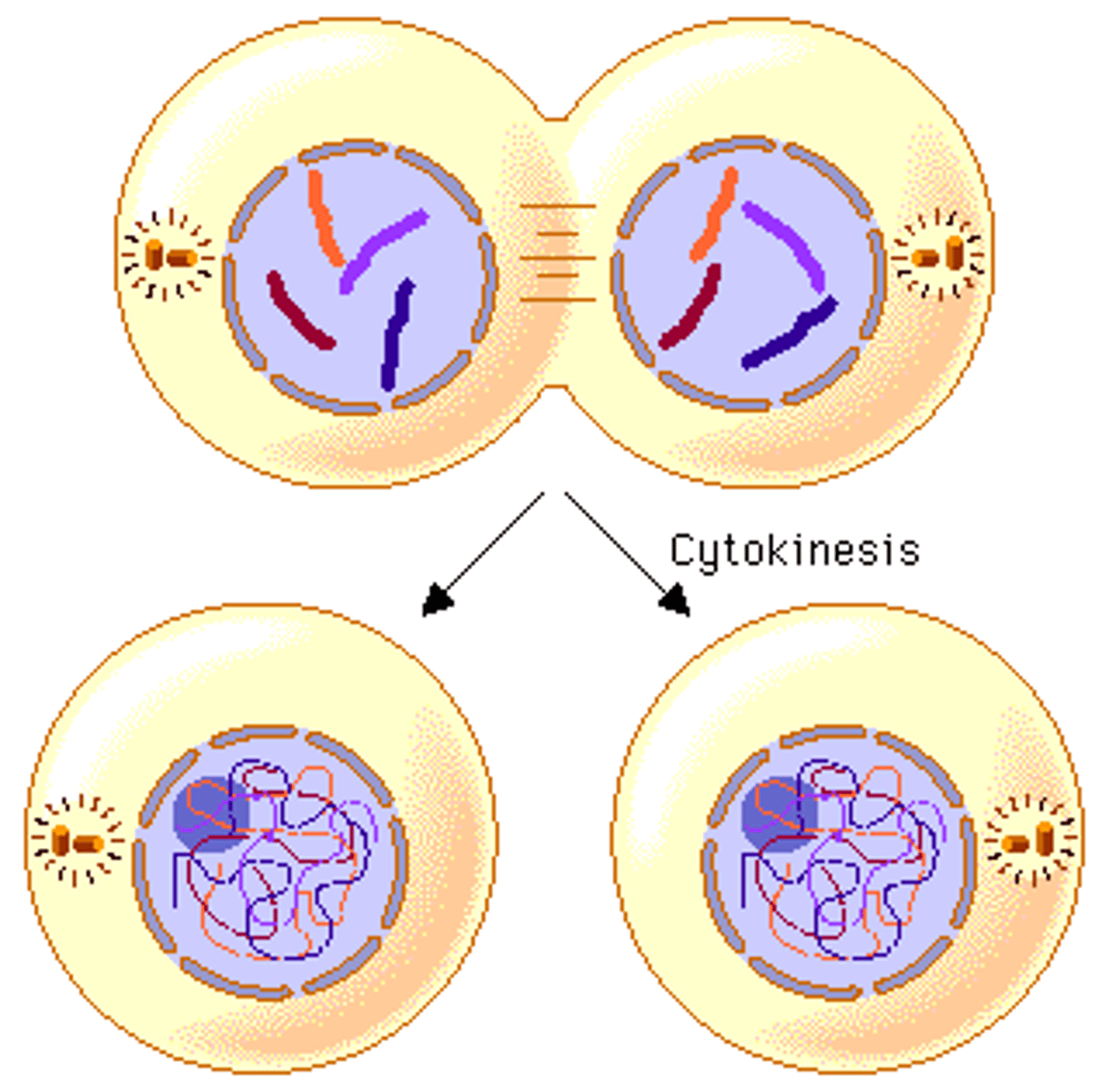
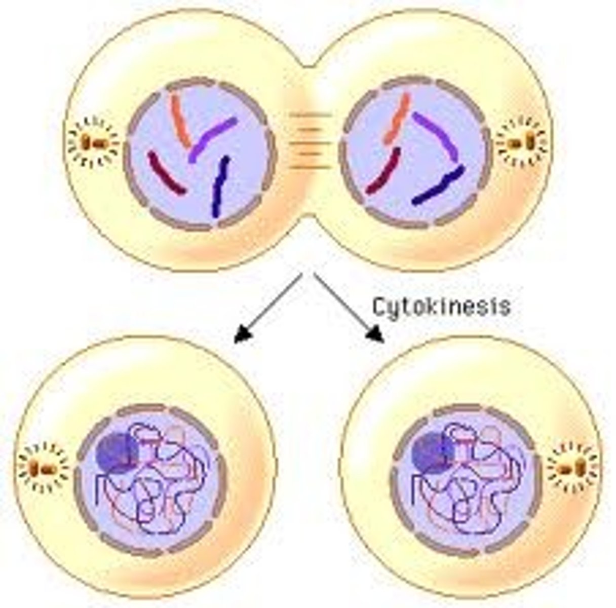
Cytokinesis
This is the separation of the cytoplasm and the organelles so that each daughter cell will have a sufficient amount to survive completely on its own
Number of division that are possible for human somatic cells
Each cell undergoes a finite number of divisions before programmed death
- human somatic cells: usually between 20 and 50, after that cell can no longer divide continuously
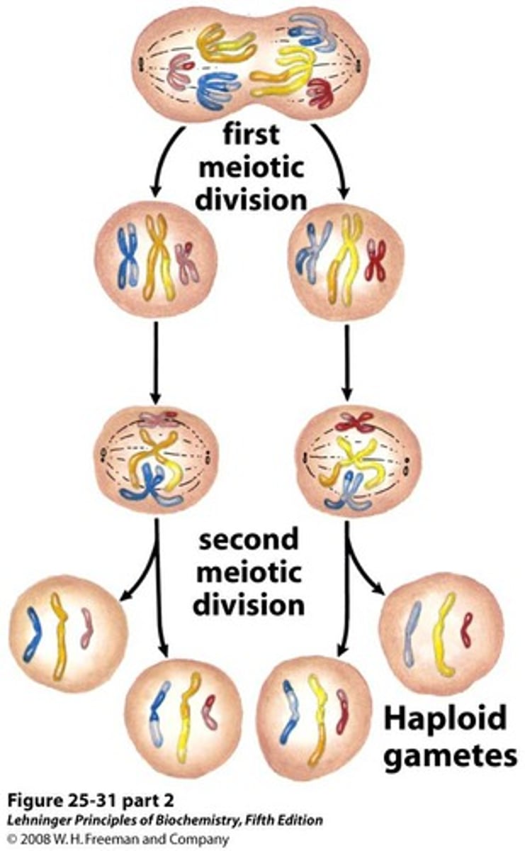
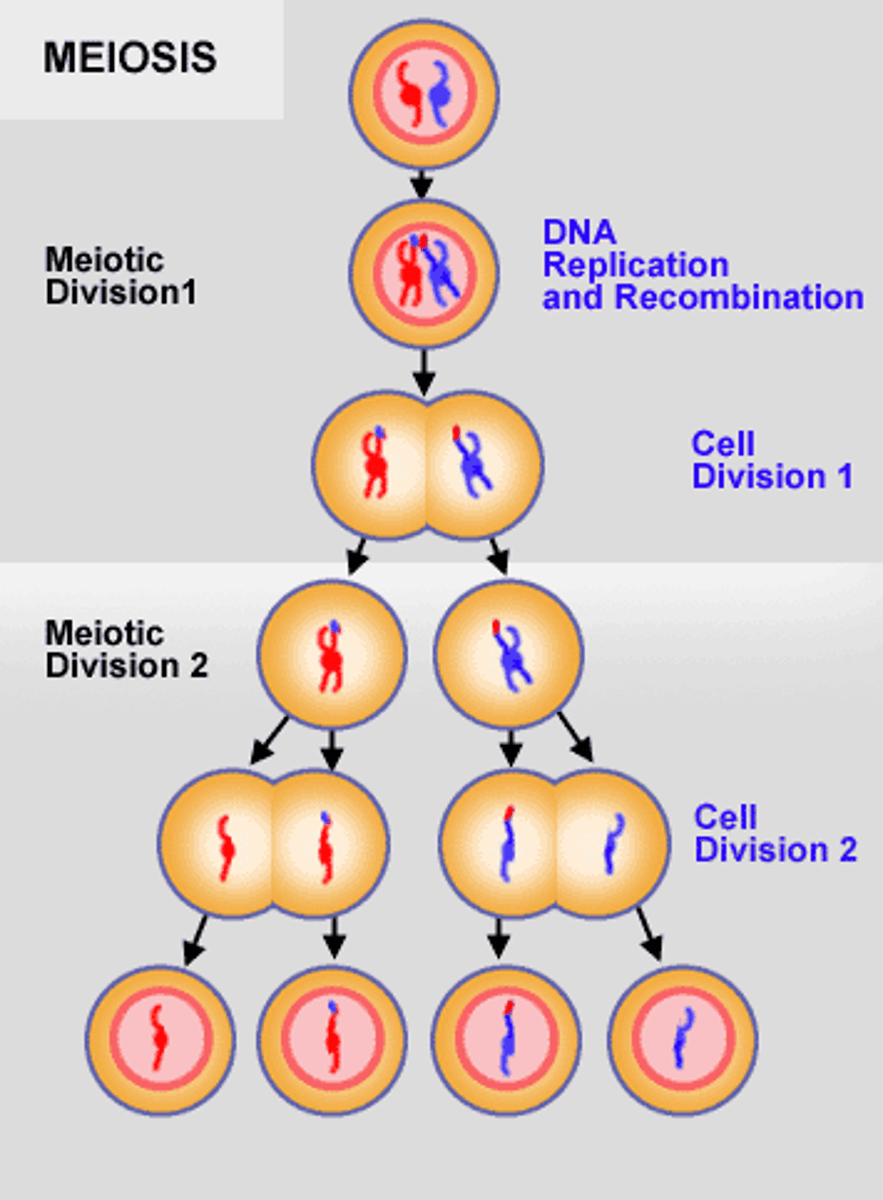
Where does meiosis occur/ the types of cells it occurs in
Meiosis occurs in gametocytes (germ cells) and results in four nonidentical sex cells called gametes

Differences between mitosis and meiosis
Similarities: genetic material must be duplicated, chromatin is condensed forming chromosomes, microtubules emanating from centrioles involved in dividing genetic material
Whereas mitosis consists one round of replication and division:
Meiosis consists one round of replication followed by two rounds of division
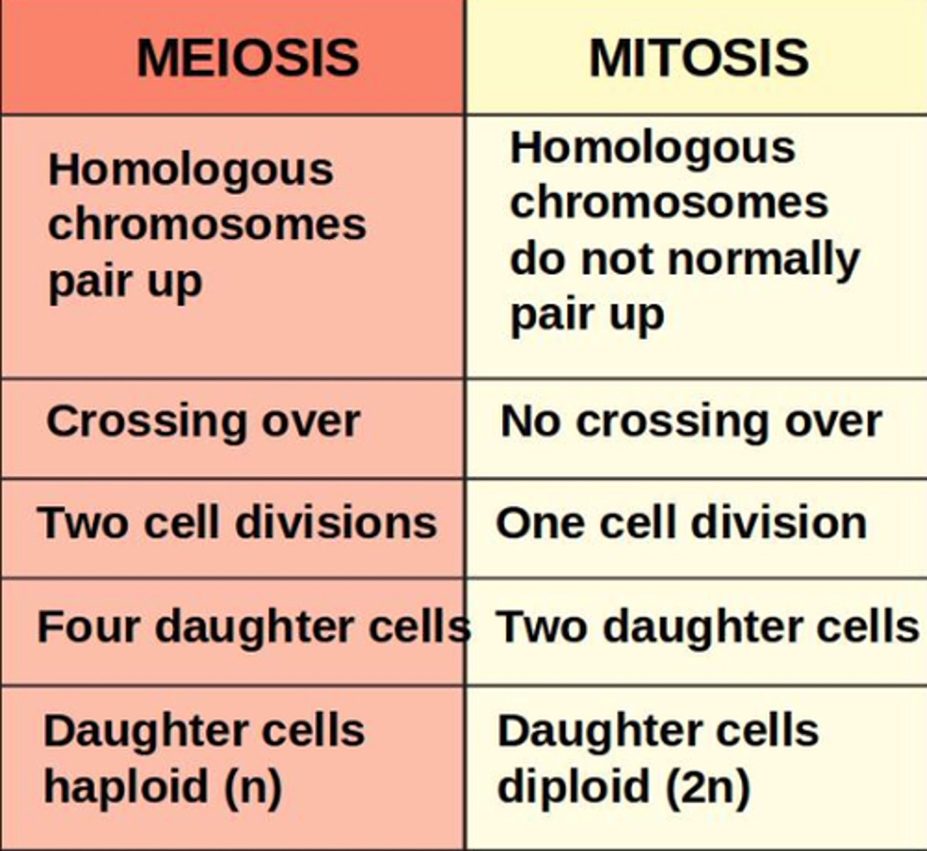
Reductional division
This is in meiosis I and it results in homologous chromosomes being separated and generating haploid daughter cells
- Reducing from two copies of each chromosome to one

Equational division
Meiosis II division. Similar to mitosis it results in the separation of sister chromatids
- Number of copies of chromosomes remains the same
Homologous pairs of chromosomes
Each contain pair contains a chromosome that is inherited from each parent
These are considered separate chromosomes
- Ex: maternal chromosome 15 and paternal chromosome 15
There are 23 pairs of homologous chromosomes giving the 46 total chromosomes
Also good to note, whereas, sister chromatids are identical strands of DNA that are connected at a centromere
Count after the S phase (replication)
There are 92 chromatids that are organized into 46 chromosomes that are then organized into 23 homologous pairs- this is also the count that is present at the start of meiosis I
Prophase I
1. Chromatin condenses into chromosomes
2. The spindle apparatus forms
3. The nucleoli and nuclear membrane will disappear
4. First major difference b/w meiosis and mitosis:
- Homologous chromosomes come together and intertwine in a process called synapsis (tetrad formation) - when crossing over can occur
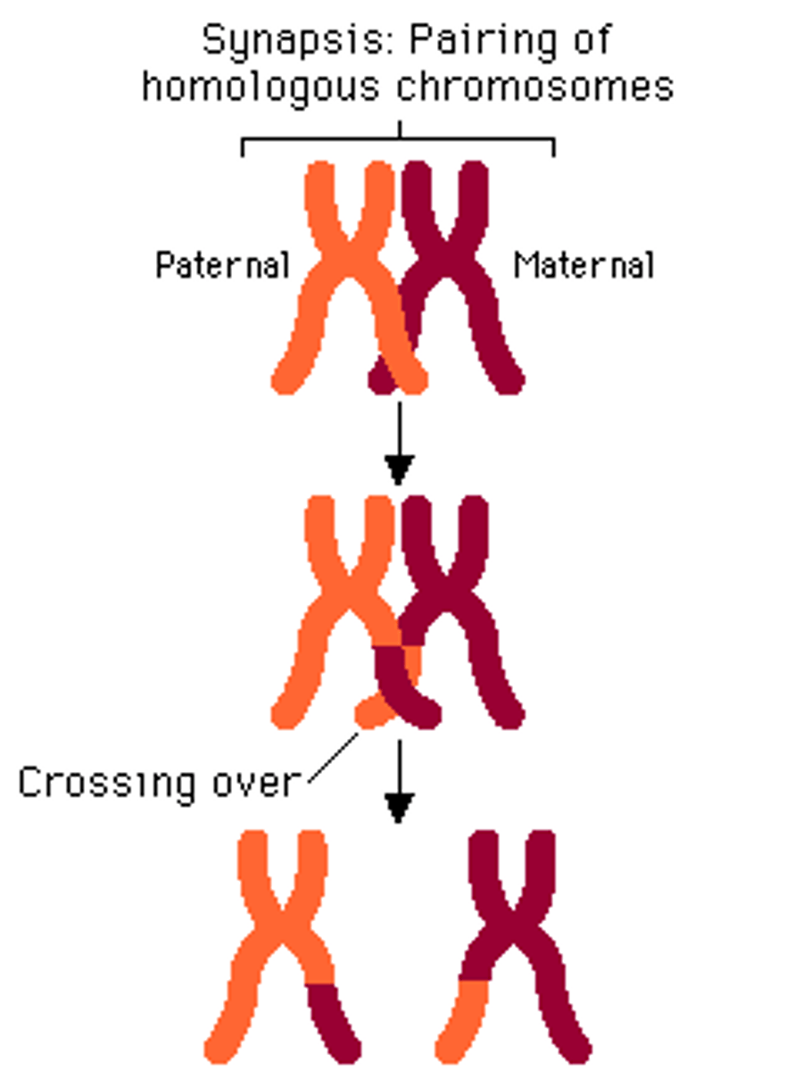
Synapsis
Process where the chromosomes will come together and intertwine
At this point each chromosome will consist of 2 sister chromatids so each synaptic pair will consist of 4 chromatids
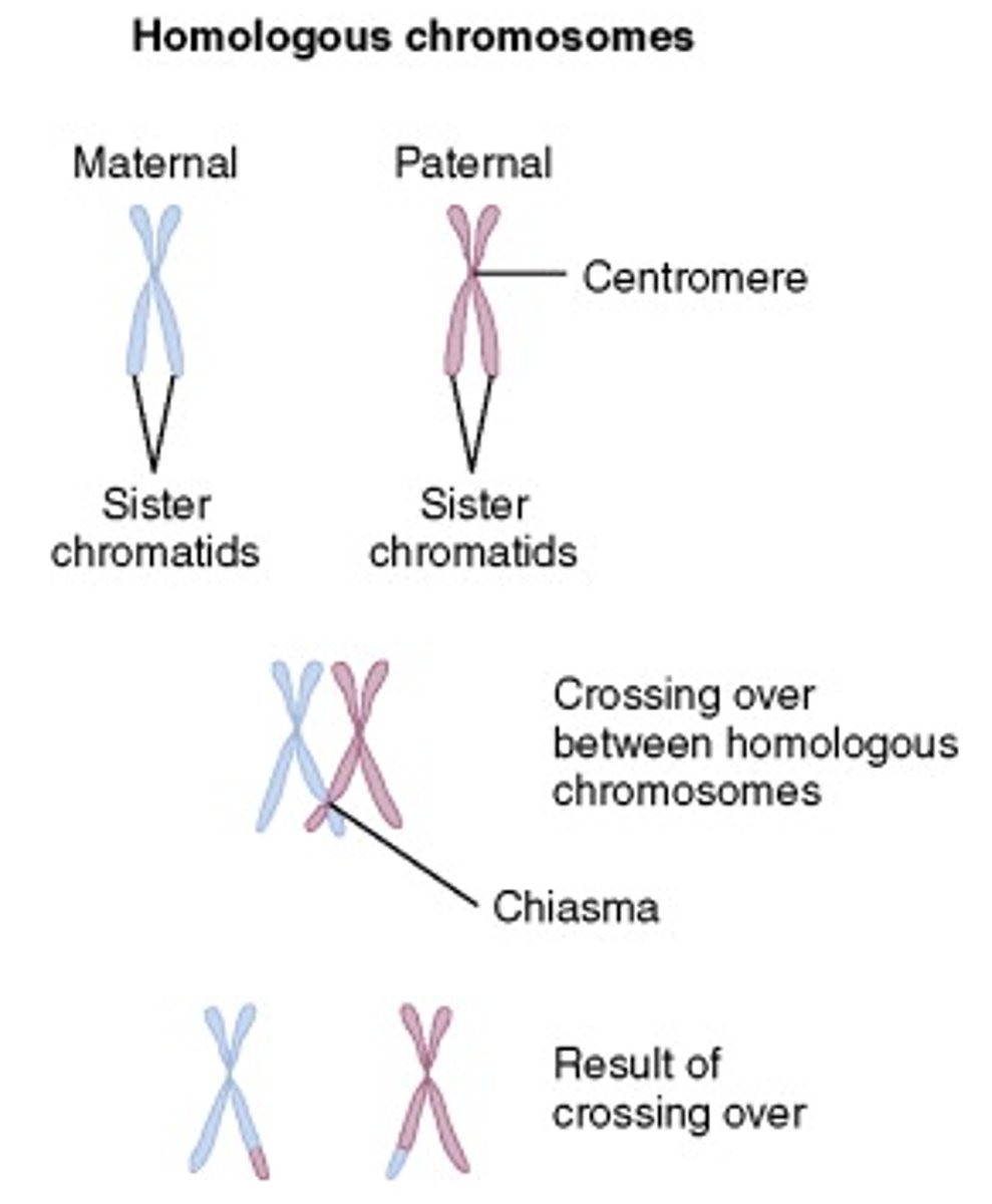
Tetrad
This is where the 4 sister chromatids come together and are held together by a group of proteins known as the synaptonemal complex at the chiasma, the point of contact
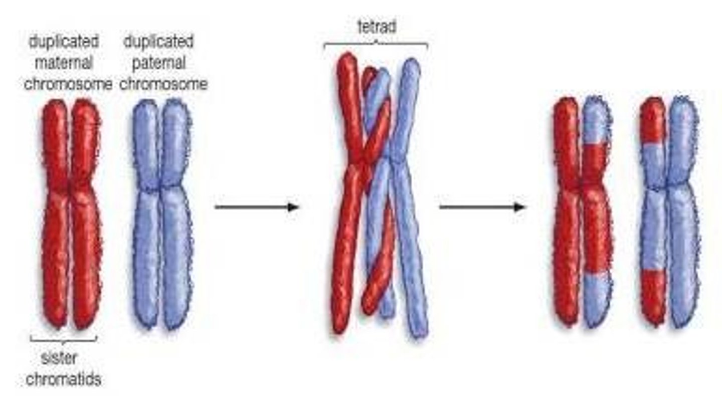
Chiasma/chiasmata (pl.)
This is where the homologous chromosomes of a tetrad come together, the point of contact
- the synaptonemal complex holds the 4 tetra together at this point
- the chromatids of homologous chromosomes may break at this point of contact
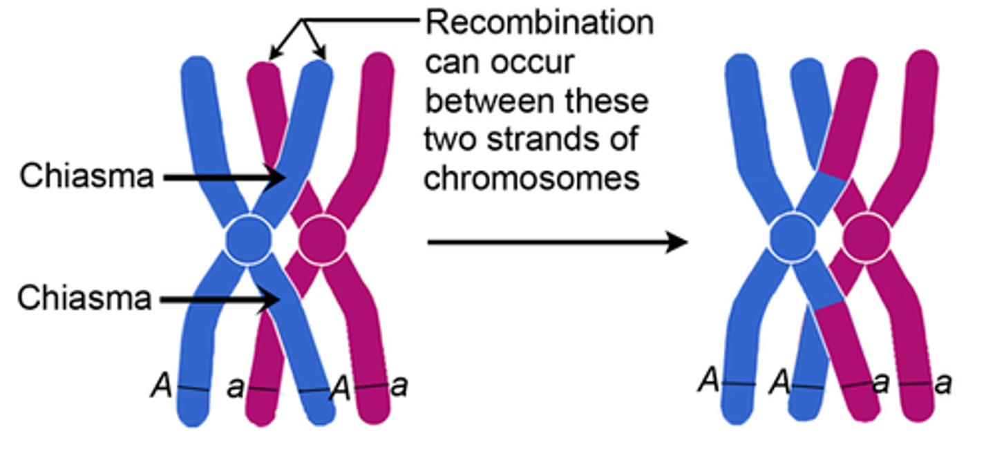
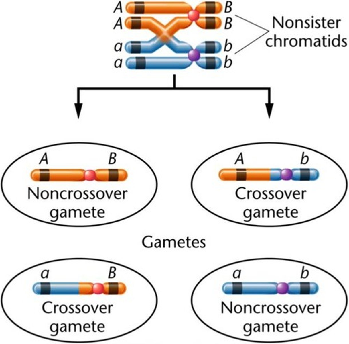
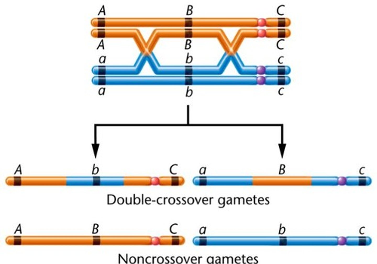
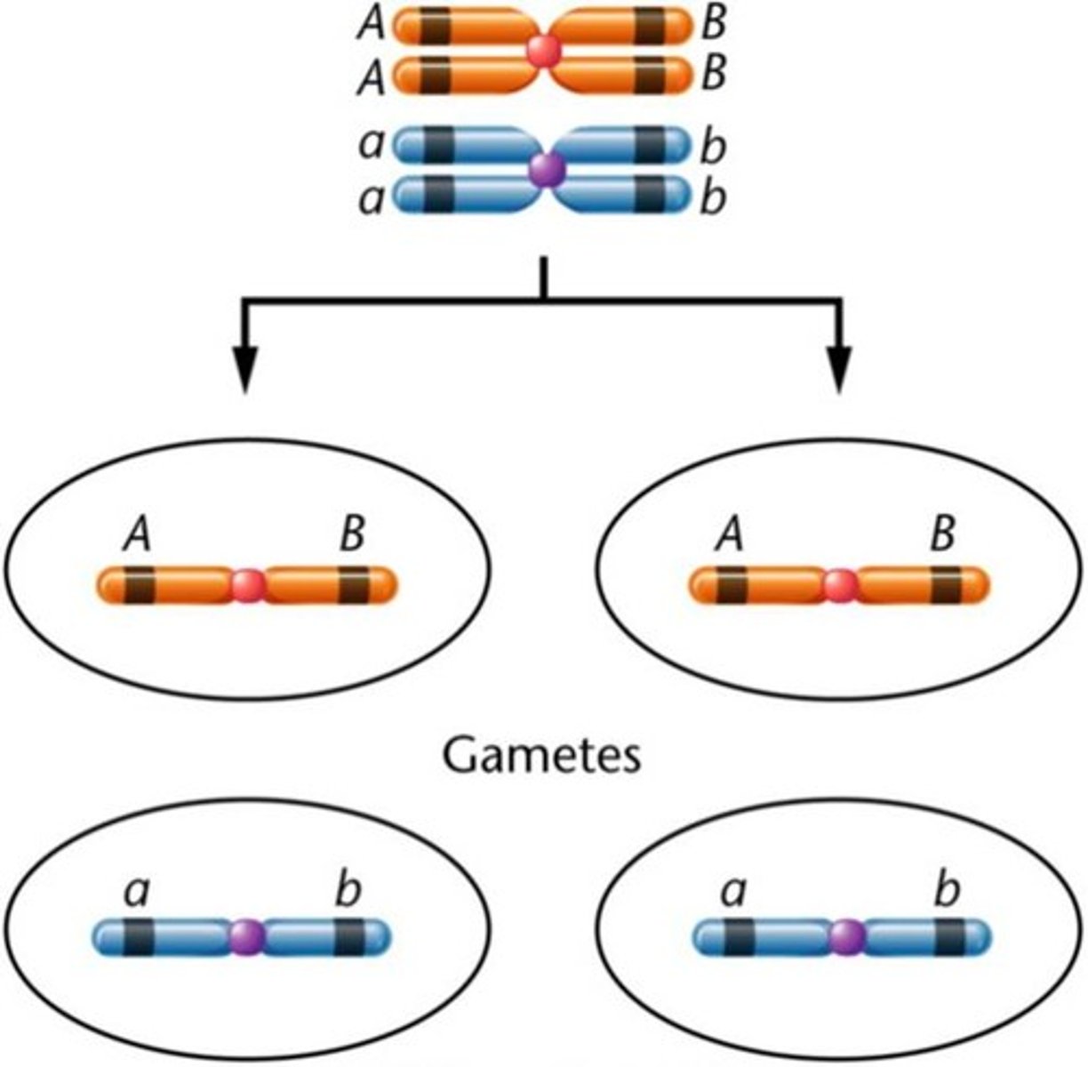
Crossing over
This is the process where the homologous chromosomes are together and they will exchange equivalent pieces of DNA
- number of crossovers can be single crossover or double crossovers
- Note that this is only between the homologous chromosomes and not the sister chromatids because the latter are identical and will not produce any change
This is the event that allows for genetic diversity in humans, allowing the offspring to have a unique pool of alleles that arose from the random mixture of maternal and paternal origin
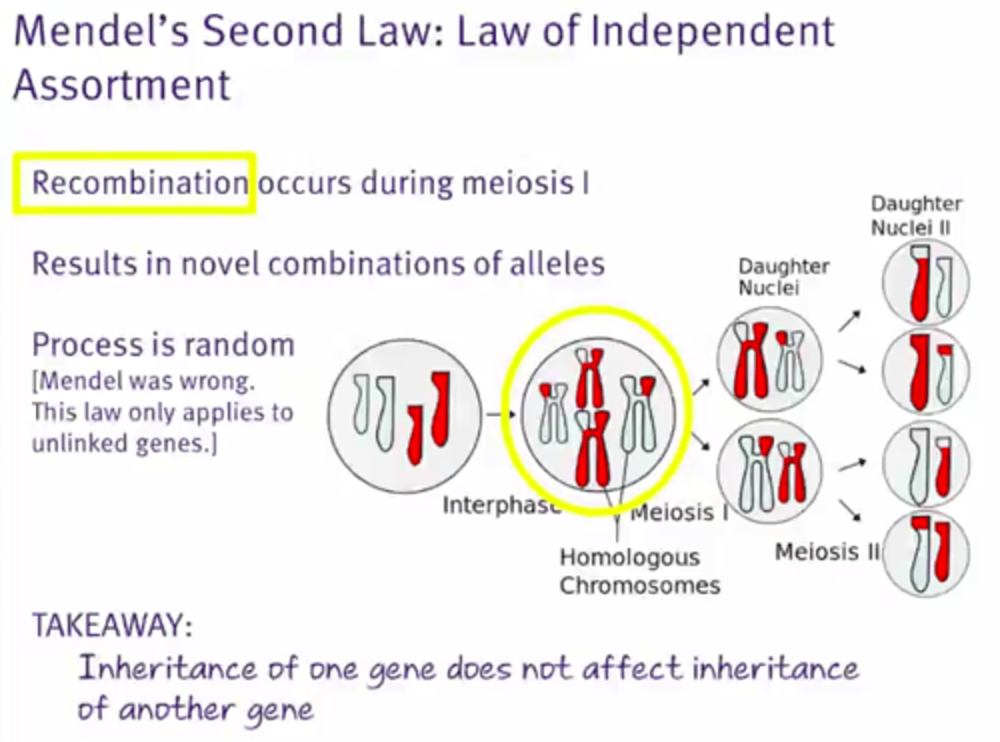
Mendel's second law of independent assortment
This states that the inheritance of one allele has no effect on the likelihood of inheriting certain alleles for other genes
This is because of crossing over
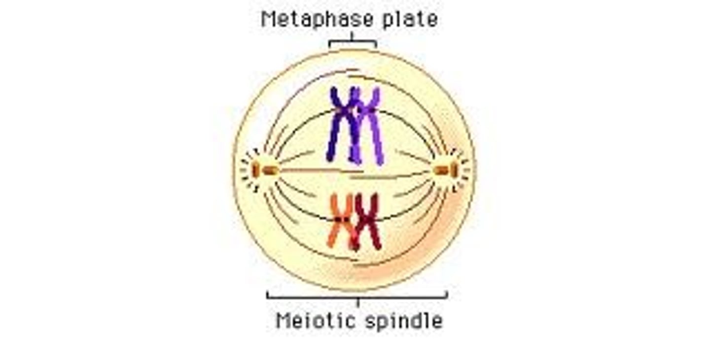
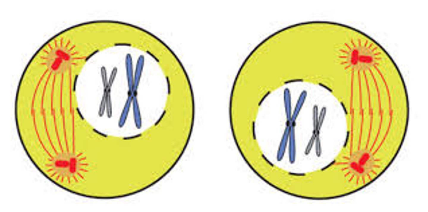
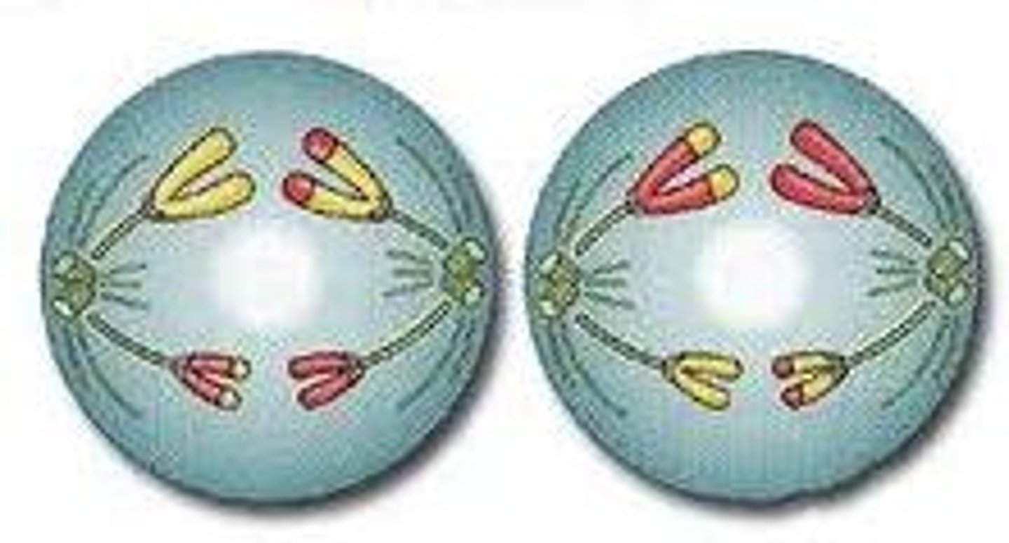
Metaphase I
The homologous pairs (tetras) of the chromosomes will align at the metaphase plate and each pair attached to a separate spindle fiber by its Kinetochore
Note that it is the homologous chromosome tetrad that lines up as a single unit in metaphase for meiosis I
Anaphase I
The homologous pairs of chromosomes will separate and are pulled to the opposite poles of the cell
- process called disjunction
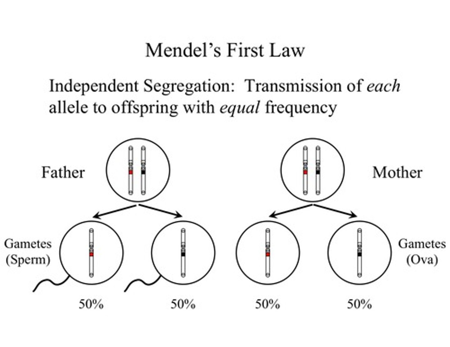
Mendel's first law of segregation
This is accounted for by the disjunction that occurs during anaphase I
- the chromosome of maternal origin or paternal origin from the particular homologous can end up in either daughter cell with respect to the parental origin
Segregation
This occurs in anaphase I and it is the separating of the 2 homologous chromosomes
Telophase I
This is where a nuclear membrane forms around each nucleus
At this point each nucleus still consists of 2 sister chromatids that are joined at the centromere
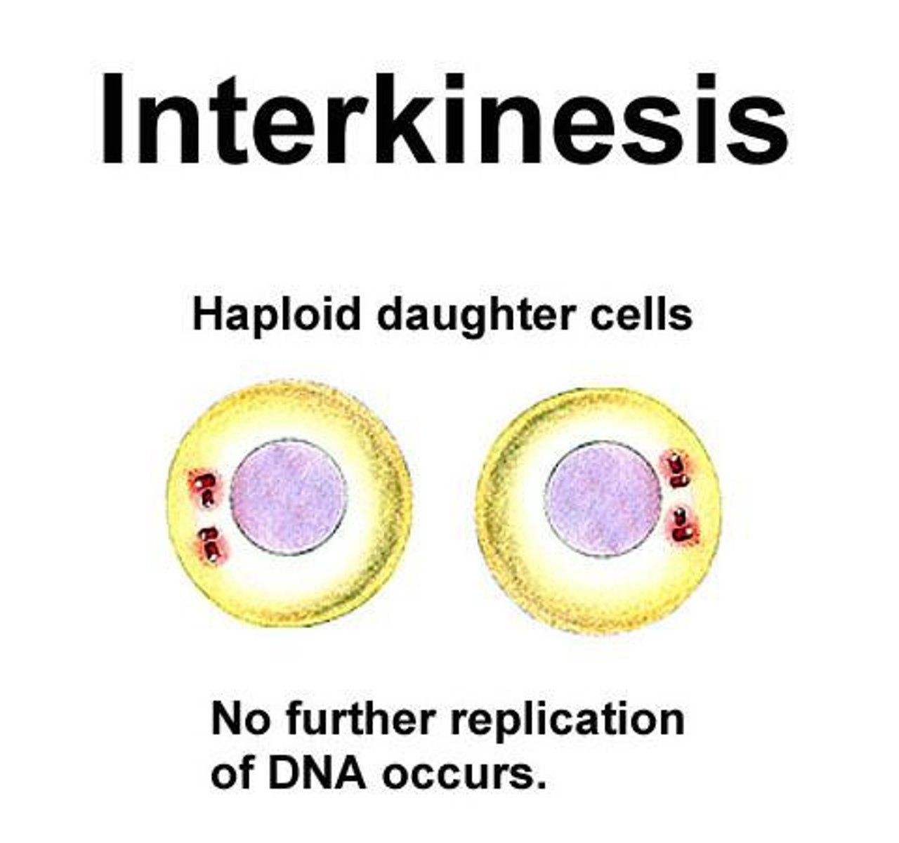
The cells are now haploid
- once the homologous chromosomes separated and there will only be 23 chromosomes that are found in each daughter cell (n instead of 2n)
Interkinesis
This is the short rest period in between the 2 meiosis phases where chromosomes will partially uncoil
Important note b/t mitosis and meiosis
It is important to note that a major difference between meiosis I and mitosis is that the daughter cells in meiosis will have their ploidy be halved and they will be n while in mitosis the ploidy will never be halved or changed at all
Single crossovers
This is when one cross over event occurs
Double cross overs
This is when 2 cross over events occurs
Genetic recombination
This is the process of changing the genes that are involved/ crossing over, this is the recombination of the genes
- unlinking linked genes

Linkage of genes
These are genes that tend to be inherited together and crossing over can cause genes to become unlinked
Linkage - tendency of genes to be inherited together; the further away genes are from each other on the chromosome, the less likely they are to be inherited together
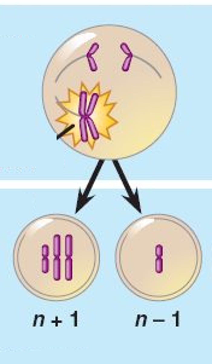
Nondisjunction
- during anaphase I or II of meiosis homologous chromosomes or sister chromatids will fail to separate
- one resulting gametes will have 2 copies of a particular chromosome and the other gamete will have none
- subsequently during the fertilization the resulting zygote may have too many or too few copies of the chromosome
Meiosis II
This is very similar to mitosis in that sister chromatids rather than homologous chromosomes will be separated from each other
Prophase II
1. Nuclear envelope dissolves
2. Nucleoli disappear
3. Centrioles migrate to the opposite poles
4. The spindle apparatus will begin to form
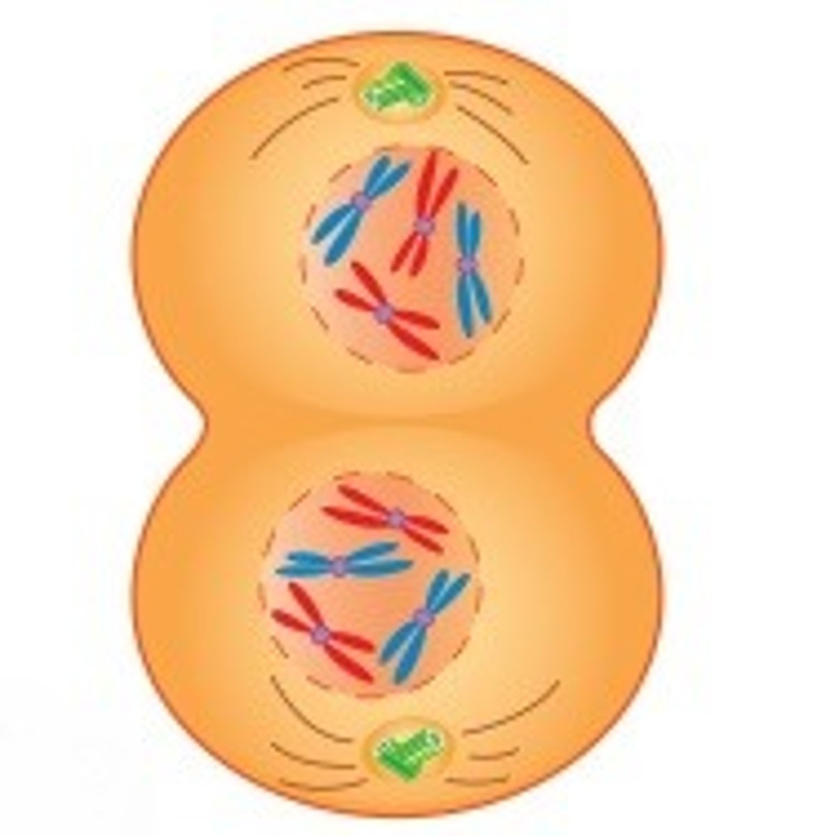
Metaphase II
The chromosomes will line up on the metaphase plate
Anaphase II
the centromeres will divide and separate the chromosomes into sister chromatids--they will be pulled to the opposite poles by spindle fibers
Telophase II
1. A nuclear membrane will form around each new nucleus

Cytokinesis
This is when two daughter cells are formed
Number of cells that are present after the completion of meiosis II
There are up to 4 haploid daughter cells that are produced per gametocyte
Mitosis key concepts
1. It stays diploid the whole time because it only ever deals with sister chromatids
2. Occurs in all dividing cells
3. The homologous chromosomes will never line up/pair
4. There is no crossing over that happens
Meiosis key concepts
1. It goes from diploid to haploid
2. This only occurs in sex cells
3. The homologous chromosomes will align on the opposite sides of the metaphase plate
4. Crossing over can occur

Biological sex
This is determined by the 23rd pair of chromosomes
Female: XX
Male: XY
X chromosome
This carries a sizeable amount of genetic information
Sex linked disorders
Sex-linked is X-linked
- mutations in genes of X chromosome
Hemizygous
- males are considered to be hemizygous with many of the genes that are on the X chromosome because they only have one copy
- This means that a male with a diseased allele on the X chromosome will necessarily express the allele
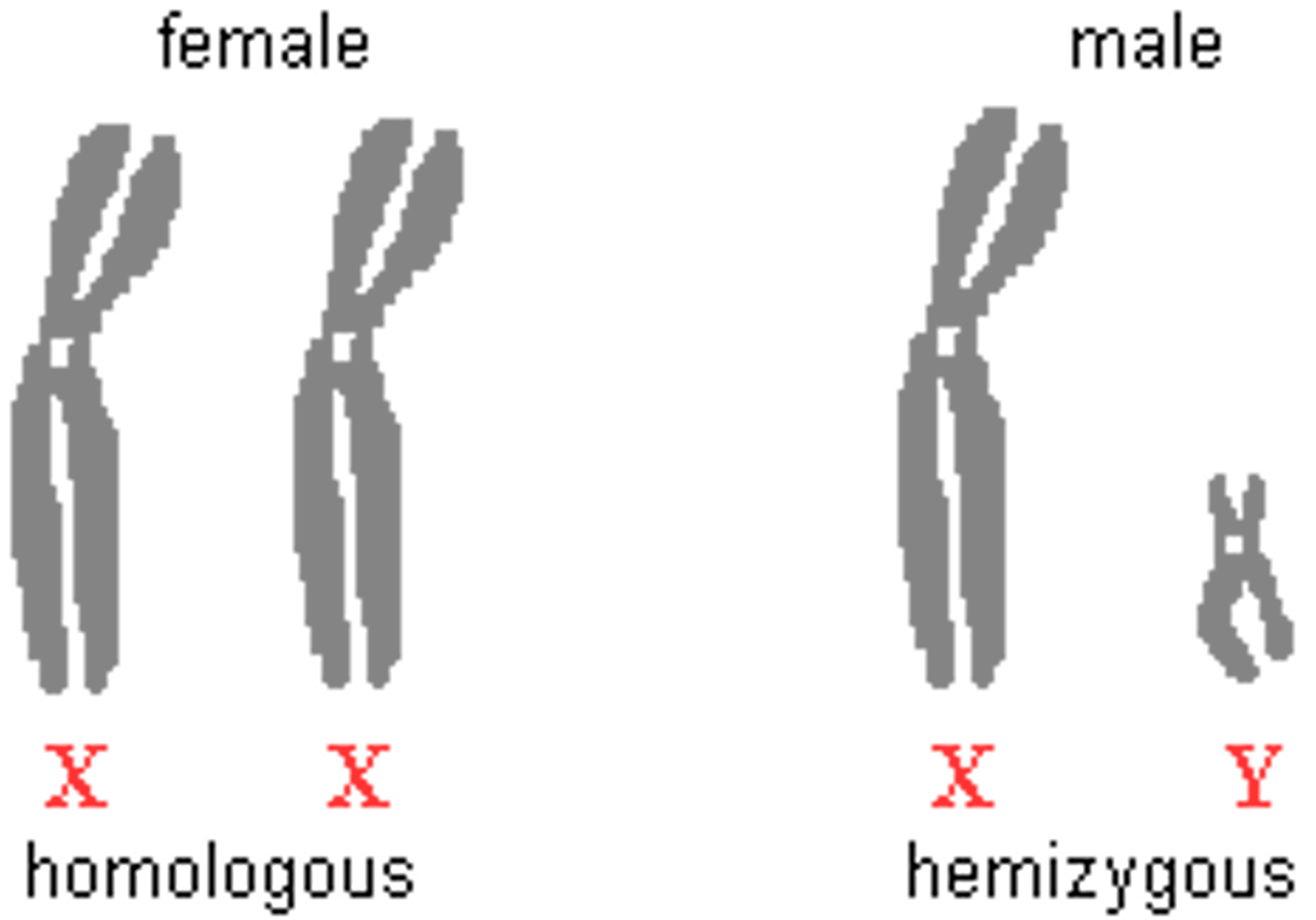
Females and X linked disorders
They can be homozygous or heterozygous with respect to the genes that are on the X chromosome because they have 2 copies of the X chromosome
X linked disorders are mostly ______________ inherited
recessively
- Thus females express these disorder far less frequently than males
Carriers
These are females that are carrying a diseased allele on an X chromosome and they are not exhibiting the disease
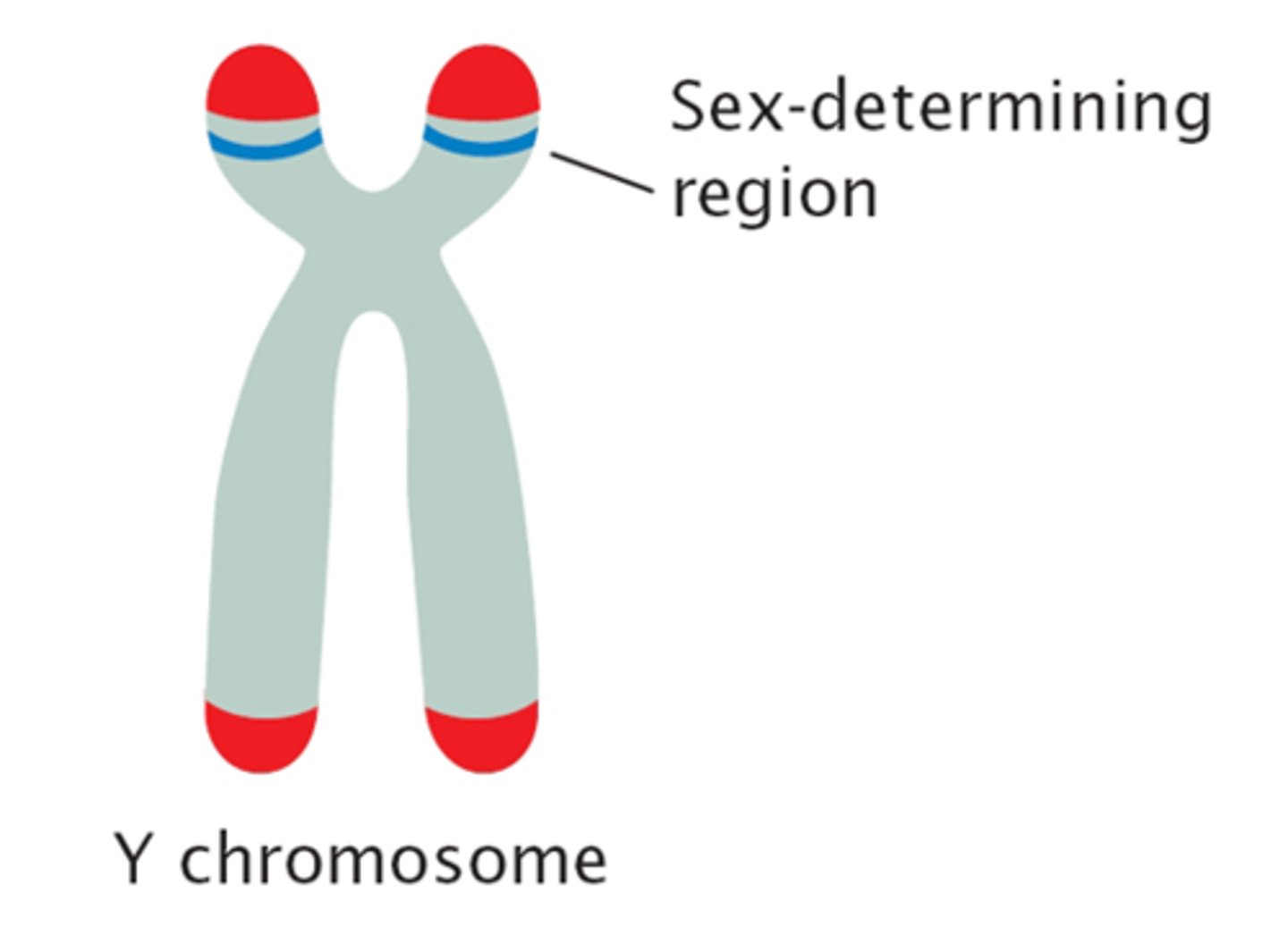
SRY
This is the sex determining region on the Y chromosome and codes for a transcription factor that initiate testis differentiation and thus the formation of the male gonads
- Thus in the absence of the Y chromosome all zygotes will be female
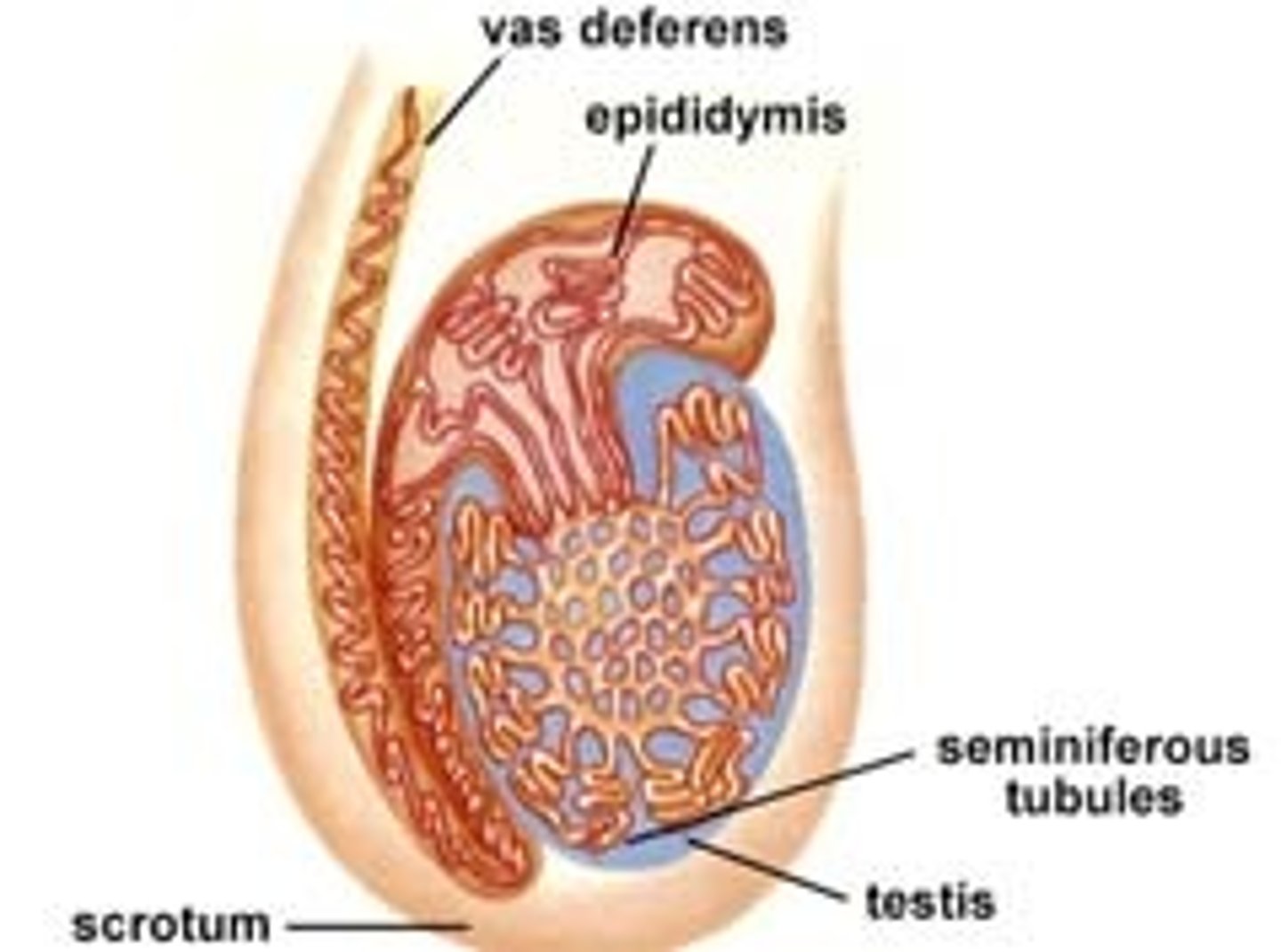
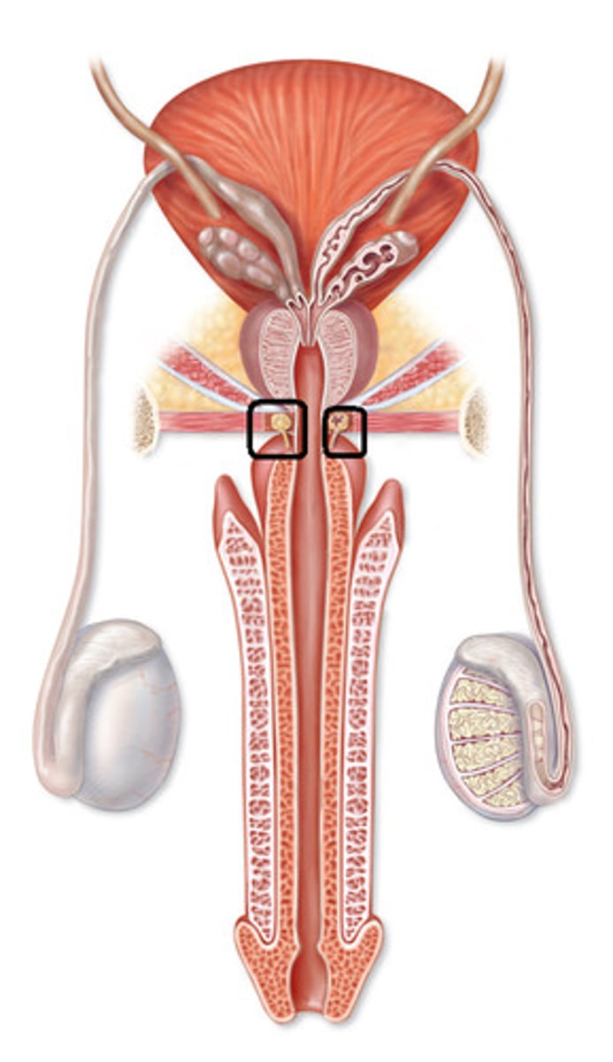
Testes
These are the what the primitive gonads develop into
2 functional components of the testes
1. Seminiferous tubules
2. Interstitial cells of Leydig
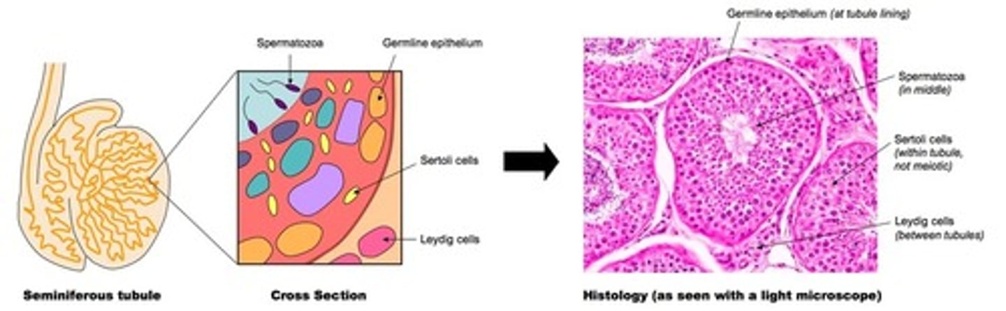
Seminiferous tubules
These produce sperm
They are highly coiled cells
Sertoli cells
These are cells that will nourish sperm in the seminiferous tubules

Leydig cells
These will secrete testosterone and other male sex hormones (androgens)
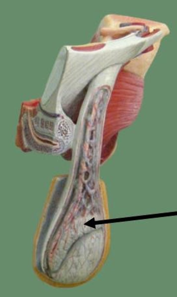
Scrotum
This is the external skin pouch that the testes are located in that hangs below the penis
This maintains a temperature of 2-4 degrees Celsius lower than the rest of the body- there is a layer around the vas deferens (also called ductus deferents) that can raise and lower the testis to maintain the proper temperature for the sperm development
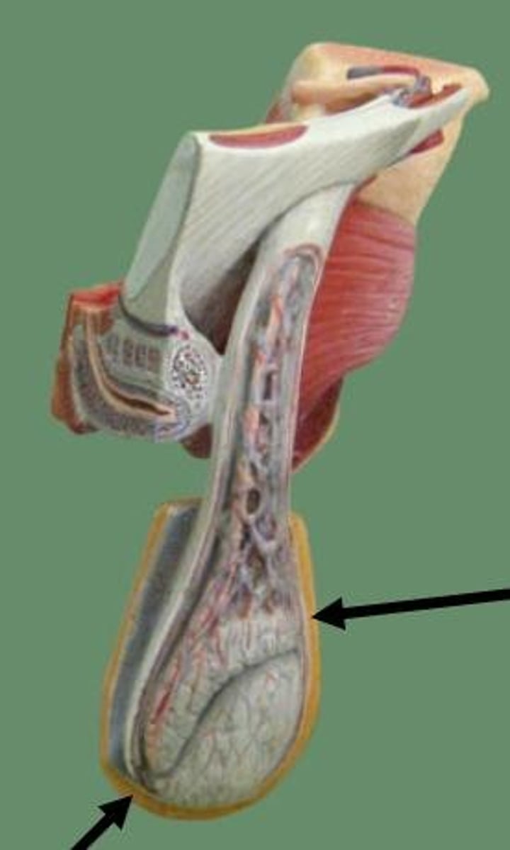
Epididymis
This is where the sperm are stored until ejaculation and where their flagella gain motility
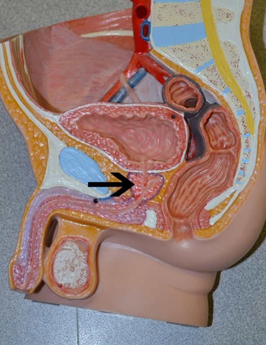
Ejaculation
- The sperm travel through the vas deferents then to the ejaculatory duct at the posterior edge of the prostate gland
- the two ejaculatory ducts will then fuse to form the urethra which will then carry sperm through the penis to exit the body
The reproductive and the urinary systems will share a common pathway in males
Seminal fluid
As the sperm passes through the reproductive tract they will be mixed with seminal fluid that is produced through a combined effort by:
1. Seminal vesicles
2. Prostate gland
3. Bulbourethral gland
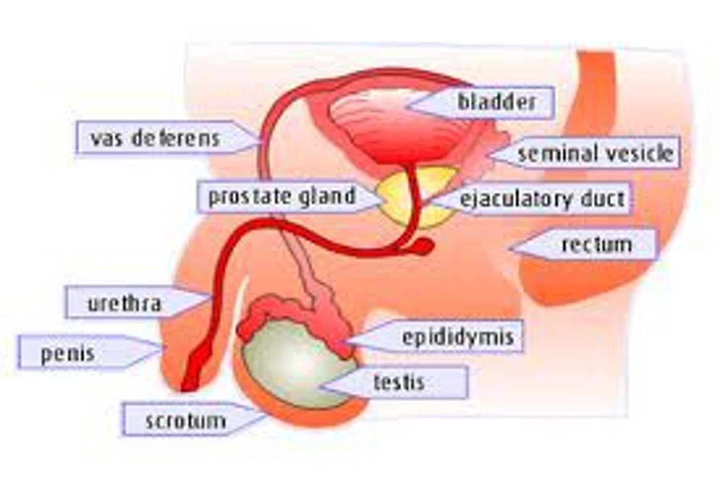
Seminal vesicles
These contribute "fructose" to nourish the sperm
and it gives mildly "alkaline" properties to the sperm
so that they will be able to survive the relatively acidic environment of the female reproductive tract
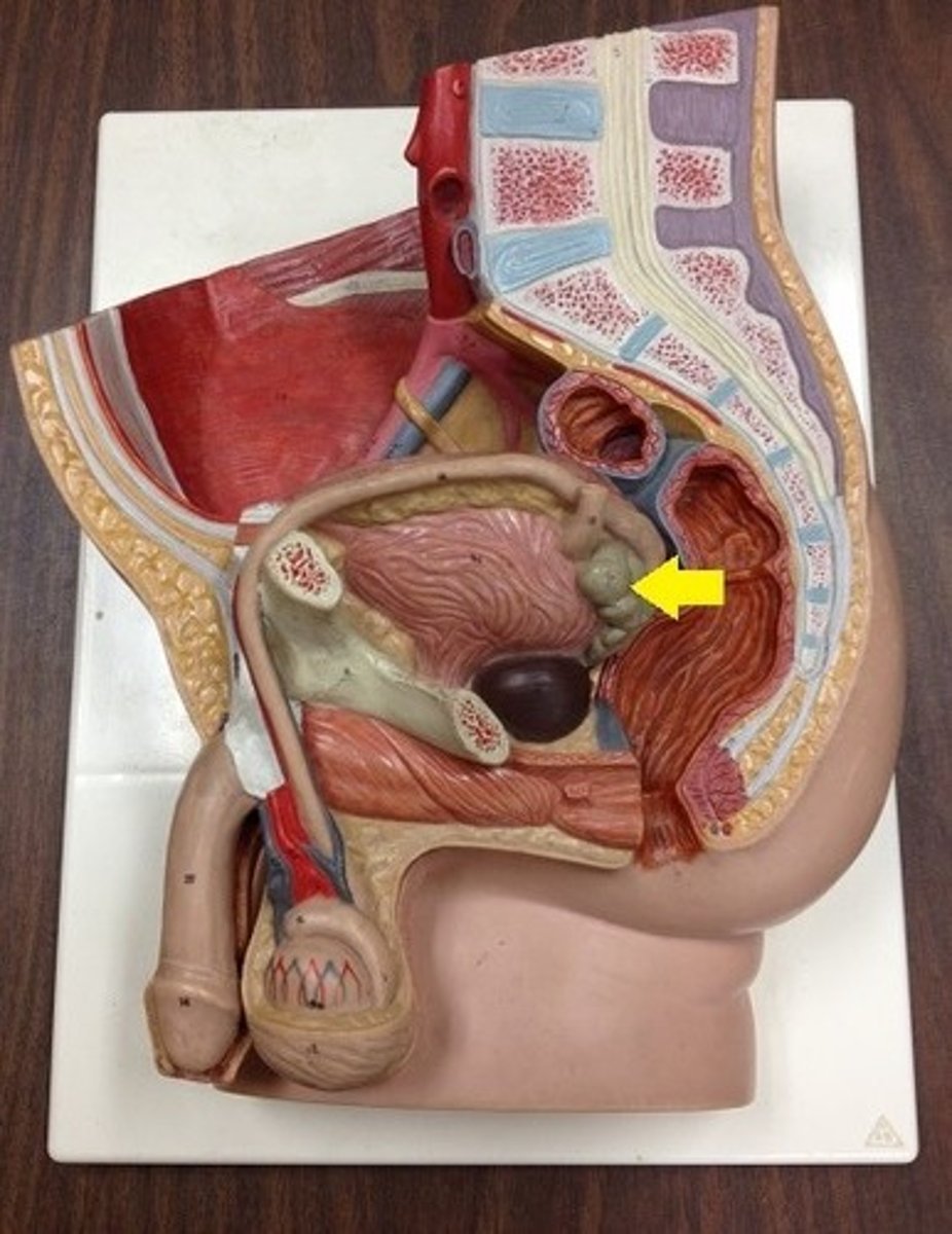
Prostate gland
This contributes alkaline properties to survive the acidic environment of the female reproductive tract
Bulbourethral (Cowper's) gland
This produces a clear viscous fluid that cleans out any remnants of urine and lubricates the urethra during sexual arousal
Semen
This is the combination of sperm and seminal fluid
Mnemonic to remember the pathway of sperm through the reproductive tract
Seve (n) up
1. Seminiferous tubules
2. Epididymis
3. Vas deferens
4. Ejaculatory duct
5. Nothing
6. Urethra
7. Penis
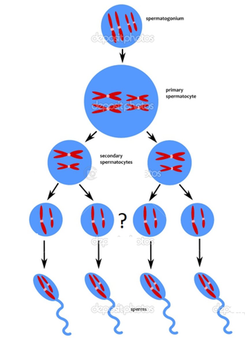
Spermatogenesis
This is the process of sperm formation that occurs through meiosis that happens in the seminiferous tubules

Spermatogonia
This is the diploid stem cells that are in the seminiferous tubules that will go through meiosis

Primary spermatocytes
These are what result after the Spermatogonia first replicate their genetic material (S Stage)
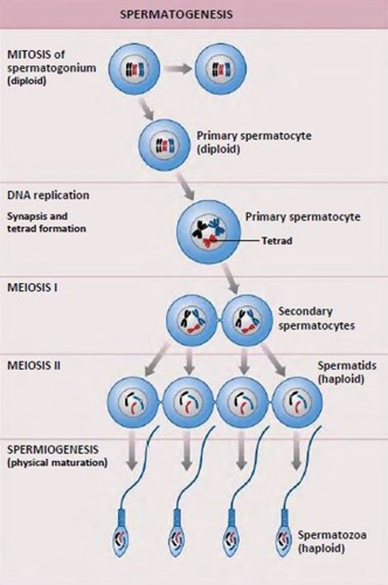
Secondary spermatocytes
This is what results after the first meiosis division of Primary Spermatocytes and it is haploid
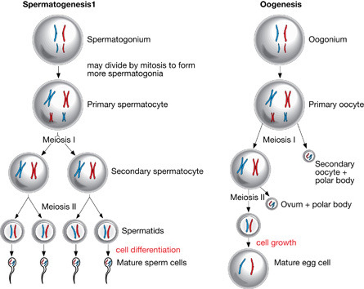
Spermatids
These are the result of the second meiotic division of the secondary spermatocytes that will result in the 4 haploid spermatocytes cells