Cell Bio Exam 4
1/125
There's no tags or description
Looks like no tags are added yet.
Name | Mastery | Learn | Test | Matching | Spaced | Call with Kai |
|---|
No analytics yet
Send a link to your students to track their progress
126 Terms
Cytoskeleton
A network of protein fibers that extends throughout the cytoplasm
Which of the following is false about the cytoskeleton?
A. Provides mechanical strength and helps maintain the cell’s shape
B. Enables cell movement and motility
C. Acts as a network for transporting organelles, vesicles, and molecules within the cell
D. Organizes and separates chromosomes and helps divide the cytoplasm
E. Facilitates cell adhesion and signal transduction
All of the above
What are the three main cytoskeletal filaments
A. Microtubules, myofibrils, intermediate filaments
B. Myofibrils, intermediate filaments, microfilaments
C. Microfilaments, sarcomeres, myosin
D. Microtubules, Intermediate filaments, microfilaments
D. Microtubules, Intermediate filaments, microfilaments
T/F: The subunit of a microtubule is made up of an alpha and beta tubulin
True
T/F: The subunit for Intermediate filaments is a heterogeneous family of fibrous proteins
True
T/F: The subunit for Microfilaments is actin
True
Intermediate filament structure
Ropelike
~10 nm diameter
Very flexible
Good tensile strength
Deform under stress, do not rupture
Intermediate filament examples
Nuclear lamina meshwork
Keratins
Microtubule structure
Hollow tubulin cylinders
~25 nm outer diameter
More ridgid
Rupture under stress
Minus (-) end attached to the microtubule organizing center (centrosome)
T/F: Microtubules serve as tracks for kinesin and dynein in vesicular transport.
True
Microfilament (Actin filament) structure
Helical polymers of actin
Flexible
~7 nm diameter
Most concentrated just inside plasma membrane
Organization: linear bundles, 2D networks, 3D gels
If you make a cross-section across an intermediate filament, how many monomers would you typically see in that section?
32 (actual range 16-48)
When the intermediate filament is found in epithelial cells
A. What is the subtype/major class
and
B. is this cytoplasmic or nuclear
A. keratin filaments
B. Cytoplasmic
When the intermediate filament is found in connective tissue, muscle, and glial cells
A. What is the subtype/major class
and
B. is this cytoplasmic or nuclear
A. vimentin and vimnetin-related filaments
B. Cytoplasmic
When the intermediate filament is found in nerve cells
A. What is the subtype/major class
and
B. is this cytoplasmic or nuclear
A. Neurofilaments
B. Cytoplasmic
When the intermediate filament is found in all animals cells
A. What is the subtype/major class
and
B. is this cytoplasmic or nuclear
A. Nuclear lamins
B. Nuclear
Keratin filaments (Cytoplasmic IFs)
Most diverse IF
Present in all epithelial cells (tissue surfaces and linings)
Provide high tensile strength through lace-like networks
Make up hair, nails, feathers, hooves, horns
How do Keratin filaments connect to adjacent cells?
A. Directly through desmosomes
B. Indirectly through desmosomes
C. Directly with no intermediate
B. Indirectly with no intermediate
B. Indirectly through desmosomes
What happens at desmosomes?
Cadherin molecules connect to keratin filaments
Cadherins then bind to each other in extracellular space
T/F: Cytoplasmic IFs do not provide mechanical strength
False
T/F: A mutant form of keratin makes skin less prone to blisters
False
Nuclear Lamina
Made of special IFs called Lamins
Support the nuclear envelope
Provides attachment points for chromosomes
T/F: During interphase, lamins play an important role in gene expression regulation by tethering euchromatin to the nuclear periphery.
False
T/F: Plectins stabilize IFs and link them to other IFs, microtubules, and microfilaments
True
Where do protein complexes bridge the nucleus and cytoplasm
The nuclear envelope
Which of the following statements is FALSE regarding the structure and function of intermediate filaments?
A. Intermediate filaments can connect cells at cell–cell junctions called desmosomes.
B. Intermediate filaments protect cells from mechanical stress because they have high tensile strength and resist stretching.
C. Intermediate filaments are constructed of identical subunits found in all eukaryotic cells.
D. Each filament is made of eight strands, and each strand is made from staggered tetramers linked end to end.
B. Intermediate filaments protect cells from mechanical stress because they have high tensile strength and resist stretching.
Microtubule (MT) structure
Stiff hollow tube made of 13 protofilaments made of tubulin dimers
Tubulin dimers: alpha tubulin, Beta tubulin. GTP bound
Beta tubulin = plus end
Alpha tubulin = minus end
T/F: Microtubules usually do not grow out from an organizing center
False
What is a microtubule organizing center?
centrosome or basal body
T/F: Microtubules typically have their plus ends extending out toward cell periphery
True
T/F: Microtubules are nucleated from gamma tubulin complexes in centrosomes, growing out the + ends
True
T/F: Microtubules display dynamic instability (rapid growth and shrinkage)
True
What is dynamic instabiliy driven by?
GTP hydrolysis
How are Microtubules grown/disassembled?
Beta tubulin is a GTPase
Multiple GTP-tubulin dimers bind together
GTP is hydrolyzed, causing weakness in the protofilament
Section of the protofilament breaks off
GDP is exchanged for GTP
Cycle repeats
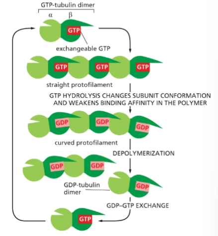
GTP ‘Cap’
When microtubules assemble too fast, the most recent Beta-tubulin may still have GTP instead of GDP
‘Cap’ favors growth
T/F: If GTP hydrolysis proceeds faster than tubulin addition, GTP cap is lost and microtubule disassembles
True
T/F: Tubulin dimers carrying GDP bind more tightly to one another than do tubulin dimers carrying GTP.
False
T/F: Colchicine arrests cells in metaphase by inhibiting MT polymerization. This property makes colchicine useful in karyotyping.
True
What does the drug, Taxol do?
Binds to filaments in microtubules and prevents depolymerization
What does the drug Colchicine/colcemid do?
Forms a complex with tubulin dimers, preventing further polymerization
What does the drug Nocodazole do?
Binds to tubulin dimers, preventing their polymerization
What are the roles of microtubules in a cell?
Intracellular transport
Cell organization
Structural support of eukaryotic cilia and flagella
Spindle assembly and function
T/F: Microtubules guide the transport of organelles, vesicles, and macromolecules (e.g. transport along the nerve axon)
True
What is intracellular transport mediated by?
Transport proteins called motor proteins
T/F: Motor proteins use kinesin and dynein to move along microtubules
True
Kinesin moves the motor protein toward the ____ end and Dynein moves the motor protein toward the ____ end
A. minus; plus
B. plus; minus
C. plus or minus; plus or minus
B. plus; minus
T/F: Kinesin and dynein movement is due to conformational changes powered by ATP hydrolysis
True
Which motor protein is used to move vesicles from Golgi apparatus to ER?
Kinesin
What do kinesin and dyein transport within a cell
organelles and vesicles
T/F: Microtubules help position organelles in the cytoplasm
True
T/F: Motor proteins arrange the ER and Golgi apparatus
True
T/F: Stable microtubules nucleated from basal bodies support cilia and flagella
True
What is the characteristic arrangement in cilia and flagella called?
9+2
_____ coordinates cilia and flagella movement
A. Flagellar dynein
B. Flagellar kinesin
C. Ciliary dynein
D. Ciliary kinesin
C. Ciliary dynein
What movement does dynein produce in an isolated doublet?
A. Microtubule Sliding
B. Microtubule Bending
A. Microtubule Sliding
What movement does dynein produce in a normal flagellum?
A. Microtubule Sliding
B. Microtubule Bending
B. Microtubule Bending
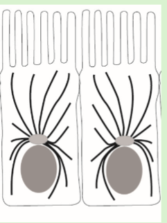
Identify the cytoskeletal structures (black lines) depicted in the epithelial cells shown
A. microfilaments
B. microtubules
C. intermediate filaments
D. nanotubes
B. microtubules
What type of cytoskeletal filaments lines the inner side of the nuclear envelope?
A. intermediate filaments
B. microtubules
C. actin filaments
A. intermediate filaments
Microfilaments (MFs/Actin filaments) structure
Monomers of globular actin proteins in a twisted two-stranded helix
Distinct polarity; more likely to be added to the + end
Actin monomer structure
Folds into two lobes
Cleft between lobes contains 1 ATP or ADP
ATP bound monomers tend to polymerize into a stable actin filament
How are Microfilaments grown/dissasembled?
Actin-ATP added to MF + end
ATP hydrolyzed to ADP, “trapping” the Actin-ADP
Actin-ADP dissociates at - end
What does the drug Phalloidin do?
Binds and stabilizes Actin Filaments
Also used for staining actin filaments
What does the drug Cytochalasin do?
Caps the + end of Actin Filaments, preventing polymerization there
What does the drug Latrunculin do?
Binds actin monomers, preventing their polymerization
What is microfilament behavior modified by?
Actin-binding proteins
What does the Nucleating protein do?
Promotes polymerization
What does the monomer sequestering-protein do?
Bind monomers, reducing change of polymerization
What % of the protein in animal cells is Actin?
A. 10%
B. 38%
C. 8%
D. 5%
D. 5%
T/F: Actin-binding proteins regulate when and where actin filaments will form and grow.
True
How many protofilaments are found in a typical actin filament?
A. 4
B. 32
C. 2
D. 9
E. 13
C. 2
What are the two roles of actin filaments?
Stabilizing and Temporary
Stable examples: Microvilli and contractile bundles
Temporary examples: Cytokinetic contractile ring and protrusions needed for cell movement
What does Cell Crawling depend on?
The cortical (cortex) actin
Actin Cortex
A thin layer of actin mesh-work that uniformly underlies the plasma membrane of the entire cell
Lamellipodium
Thin (0.1-0.3 um) and usually long (1-5 um) projection that adheres to the underlying substrate
Branched actin network
T/F: the primary energy source for cell crawling is ATP
True
Filopodia
Unbranched actin network
T/F: Actin polymerization at the leading edge of the cell results in the protrusion of lamellipodia and filopodia, motile structures that form and retract at great speed
True
A web of _____ pushes the leading edge of a lamellipodium forward
A. Actin
B. Microtubules
C. Polymerizing microtubules
D. Polymerizing actin
D. Polymerizing actin
What are Lamellipodia nucleated by?
Actin-Related Protein (ARP) complexes
What is the main structural protein in the extracellular matrix?
Collagen
What do integrins bind to?
Extracellular proteins and internal microfilaments
T/F: Integrins are transmembrane proteins that interact with the crawling cell’s environment
True
______ is an important ECM protein that mediates cell-ECM interaction
Fibronectin
How does a cell know what type of microfilament network to form?
Extracellular signals that alter the arrangement of actin filaments
What do Rho proteins do?
promote contractile bundles
What do Rac proteins do
Promote lamellipodia
What does Cdc42 do?
Promotes filopodia
T/F: Myosin I is the most complex myosin
False
Myosin I
Involved in vesicle movement
Can bind to an actin filament in the cortex
Generally moves toward the + end of actin filaments
Uses ATP hydrolysis for energy
T/F: Actin associates with myosin to form contractile structures
True
The head of Myosin I attaches to _____ and the tail attaches to ____
A. a molecule or organelle; an actin filament
B. an actin filament; a molecule or organelle
B. an actin filament; a molecule or organelle
What do muscle contractions depend on?
Interacting filaments of actin and myosin
Bipolar myosin filaments
When Myosin II molecules associate with one another
What creates a contractile force?
Movement of Myosin II along oppositely orientated action filaments
Where does the Myosin II head group walk toward?
A. + end
B. - end
A. + end
T/F: Myosin II is responsible for muscle contraction by forming cross-bridges with actin filaments and moving toward the plus end of the actin filaments.
True
Sarcomere
Contractile unit of muscle
Mofibril
repeating sarcomeres
T/F: The minus end of actin filaments attach to the Z disc in a sarcomere
True
Muscles contract by a _____ mechanism
sliding-filament