cells and tissues of the immune system
 Call Kai
 Learn
Learn Practice Test
 Spaced Repetition
 Match
Match Flashcards
 Knowt Play
1/45

There's no tags or description
Looks like no tags are added yet.
Name | Mastery | Learn | Test | Matching | Spaced | Call with Kai |
|---|
No analytics yet
Send a link to your students to track their progress
46 Terms
granulocytes
neutrophils
eosinophils
basophils
mononuclear cells
monocytes
lymphocytes
phagocytosis of bacteria
neutrophls
monocytes
destruction of parasites
eosinophils
basophils (tissue mast cells)
immunity to viruses
lymphocytes
primary lymphoid organs
bone marrow
thymus
lymphocyte development
secondary lymphoid organs
lymph nodes
spleen
malt (eg peyers patches)
lymphocyte response to infection
lymphocyte development
the bone marrow is the source of all lymphocytes
thesedifferentiate from lymphoid precursors into B or T cells
B cells remain in B
Tcells must leave B and goto thymus
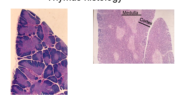
thymus
cranial to the heart
lobulated
each lobule has a darker cortex and lighter medulla
T cell development in the thymus
must generate a unique antigen receptor (TCR)
mus decide whetehr to become a CD4 or CD8
immature t cells in the thymus are initially double positive (CD4 and CD8)
if the tcr perfers mhc class 1 they become cd t cell
if the tcr prefers mhc class ii they become cd4 t cell
T cell selection in the thymus
some tcr fail to interact with mhc molecules and are therefore useless
some tcr recognise peptides from self antigens and are potentially dangerous
the thymus assesses t cells and disposes of those that ae unreactive or autoreactive
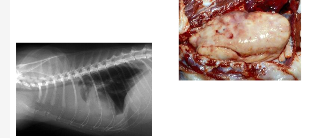
thymic lymphoma
neoplasia of thymic kymphocytes
associated with FeLV infection
anorexia, weight loss, dyspnoea
bursa of fabricius
found in birds
cloacal tonsil
primary lymphoid organ for maturation and differentiation of B cells
bone marrow in mammals
bursa in birds
reaches maximum size around 2 weeks after hatching and then undergoes gradual involution
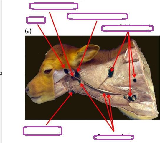
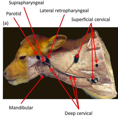
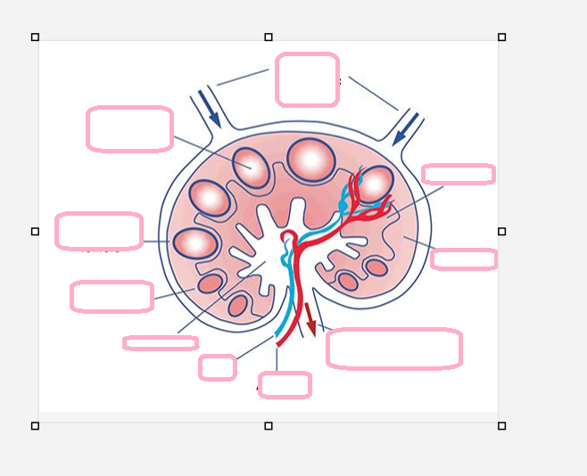
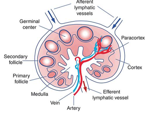
where are t cells and b cells in the lymph node
t cells in paracortex
b cells in follicles
lymph node function
filters tissue fliod (lymph) and traps foreign organisms
lymphocytes enter via blood vessels to inspect th anitgens present
the LN provides an env for recognition of antigens by lymphocytes
also provide the right conditions for lymphocytes activationa nd differentiation
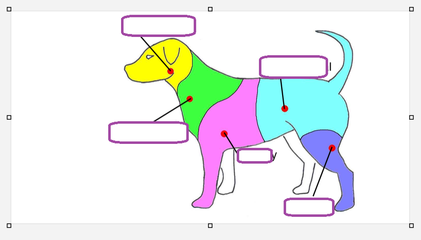
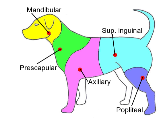
what do the efferent lymphatics join to form
thoracic and tracheal duct
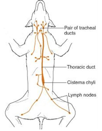
where do the thoracic and tracheal ducts empty
cranial vena cava
what are the 2 routes
free antigen is swept into LN in affernt lymph and is trapped
specialist antigen presenting cells in hte tissues eat antigen then migrate via the afferent lymph to the LN paracortex
dendritic cells
differentiate from circulating monocytes
immature dc usually located at epithelial surfaces where they eat antigen
migrate via afferent lymphatics to LN during which times they process antigen
take up residency in the LN paracortex where they present antigen to naive T cells
lymphadenopathy
enlargement of lymph node
infection- reactive lymph node
neoplasia- lymphoma
what can be taken to see what is happening inside the LN
aspirate
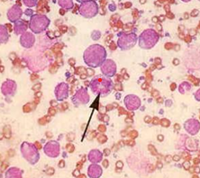
lymphoblastic lymphoma
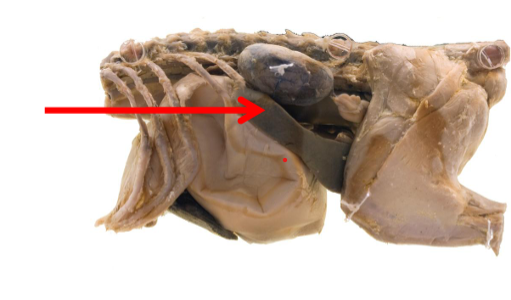
what is this
spleen
in the abdominal cavity
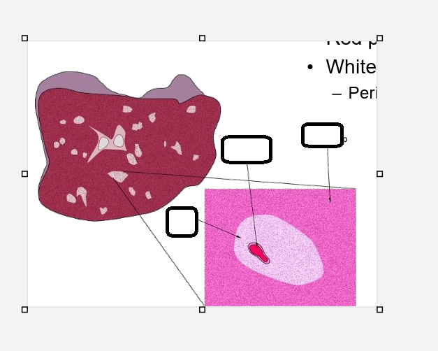
spleen structure
white pulp for haemotological functions
white pulp for immunological functions
periarteriolar lymphoid sheaths
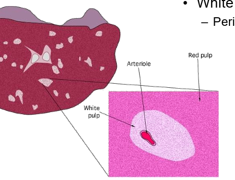
spleen function
haematopoietic organ in foetal life
sore of rbc and platelets
removal of particulate material from bloodstream
foreign organisms
old rbc
immune responses to blood borne pathogens often transmitted by arthropod vectors
what can the spleen be affected by
haemangiosarcoma in german sheperd dogs
neoplasia
muscosal associated lymphoid tissue
respiratory tract- bronchial associated
git- gut associated
tonsils are aggregates of lymphoid tissue in the pharynx that initiate immune responses entering the respiratory or alimentary tracts
initiate immune responses to organisms at muscosal surfaces
peyers patches
found predominantlyin the ileum at the ileocaecal junction
large aggregates of lymphoid tissue found in the walls of the small intestine particularly the ileum and ileocaecal junction
consist of lymphoid follicles of densely packed lymphocytes predo B cells
overlying epithelium containsmicrofold cells
important in transferring antigen from the intestinal lumen to the lymphoid tissue underneath
B cells which are stimulated with antigen differentiate into plasma cells which secrete IgA which is exported across the mucosal surface into the lumen to prevent pathogens from attaching to mucosal epithelial cells
reactive lymph node
in a healty animal lymph nodes contain primary follicles
when there is an infection lymph become activated and multiply and enlarge
B cells begin to proliferate and differentiate into plasma cells
this can be seen histologically as a central lighter region called the germinal centre
this is the secondary follicle
following activation the mature plasma cells migrate to the medullar cords where they begin secreting antibody into efferent lymph which reaches the blood
what are lymph nodes surrounded by
a capsule that is penetrated by many afferent lymphatic vessels
path of lymph
lymph is filtered as it moved through the lymph node then leaves through a single efferent lymphatic vessel located at the hilius
lymph travels through a network of lymphatic vessles returning to the circulation via the trachea and thoracic duct
blood vessels also enter and leave the LN from the hilius
within the paracortex blood vessels form high endothelial venules that function to allow lymphocytes to exit the bloodstream and enter the lymph tissue to look for antigen
structure of the lymp
divided into a peripheral cortex and centra medulla
paracortex in between
b cells are located in the cortex and arranged into follicles
t cells are found scattered loosely in the paracotex
medulla contains many macrophages and plasna cells
in pgs the lymph node structure is inverted with the medulla surrounded by a central cortex
what are the antigen presenting cells in the paracortex called
dendritic cells
they present the antigen to t cell
where is the spleen located and what is the structure of it
located in anterior left quadrant of the abdomen
surrounded by a fibrous or fibromuscular capsule
red pulp functions as teh erythrocyte and platelet store
white pulp is rich in lymphocytes and is where immune responses occur
located around blood vessels forming peri arteriolar lymphnatic sheaths
marginal zone between red and white is rich in macrophages for trapping particulate material and antigens
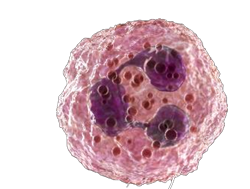
neutrophil
polymorphonuclear leukocytes
important in the acute inflammatory response to infection and play a key role in phagocytosis
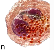
eosinophils
play a major role in inflammation
selectively recruited to tissue during parasite infections
desgined to degranulate and release their biologically active molecules onto the surface of pathogens
cell numbers often raised in parasite infection and allergic disease
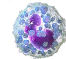
basophils
contribute to inflammation
mast cells are widespread throughout the tissues and are prominent around the epithelial surfaces
cytoplasmic granules contain inflammatory mediators including histamine
their degranulation results in a potent inflammatory response.
important in parasite defence
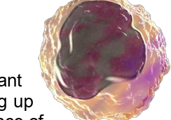
monocyte
produced in the bone marrow and circulate before leaving the blood to differenitate into ither tissue macrophages or dendritic cells
function primarily as phagocytic scavenger cells and are important for bacterial infections
professional antigen presenting cells
pick up antigen in infected tissues then move to LN to alert T cell
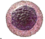
lymphocyte
adaptive immunity cells
B and T
circulate in blood and lymph and go to different secondary lymphoid tissues as part of immune surveillance
important for virus
how to evaluate wbc in a sample
use a tube containing anti coagulant eg EDTA, heparin
if you spin the sa,ple the cells are in the pellet and the clear liquid is plasma
if you dont use anti coagulant and allow blood to clot the clear liquid is serum which can be used to evaluate the soluble mediators of immunity
thymus
located in the anteiror mediastinum of the thoracic cavity
size varies with age
increases in size from birth to puberty
then it undergoes involution and is replaced by adipose tissue
arranged in lobules with an outer more densely cellular cortex and an inner less cellular medulla
immature t cells leave the bone marrow and enter the subcapsualr region where they proliferate before migrating through the cortex and medulla where they are assessed for suitability ebfore they are allowed to exit into the ciruclation as functional naive t cells
problems with TCR
they are made through random re arrangement of variable genes
some express receptors that fail to engage with MHC which is useless to the immune system and these cells are eliminated
some receptors are created tha inadvertently recognise self antigen, and these autoreactive T cells must also be eliminated otherwise they would attack healthy tissues after leaving the thymus