Concepts M1- Extraoral and Intraoral Exams
1/71
There's no tags or description
Looks like no tags are added yet.
Name | Mastery | Learn | Test | Matching | Spaced | Call with Kai | Chat |
|---|
No analytics yet
Send a link to your students to track their progress
72 Terms
Rationale for EIO exam
A: ASSESS for ABNORMALITIES
B: Establish BASELINE for comparison of future findings
C: Screen for CANCER
D: Identify DEVIATIONS from normal
Oral cancer
Accounts for 3-4% of cancers
> early detection can lead to 90% cure rate
Principle method of oral cancer detection
Observation by the dental professional
Common sites for oral cancer
- Lateral border of the tongue
- Oropharynx, hard and soft palate
- Lower lip
- Floor of the mouth
Standard of care importance
Failure to perform and diagnose oral cancer screening can be considered malpractice
Hygienist responsibilities
- Complete a thorough oral exam
- Ask about tobacco and alcohol use
- Inform pt about their risky habits to oral cancer
- Follow through on referrals to specialists
Assessment techniques
- Observation/ inspection
- Palpation
- Auscultation
- Olfaction
Observation/ inspection
Act of viewing and watching the client to collect data
Palpation
Act of using the sense of touch to collect data
Auscultation
Act of listening to and detecting body sounds in order to determine variations normal
Olfaction
Act of sensing body odors to detect variations from normal and potential disease
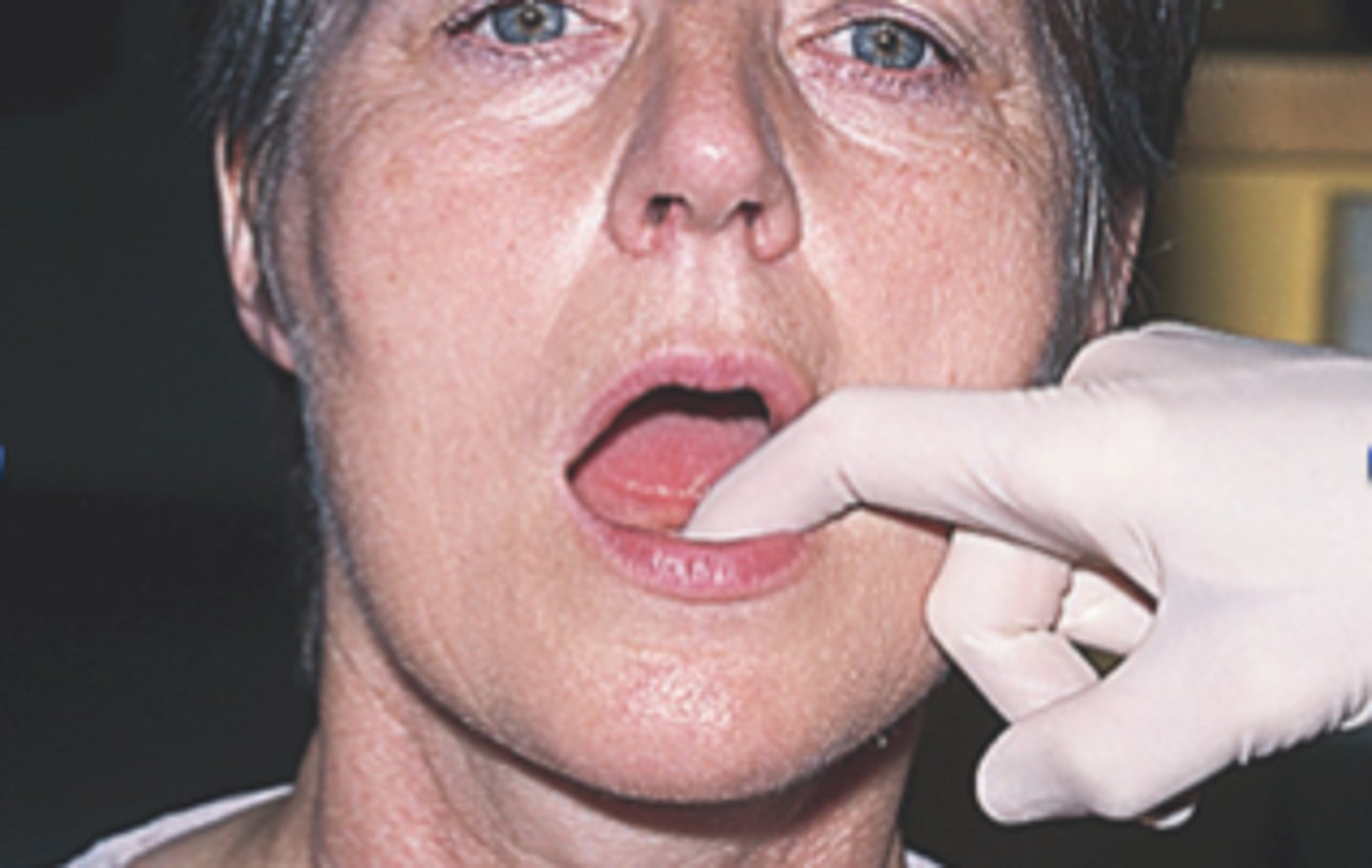
Digital palpation
Use of a single finger to move or press against tissue such as palate and alveolar ridge
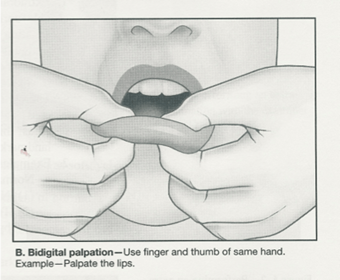
Bidigital palpation
Use of one or
more fingers and
thumb to move or
compress tissue such as
cheeks, tongue, lips
Bimanual palpation
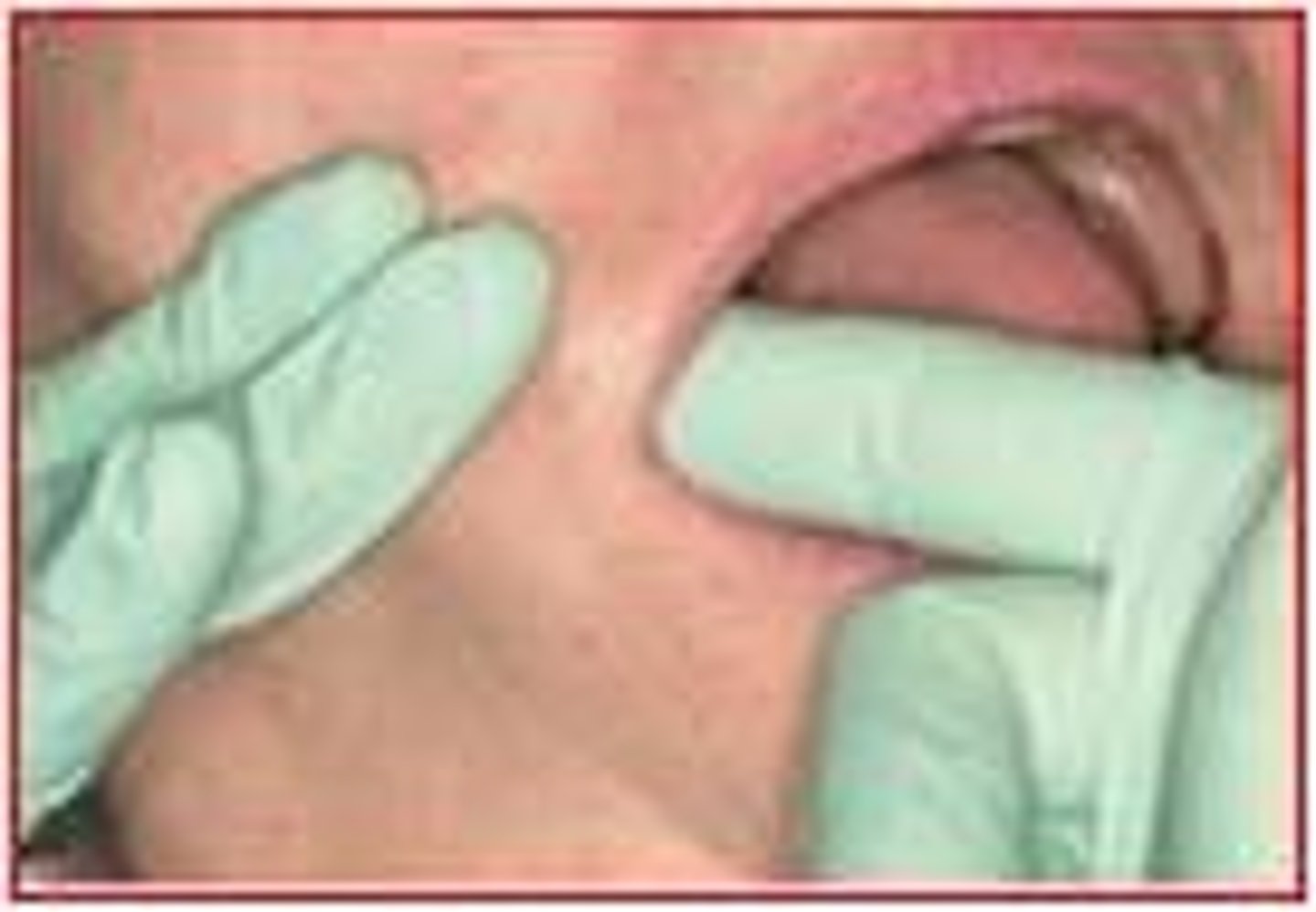
Use finger of one hand and the thumb of the other hand simultaneously to move or compress tissue
Bilateral palpation
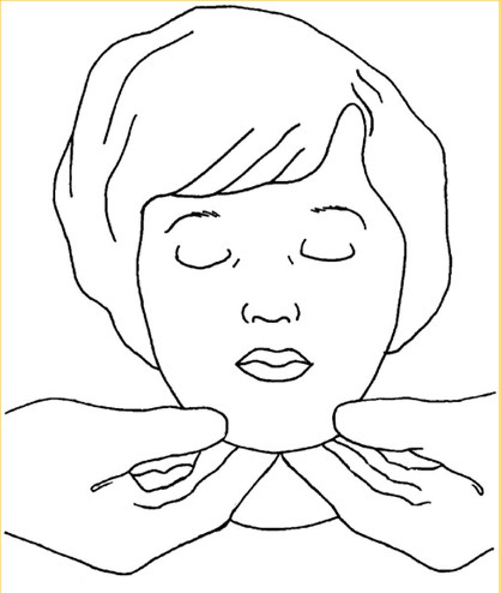
Use fingers of both hands simultaneously to move or press on contralateral sides of the head/neck, checks for symmetry
Thyroid gland
Secretes thyroid hormone that controls the body's metabolic rate
> check for nodules during exam
Thyroid gland location
- Middle of lower neck over trachea
- Shaped like a bow tie
Goiter
Enlarged thyroid gland due to iodine deficiency
Graves disease
Autoimmune disease, HYPERthyroidism
- gittery, hyperactive
- "thyroid" eye disease- bulging eyes
- can lead to thyroid enlargement
Hypothyroidism symptoms
Hashimoto's disease for example
- weight gain
- faint when tired
- often cold
Lymphatic system
- Network of lymph nodes connected by vessels, which plays a role in defense against infection
Lymph fluid
Carries nutrients and waste between body tissues and bloodstream
Lymph node function
Filter and trap bacteria, fungi, and waste
Lymph nodes
Vary in size from head of pin to baked bean
> 400-700 in the body
> 170-300 in neck
Lymph node major chains
in anterior and posterior of neck and under chin
Lymphadenopathy (enlarged lymph node)
Occurs when infected, if there is an inflammatory condition, or cancer
Lymph node enlargement due to a virus
½ to 1 inch
Lymph node enlargement due to bacterial infection
over 1 inch
Cancer can lead to
Painless lymph nodes
Normal lymph nodes are
Undetectable
Infected lymph nodes
- Firm
- Tender
- Enlarged
- Freely movable
- Swollen grape
Extraoral exam
Client is upright
- Have patient remove glasses
- Visually observe the symmetry of head, neck, eyes, nose, mouth, ears and complexion
Things to look for in an extraoral exam
- Lump
- Swelling
- Moles
- Freely movable node
- Fixed nodule
- Asymmetry
Other things to look for in an extraoral exam
- Irregular shape
- Firm or hard consistency
- Tender areas
- Red or discolored area
- Wound, bruise, scar
Cursory exam
Tongue blade used, move cheeks side to side look and back of throat before going in with hands in mirror
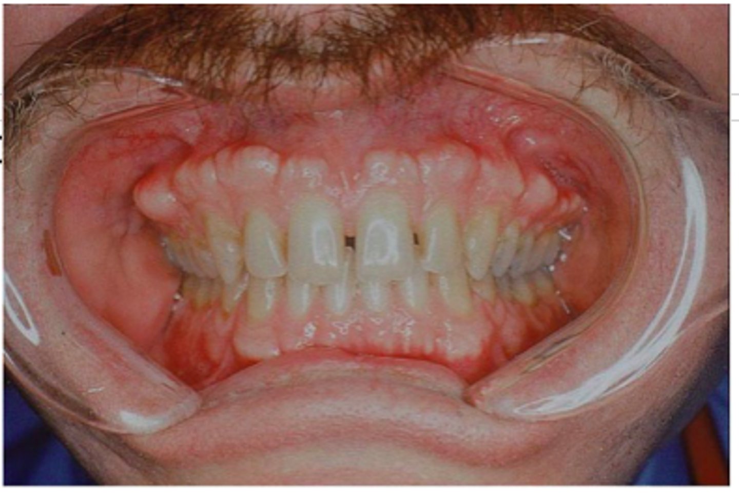
Intraoral exam
Client in a supine position
- Begin by using a tongue depressor to do a cursory examination of the oral cavity
- Follow cursory exam with initial visual exam using the mirror
Cursory exam purpose
Quick look into the mouth to notice dentures, partial orthodontics for example to be aware
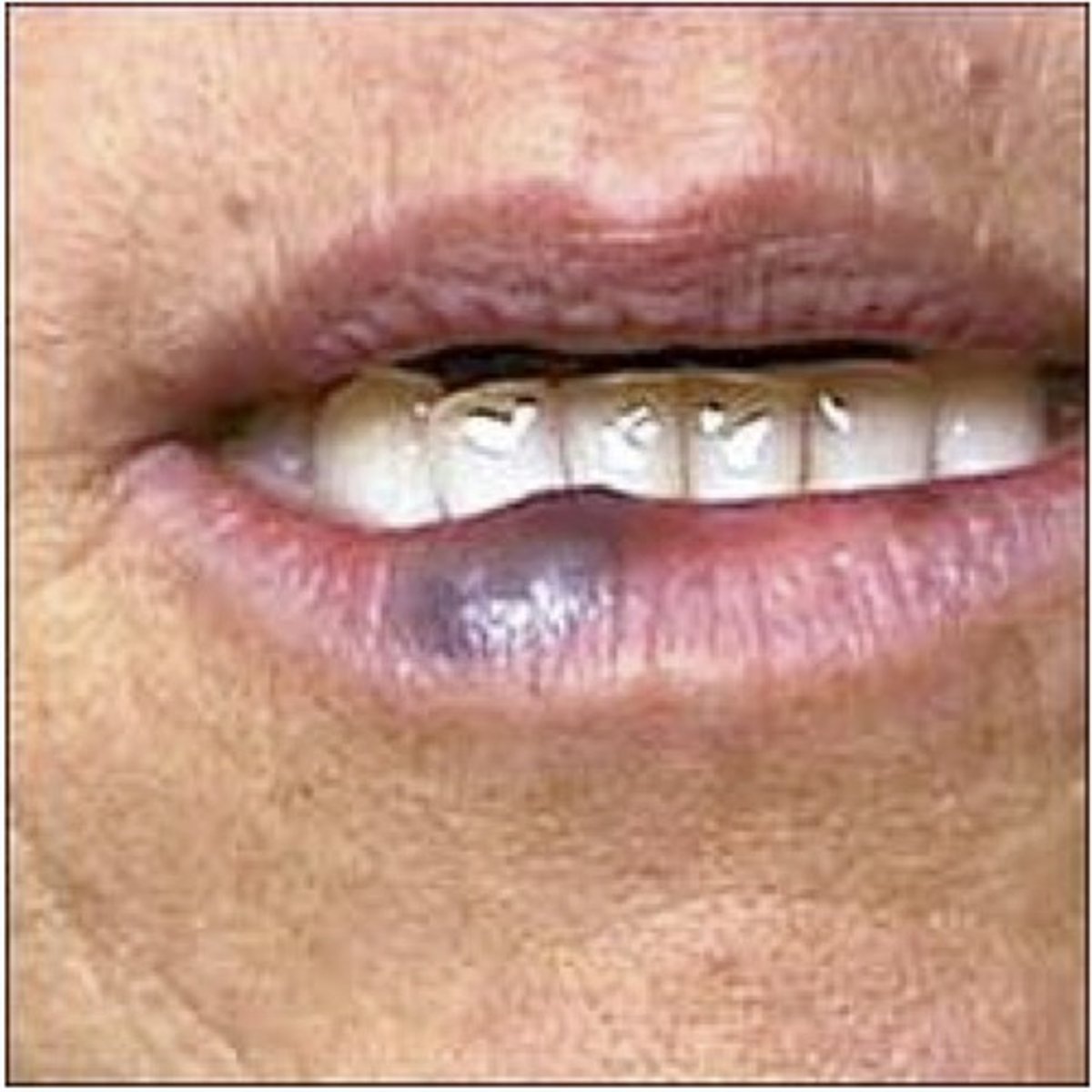
Varicosity
increased prominence of superficial veins
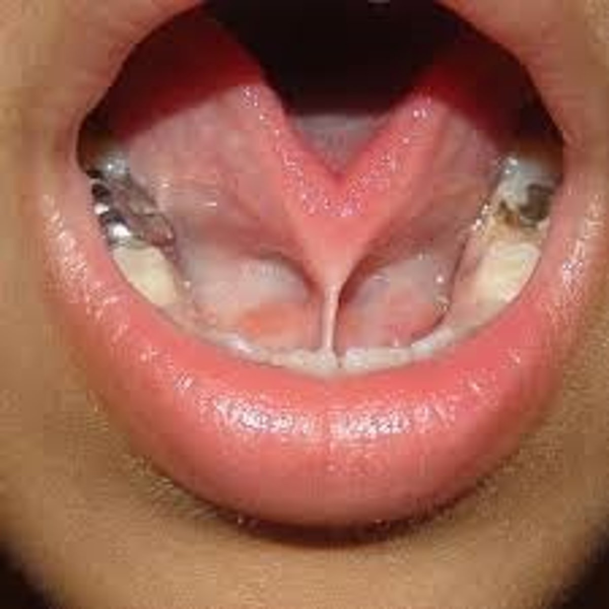
Epithelial tag on frenum
variation of normal due to chronic irritation or how frenum rests
Exostosis
bony growth (benign) arising from the surface of bone
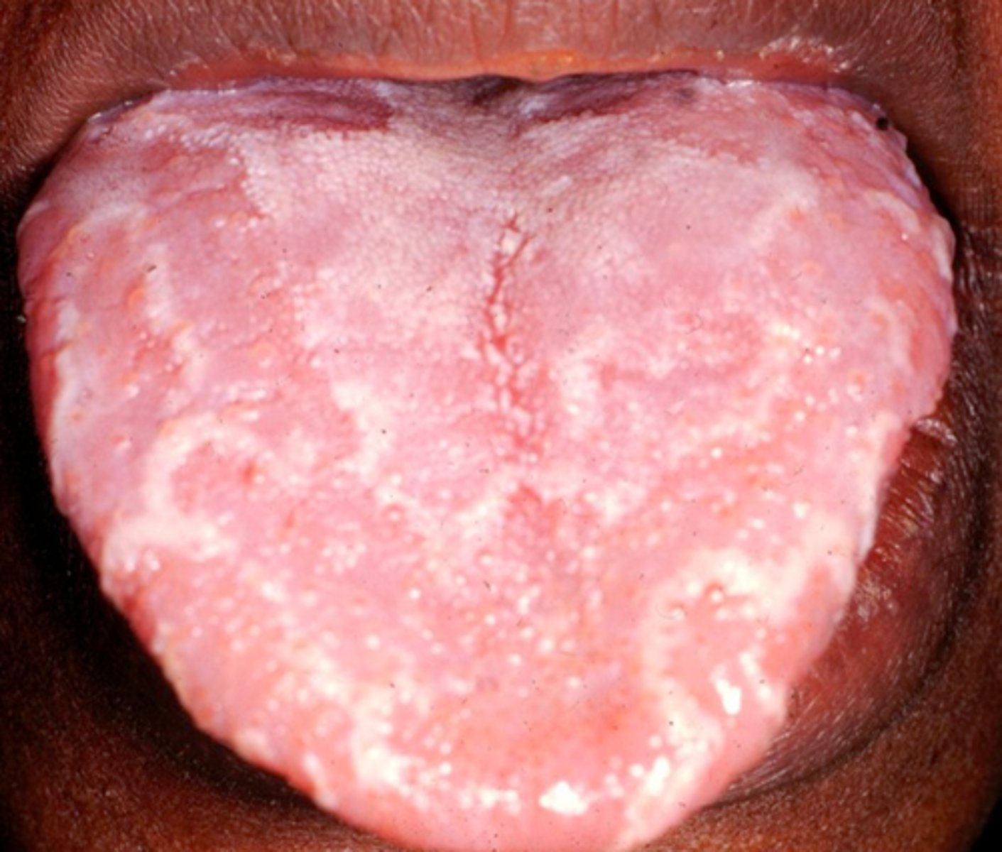
Leukoedema
deviation from normal
- benign, milky, bluish-white opaque appearance of the buccal mucosa that occurs commonly in black African Americans
Frictional keratosis
usually due to cheek biting, and is thicker than linea alba
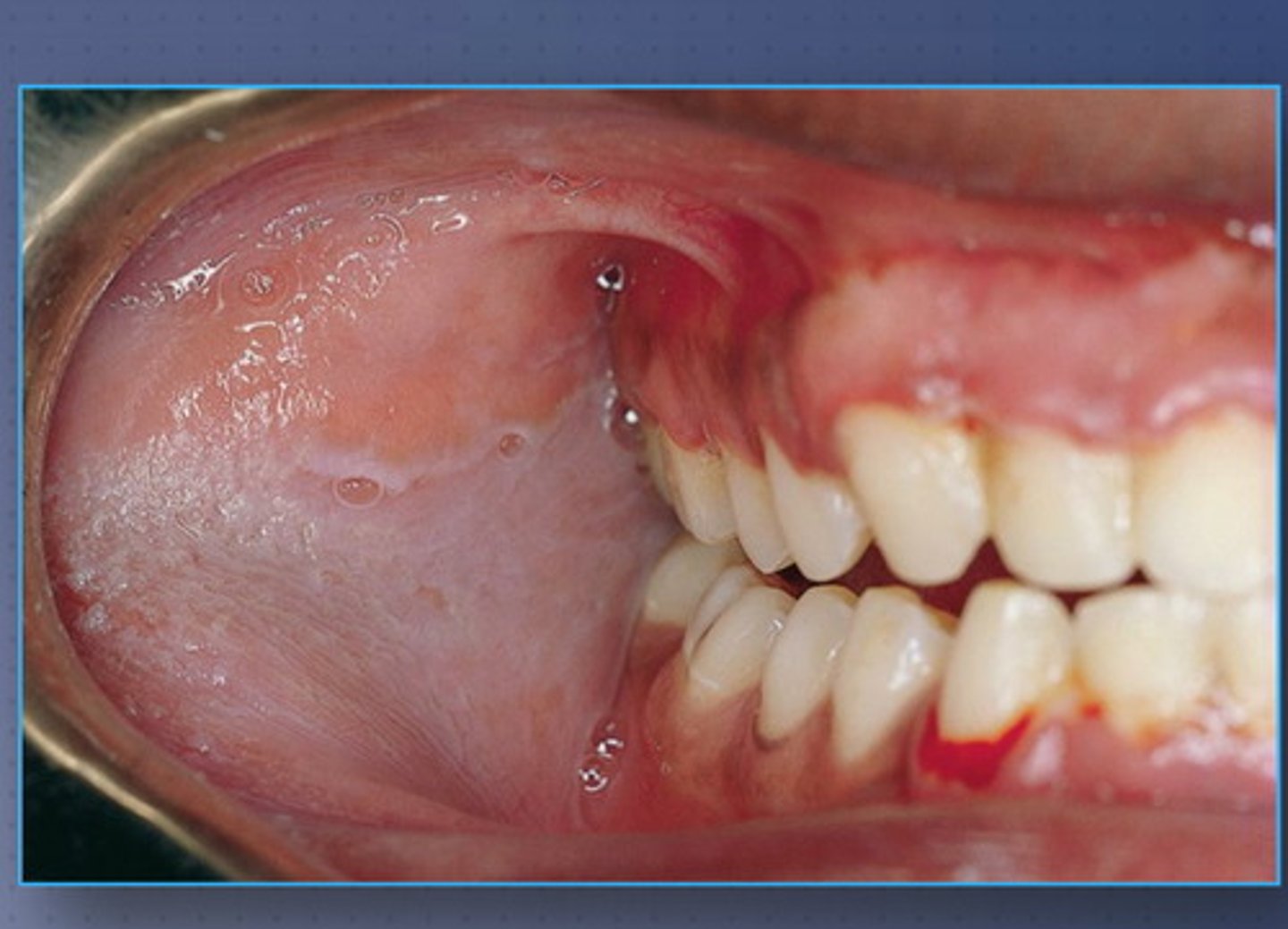
Torus palatinus
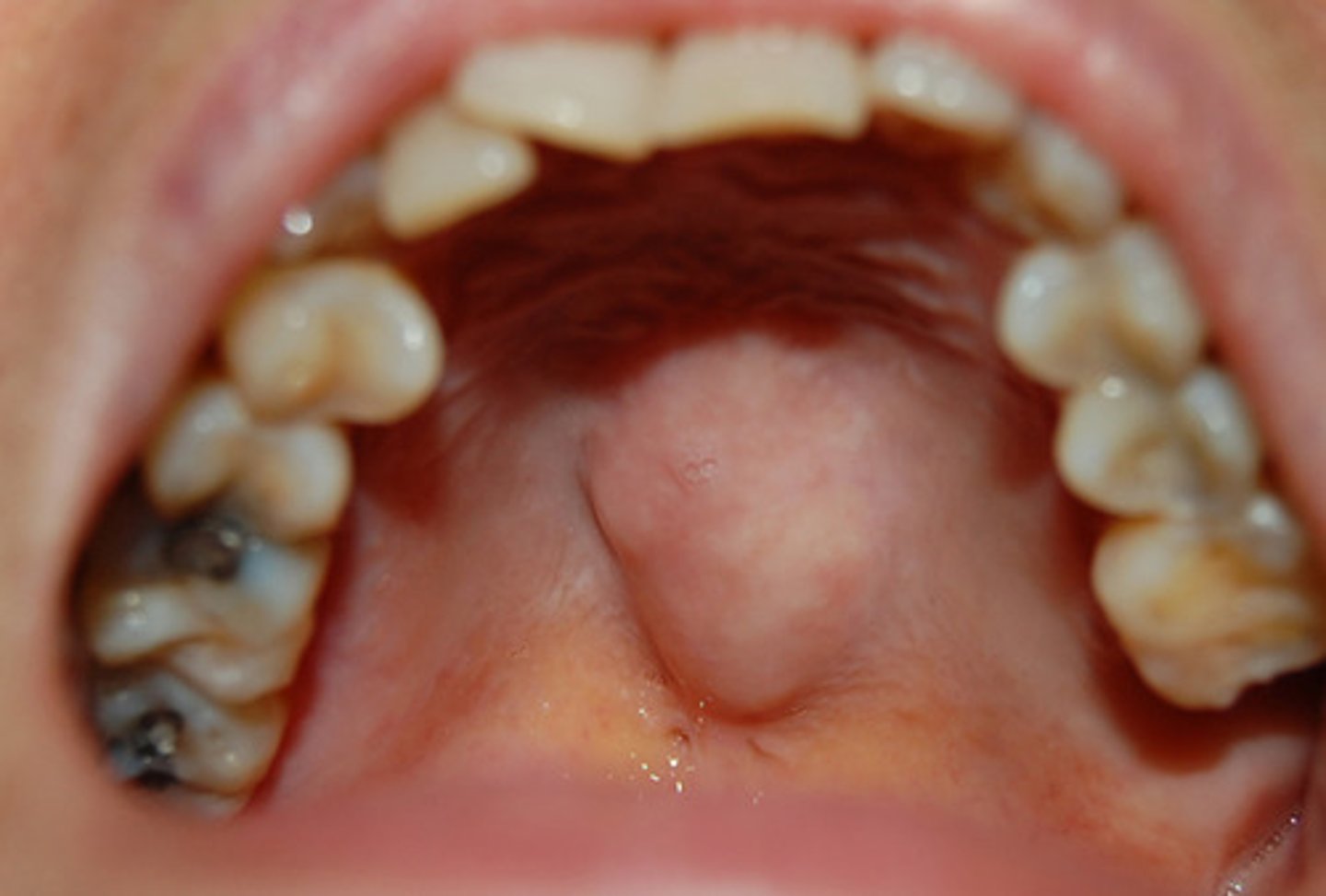
Tori can
interfere with x-ray when placing film in the mouth
Nicotine stomatitis
usually causes by the heat from smoking, vaping, or even from very hot liquids
- inflammation of the minor salivary ducts of the soft palate
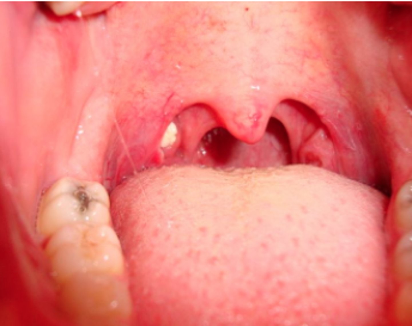
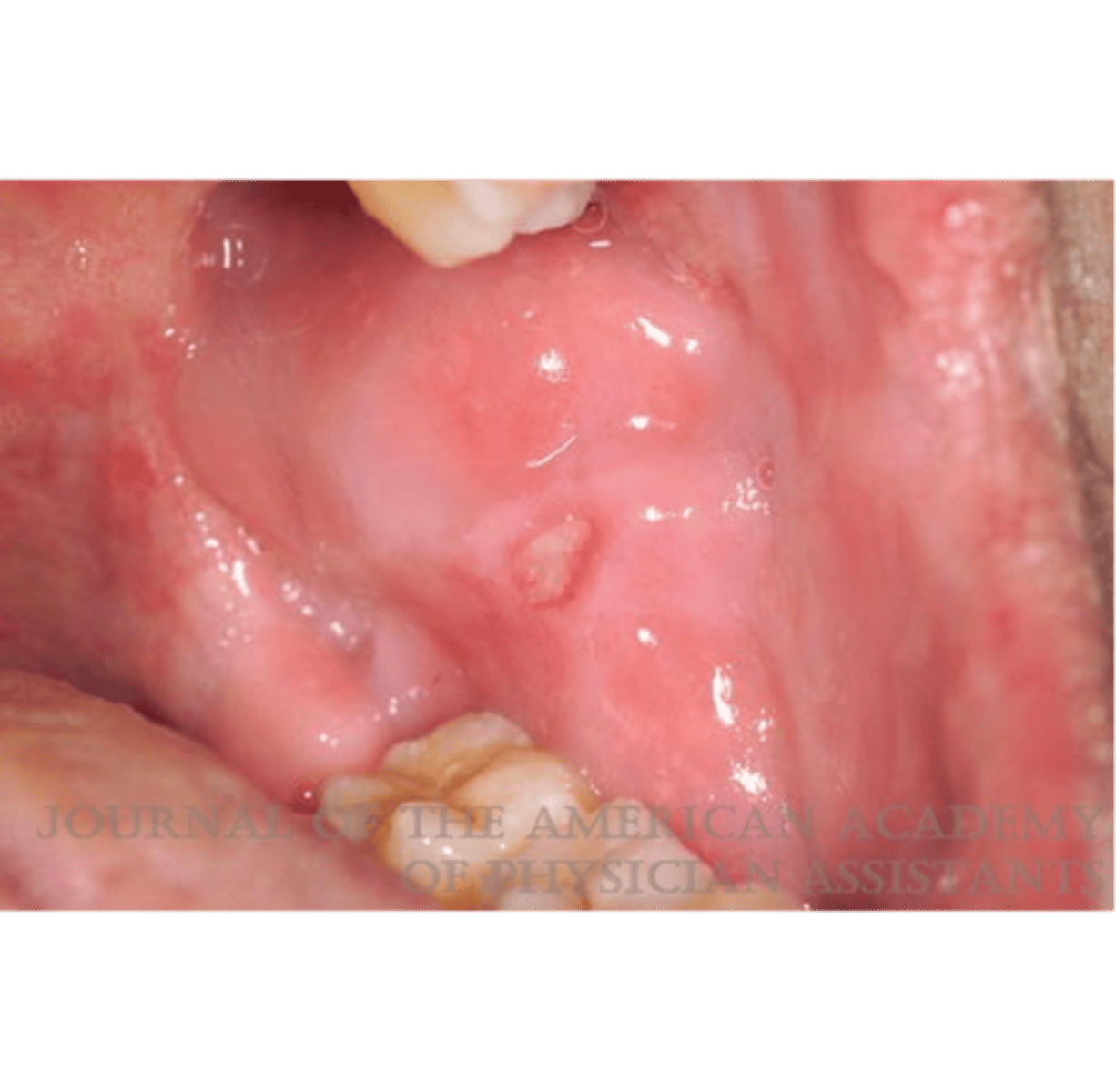
Inflamed palatine tonsils/ exudate
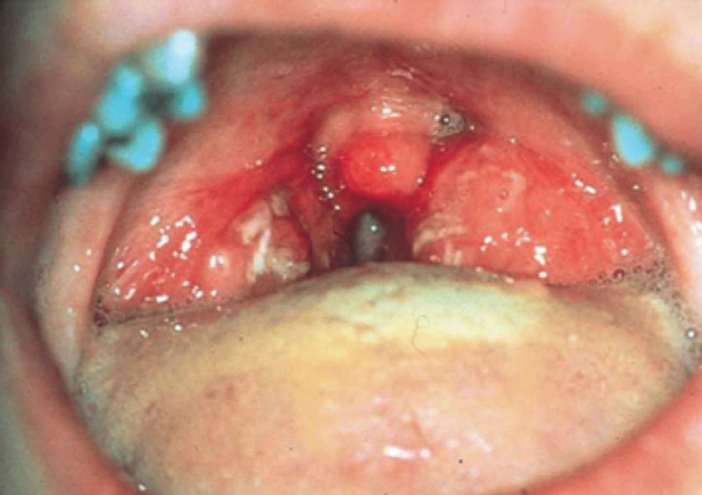
Edematous
abnormal accumulation of fluid in the tissues resulting in swelling
Bifurcated uvula
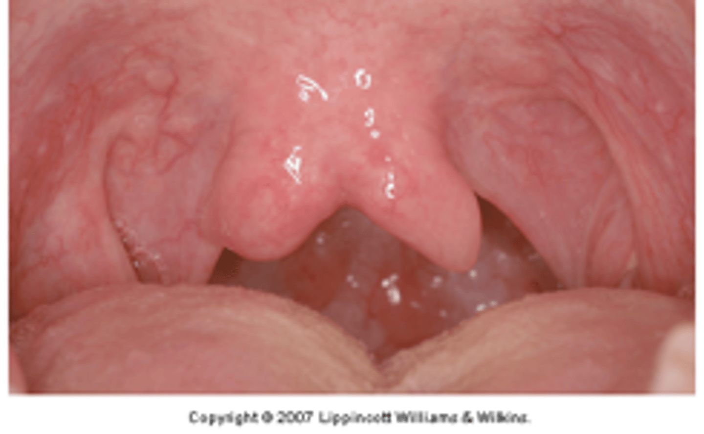
Tonsil stones (tonsilloliths)
- debris trapped in crypts
- food can get in between and then calcify
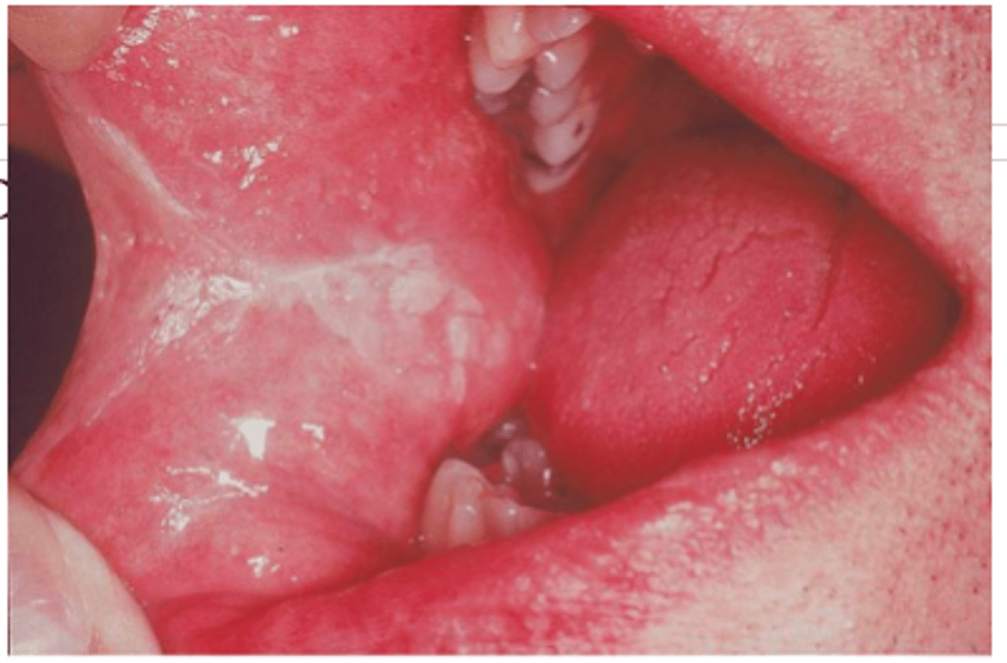
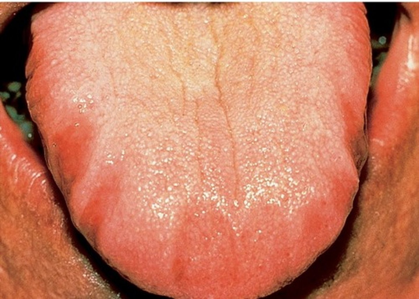
Geographic tongue
AKA benign migratory glossitis
Scalloped tongue
due to how the tongue rests against the teeth, such as when the tongue is larger than the arch
- can become more prominent with age
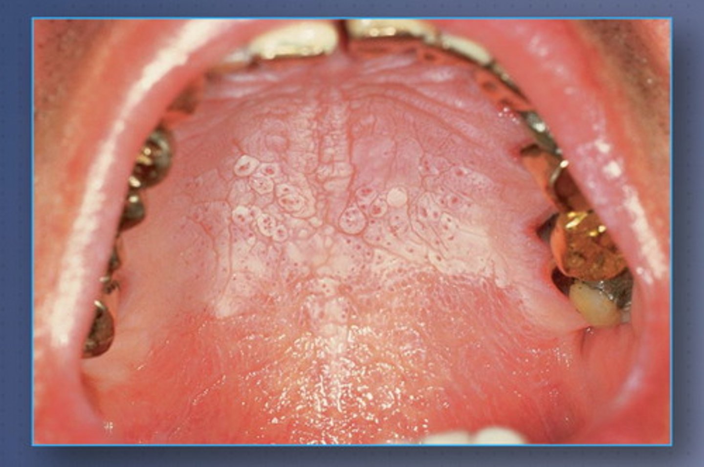
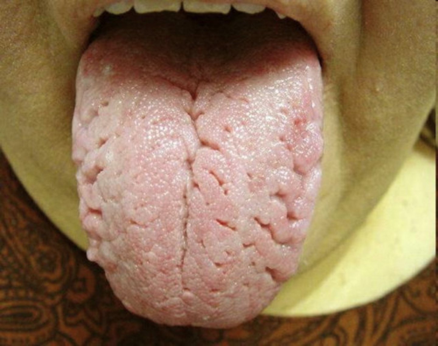
Fissured tongue
cracks in tongue
> more common in older individuals, those with xerostomia
Ankyloglossia
"tongue-tied" short lingual frenum
- if significant enough, can be surgically corrected in childhood
- can interefere with a baby's ability to feed and nurse
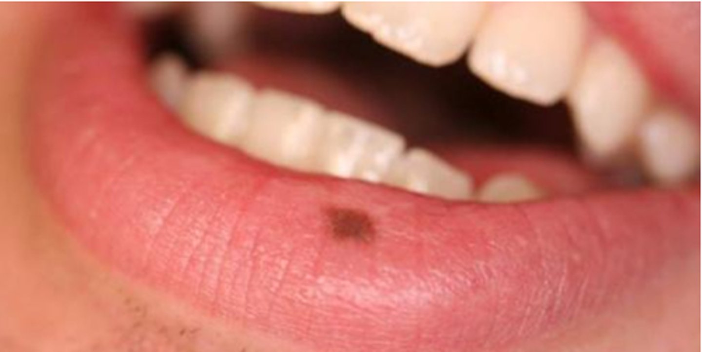
Macule
flat, colored spot on the skin (ex: freckle)
- circumscribed, nonraised area of epidermis altered in color from its surroundings
Patch
a flat, discolored area on the skin larger than 1 cm
- circumscribed pigmented or textured area larger than a macule
Erosion
denudation of epithelium above the basal cell layer
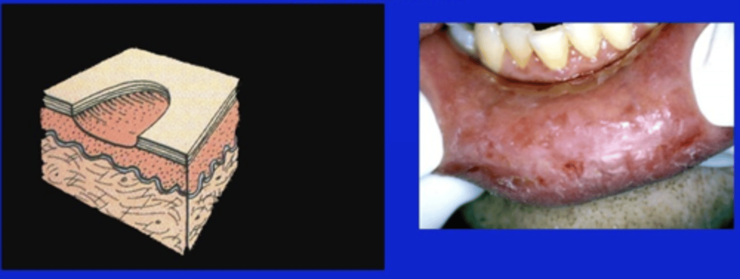
Ulcer
loss of epithelium that extends below the basal cell layer
ex. canker sore
Papule
elevated, solid lesion less than 1 cm in diameter
Plaque
flat, raised area larger than 1 cm in diameter
Nodule
raised, solid mass that has the dimension of depth and is less than 1 cm in diameter
Tumor
solid, raised benign or malignant mass that has the dimension of depth and is larger than 1 cm in diameter
Vesicle
circumscribed, fluid-filled skin elevation less than 1 cm in diameter
Pustule
vesicle filled with purulent exudate
Bulla
fluid-filled mucocutaneous elevation greater than 1 cm in diameter
Regarding to history of the lesion, ask
if patient is aware, how long its been there, change in size and appearance, symptoms
Lesion location descriptors
- Localized
- Generalized
- Single lesion
- Multiple lesion
Lesion characteristics
- Size
- Shape
- Color
- Surface texture
- Consistency (soft, hard, firm)
Attachment of raised lesions
sessile or pedunculated
Sessile
such as a mole
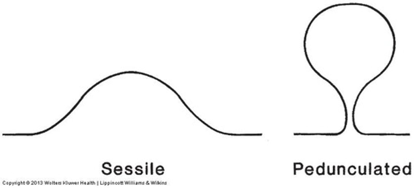
Pedunculated
such as epthelial tag

Excisional biopsy
entire lesion is removed to send for biopsy
Incisional biopsy
section of the lesion is removed