Skin structure and function 1,2,3
1/105
There's no tags or description
Looks like no tags are added yet.
Name | Mastery | Learn | Test | Matching | Spaced | Call with Kai |
|---|
No analytics yet
Send a link to your students to track their progress
106 Terms
How does the development of skin start embryologically?
initally one layer of ectodermal cells overlying a dermis of loosely arranged mesenchymal cells
what does the ectoderm divide into in skin development?
Two layers:
basal cell layer = stratum germinativum
outer layer = periderm
What is the name of the layer between the stratum germinativum and periderm?
stratum intermedium
What cell types further the embryological development of the 3 layers of skin?
melanocytes
Langerhans cells
Where do these cells originate?
melanocytes
langerhans cells
neural crest origin
bone marrow origin
How does the dermis develop embryologically into its adult structure?
increase in thickness and number of fibres
mesenchymal cells develop into fibroblasts
collagen precedes elastin fibres
ground substance accompanies them
histiocytes and dermal melanocytes are worth noting
nerves and blood vessels also develop
How does the subcutis develop?
lipocytes in the second half of gestation
Which cells divide in order to develop the multi-layered epidermis?
basal cells
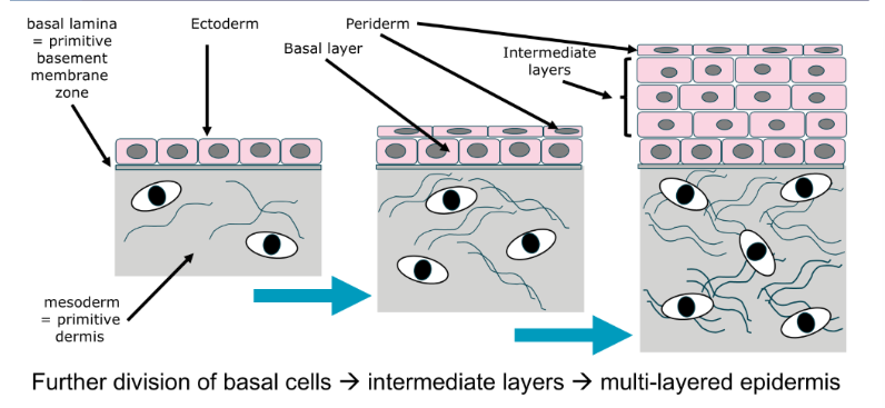
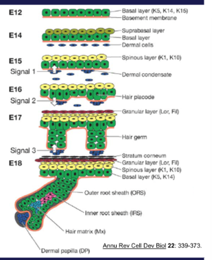
What are these drawings showing the development of?
embryology of the skin
How does the skin develop during mid-pregnancy?
baal layers give rise to the layers of the stratified squamous epithelium
periderm lost
mesodermal cells differentiate into connective tissue cells
what do we call the cells of the epithelium?
keratinocytes
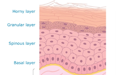
Name the 4 layers of the epidermis
basal layer → spinous layer → granular layer → horny layer
How do hairs form embryologically
epidermal basal cells divide into the dermis, this forms the hair bud/ped
groups of mesenchymal cells (dermal papilla) project into the tip of the bud
the epidermal cells grow around the papilla, forming the hair bulb
What are 3 things keratinocytes need in order to perform their function?
strength
attachment to each other - to prevent being torn apart from eachother
attachment to dermis underneath - so they don’t get riipped off
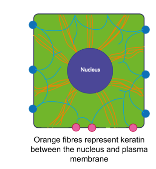
What are keratins
intermediate filament forming proteins that provide mechanical support
What do keratins link?
cell nuclear membrane to plasma membrane at desmosomes
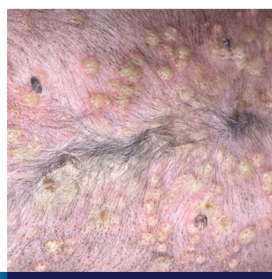
What is palmopalmar hyperkeratinosis
genetic defect which affects keratin 16 - in epidermis of foot pads
causes splitting and main from 4 mo of age
no cure
what is the function of desmosomes?
anchor the keratinocytes together
3 functions/links of desmosomes/
they act as a strong link b/w keratocytes, linking keratin intermediate filaments.
provide intracytoplasmic signalling
may be involved in congenital and autoimmune disease
Why is it important to understand the role of desmosomes?
skins shouldn’t have cracks in it, if the cells are being torn apart for some reason/not holding together, indicates something is going wrong
What is pemphigus foliaceus
an immune-mediated disease with antibodies directed against desmocollin-1 (dogs) and desmoglein-1 (other species)
forms prominent pustules
What are hemidesmosomes
filaments from dermal collagen that anchor epidermis to dermis
What does the number and complexity of molecules involved in hemidesmosomes link to?
a large number of congenital and autoimmune diseases
What are the epidermal basement membrane ultrastructures
keratin filmanets w/in cells
hemidesmosomes
anchoring filaments
lamina densa
anchoring fibrils
what is epidermolysis bullosa acquisita
immune-mediated disease with antibodies directed against collagen VII
can lead to ulceration e.g. on ear flap
Do we get different forms of epidermolysis bullosa?
yes
congenital abnormalities link to others
e.g. dystrophic or junctional EB
bullous pemphigoid antigens 1+2 and plectin are important
What ISN’T there in the epidermis?
blood vessels
What are the layers of epidermis top to bottom?
horny layer
stratum corneum
stratum granulosum
stratum spinosum
stratum basale
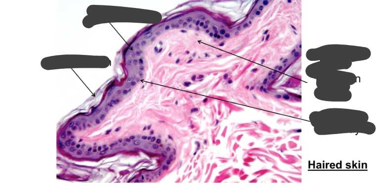
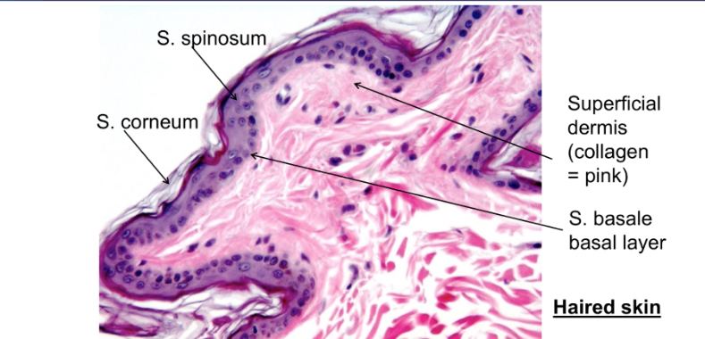
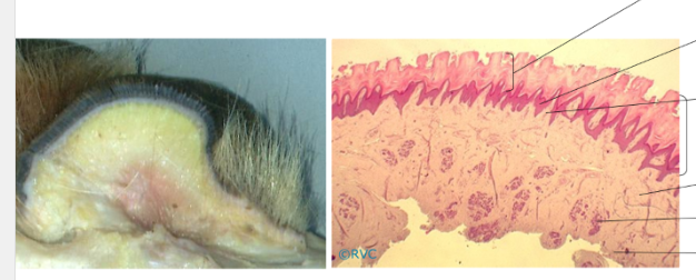
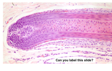
Label this histological image of the skin
Left: s. spinosum too, s. corneum lower
right: Superficial dermis = pink, lower = s.basale layer
In which part of the body do we see a much thicker layer of epidermis?
foot pads
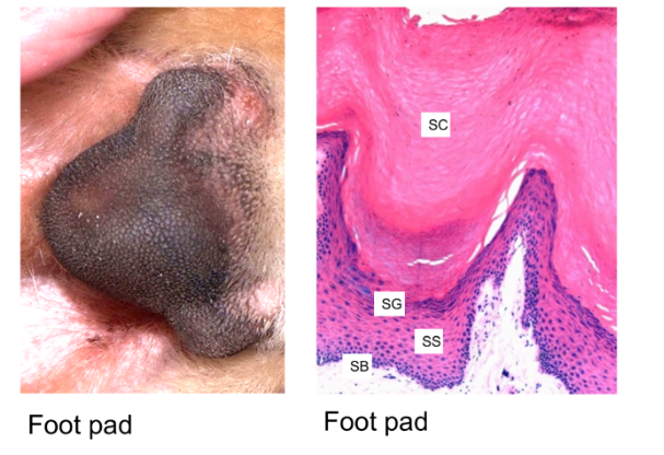
Identify the layers, top to bottom
stratum corneum
epidermal papillae
dermal papillae
epidermis (brackeet)
dermis
sweat glands
hypodermis
Identify the general skin thickness of:
dog and cat
cattle
sheep
goat
pig
horse
0.5-7mm
6mm
2.6mm
2.9mm
2.2mm
1.7-7.7mm
which 2 species have rete ridges
pigs
horses at mane and tail base
Keratinocytes:
embryological origin
features
same in whole body?
how do desmosomal proteins change in layers
epithelial origin
complex internal cytoskeleton
no, the keratin differs
under influence of calcium
What do keratinocytes produce?
extracellular lipids - ceramides, cholesterols, fatty acids - from the golgi apparatus in the granular layer → lipid lamellae
How can lipid/protein metabolism dysfunction show itself in the skin?
change secretion function → change structure and function of skin
What is the cytoskeleton in the keratinocytes attached to ?
tight junctions
desmosomes
hemidesmosomes
Stratum basale:
structure
function
single layer of cuboidal cells
proliferation, anchoring, stem cell function (pluripotent)
Stratum spinosum:
no. cells
cell shape
prominent feature?
what does the upper most layer produce?
1-20 cells thick
polyhedral
prominent desmosomes
involucrin - part of cornified layer
Stratum basale:
type of cell found these
how do they divide?
what are they influenced by?
proliferative
one cell remains as germinative, other differentiates
growth factors and hormones, inflammatory mediators, drugs and vitamins
Stratum granulosum:
always there with haired skin?
cell structure?
secretions?
what do desmosomes do?
what are they essential for?
no, variably present in haired skin
slightly flat, shrunken nuclei, have intracellular keratohyaline granules with profilaggrin and ioricrin
lipid and enzymes are secreted extracellularly
corneodesmosin
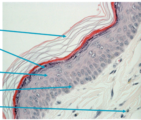
Identify the layers
stratum corneum
stratum granulosum
stratum spinosum
stratum basale
dermis
Stratum lucidum:
visible?
cell type
what do they contain?
why do they stain poorly?
not in haired skin - somewhat in hairless and thickened skin
slightly flat, shrunken nuclei
keratohyalin
increased intracellular lipids
Stratum corneum:
composed of?
what replaces the plasma membrane
what helps solidify the structure?
what do we call desmosomes, why?
anucleate flattened cells, variable thickness
cornified envelope of many molecules
hydrophilic bonding of lipids forms organised ‘mortar’
corneodesmosomes
What does flaggrin facilitate?
collapse of corneocytes into ‘building blocks’ → impermeable surface barrier
What does the breakdown of flaggrin lead to?
urocanic acid (UCA) and pyrrolidone carboxylic acid (PCA) = moisturiser and excellent UV protection
Consequence of TGM-1 mutation in jack russell terriers?
skin can’t grow properly - it’s a defect in the protein of the CE
Outline desquamation
homeostatic process - continual loss of cells
partly mediated by proteinases and glycosidases
destruction of corneodesmesome
invisible rafts of attached comeocytes fall off
What is golden retriever ichthyosis caused by?
an insertion-deletion mutation → lead to a premature stop codon
In the dermis:
what gives tensile strength?
what resist and absorb compressive forces?
what determines thickness of skin
layers?
what additional things does it contain
specific cells?
collagen and elastin
solbule polymers - proteoglycans + hyaluronan
thickeness of dermis determines skin thickness
deep and superficial
epidermal appendages, arrecter pili muscles, blood and lymph and nerves
perivascular lymphocytes, dermal dendritic cells, mast cells and fibroblasts
What causes ehler-danlos syndrome?
skin has poor strength due to a collagen defect → extra stretchy skin → more easily wounded.
With what other condition do we see similar skin fragility as with Ehler-danlos syndrome?
hyperadrenocorticism
3 layers of blood supply
deep dermal vascular plexus
mid-dermal vascular plexus
superficial dermal vascular plexus
Where in the dermis do we find:
deep dermal vascular plexus
mid dermal vascualr plexus
superficial dermal vascular plexus
interface of dermis and subcutis
level of sebaceous gland
just below epidermis
What do each of these blood plexus supply:
deep dermal vascular plexus
mid-dermal vascular plexus
superficial dermal vascular plexus
lower hair follicle and epitrichial sweat glands
arrector pili muscles, mid hair follicle and sebaceous glands
capillary loops supply epidermis and upper hair follicle
What is the lymphatic structure in the skin?
vessels and capillaries largely follow the course of blood vessels
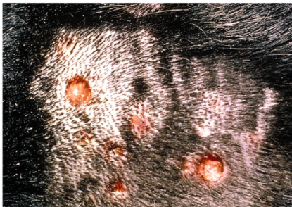
What is an example disease of blood vessels
type III hypersensitivity vasculitis (e.g. due to drug reaction)
how do sebaceous glands form?
basal alyer of germinative cells divide and differentiate → large polygonal cells with abundant vacuolated cytoplasm
What does sebum form?
triglycerides and other lipids e.g. transferrin, IgA and IgG
Functions of sebum?
lubricates hair and skin
required for normal hair shaft separation
What is sebum excreted by?
via squameous duct to hair follicle
What is a histological feature to help identify a sebaceous gland (epitrichial gland)
arector pili muscle
What is sebasceous adenitis
dog lack sebaceous glands
scale, hair breakage and follicular casts are noted
Identify 6 specialised sebaceous glands
meibomian glands
circumanal glands
supracaudal gland of dogs and cats
submental glands in cats
preputial glands in horses
infraorbital, inguinal and interdigital glands
Epitrichial glands:
associated with?
structure
structure found within the glands?
find where?
located below what?
excretes into what?
innervated?
properties of what is secreted?
hair follicles
single layer of flattened cuboidal cells
coiled and saccular/tubular
distributed throughout haired skin
sebaceous glands
sweat into piliary anal just above sebaceous gland opening
NO
pheromonal and antimicrobial
Atrichial glands:
what aren’t they associated with?
what surrounds them
structure of glands
innervation
where do we find them
hairs
single layer, flattened cuboidal cells, surrounded by myoepithelial cells - merocrine secretion
small tightly coiled glands
cholinergic nerve fibres
non-haired areas
carpus of pigs, frog and ungulates + nasolabila region of ruminants and pigs. Generally - non-hair areas e.g. nose and footpad in carnivores
What are 5 specialised atrichial glands
mammary glands
interdigital glands of small ruminants
external ear canal
nasolacrimal glands
apocrine glands of anal sac
What 2 types of hairs are there?
primary - guard hairs
secondary downy hairs
State 3 functions of hairs
insulation
signalling
physical portection
What are hairs
specialised keratinised tubular structure
What are the hair and follicle structure for omnivores and herbivoress
simple - each infundibulum contains a single hair shaft of approx same size
What type of hair follicle do we find in sheep
compound
What follicles do we find carnivores
compound
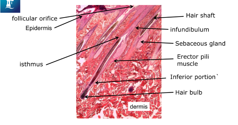
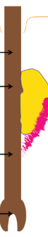
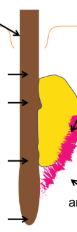
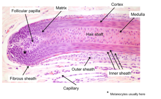
Label this diagram
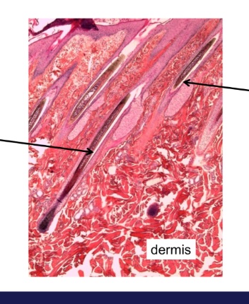
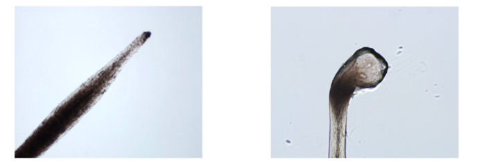
What two different stages of hair growth are these hairs in?
left = growing - see bulb
right = resting hair - shallow depth and tapering
How do the number of hairs differ in:
simple follicles
compound follicles
single
several
3 hair phases?
anagen
catagen
telogen
Compare primary and secondary hair follicles:
bulb depth
associated with?
Primary: bulb deep in dermis, have sebaceous glands, sweat glands, arrector pili muscles
Secondary - not as deep, smaller - associated sebaceous gland (MAYBE)
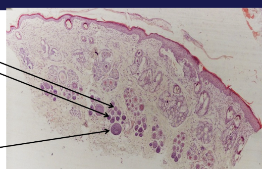
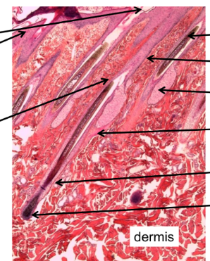
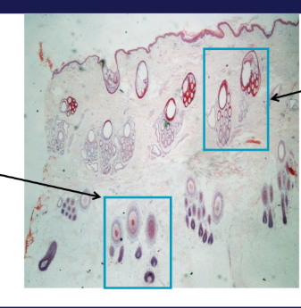
Which hairs are pirmary, which are secondary
Top = secondary
Bottom = primary (deeper in dermis)
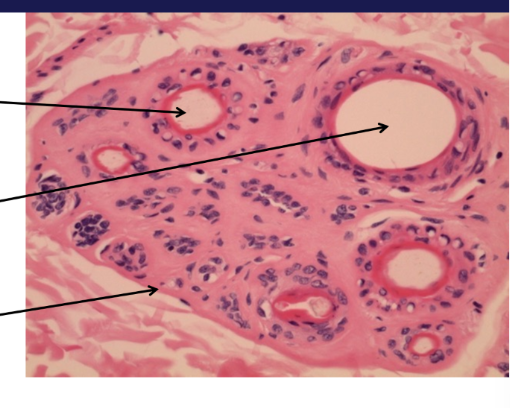
Which is primary, which is secondary
left = secondary
right = primary
Outline these phases:
anagen
catagen
telogen
exogen
growth phase, new hair produced underneath, distinctive hair bulb contains follicular dermal papilla
transitional phase, rarely seen in normal skin - feature of some diseases
process of hair being lost
What phase of growth is this hair in?
growing - note bulb shape
What phase of hair growth is this hair in
telogen phase - note small bulb
what is the isthmus
where sebaceous gland attaches divides the hair between infundibulum and inferior portion
what is the growth pattern of hair for:
angora rabbit, mohair goat, poodle
dogs, cats, horse, hedgehogs
anogenic
telogenic
what determines hair length
duration of anagen phase
what is hair cycle regulated by?
photoperiod
termperature
hormones
nutrition and general health
growth factors
drugs
What hormones regulate hair cycle?
thyroid, GH are stimulatory
oestrogen and corticosteroids are inhibitory
What phases of growth are these hairs in?
Top = telogen
Bottom = anagen
What provides blood supply to anagen hairs?
dermal papilla
What undergoes mitosis in anagen hairs?
- hair bulb epithelium
What cells provide hair pigment
melanocytes
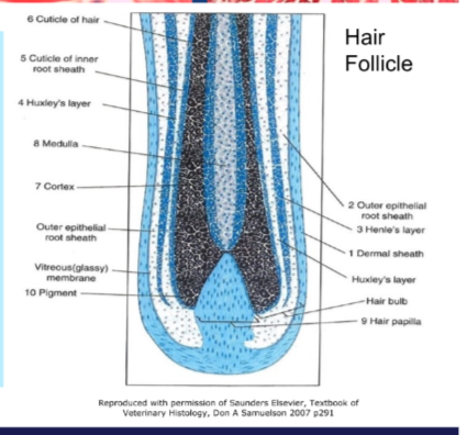
What are the 6 layers of the hair? inner to outer?
medulla, cortex, cuticle of hair
Huxley layer, Henle layer (inner root sheath)
outer root sheath (continuous with epidermis)
glassy membrane (basement membrane)
What gives the hair strength in anagen phase?
hair and IRS cuticle - fit together
what permits hair loss in exogen phase?
IRS disappears in catagen
Label
Catagen phase:
where in the dermis
how do we identify the hairs this way?
what has the hair lost
what does it develop
slow or fast?
mediated by what?
mid dermis
involution of hair bulb and dermal papilla - also upwards migration
internal root sheath
thick glassy membrane, above bulb
fast
apoptosis
Telogen phase:
where in dermis
where do we find the conical bulb
what is the hair surrounded by
what is it separated from and by?
active/dormant bulb?
what forms beneath old follicle
how do new hairs replace old ones?
mid-upper
level of attachment of arrector pili muscle
external root sheath - terminates at sebaceous gland level
separated from dermal papilla by thick basement membrane
dormant
new bulb and papilla
new bulb forms a new hair and old hair is lost
Which phases of growth are these hairs in?
Left = telogen (narrow and straight)
Right = anagen (bulb, flexible)