head and neck anatomy exam 1
 Call Kai
 Learn
Learn Practice Test
 Spaced Repetition
 Match
Match Flashcards
 Knowt Play
1/145

There's no tags or description
Looks like no tags are added yet.
Name | Mastery | Learn | Test | Matching | Spaced | Call with Kai |
|---|
No analytics yet
Send a link to your students to track their progress
146 Terms
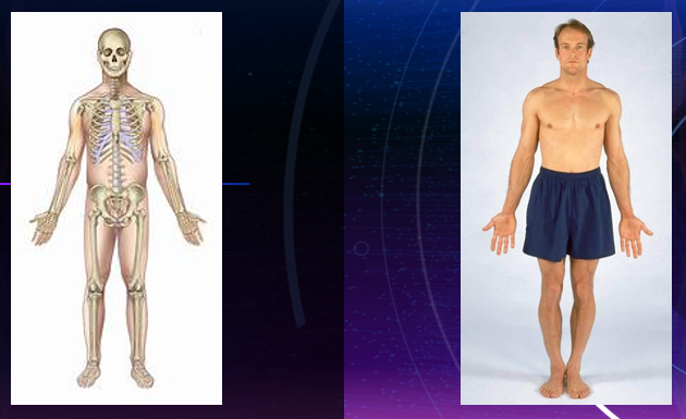
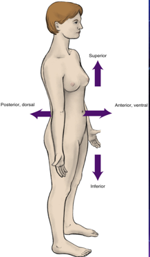
anatomic position
arms are at the sides with the palms and toes directed forward
eyes looking forward
position is assumed even when the body may be supine (on the back) or prone (face down)
the body standing erect
(erect position, arms at sides, palms and toes directed forward with eyes looking forward)
anterior
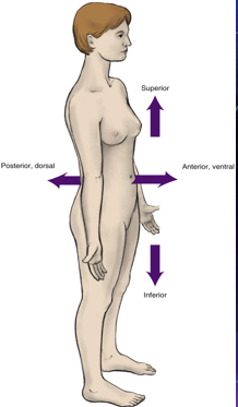
frontal of an area in relation to the entire body
front of an area
posterior
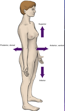
back of an area in relation to the rest of the body
back of an area
anatomic nonmenclature
system of names for anatomic structures
apex/apices
pointed end/s of conical structure
contralateral
structure on the opposite side
deep
structure located inwards and away from surface
more internal
ex: the lungs are deep to the skin
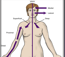
distal
area farther away from median plane
farther from the origin of a body part of the point of attachment of a limb to the body trunk
ex: the knee is distal to the thigh, or the distal surface of the tooth faces away from the median plane
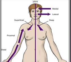
dorsal
back of area
“dorsal fin”
opposite of ventral
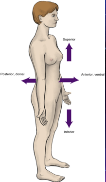
external
outer side of wall of hollow structure
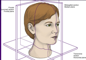
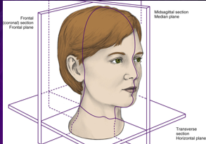
frontal plane/ coronal section
plane related to imaginary line dividing body at any level into anterior and posterior parts
splits body into ventral/ dorsal
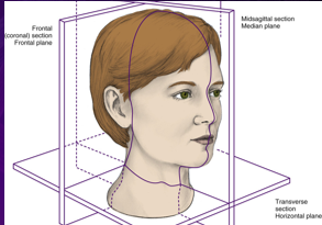
frontal section
section through any frontal plane
horizontal plane/ transverse section
plane related to imaginary line dividing body at any level into superior and inferior parts
splits body horizontally
splits body into “hamburger” style
inferior
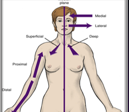
area facing away from head and toward feet
internal
inner side of wall of hollow structure
ipsilateral
structure located on the same side
lateral
area farther away from median plane
to the side
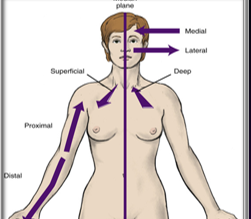
medial
area closer to median plane
considered mesial within dentition
median
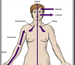
structure at median plane
exact midline
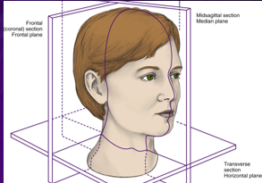
median plane
plane related to imaginary line dividing body into right and left halves
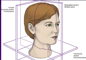
midsagittal section/ median plane
section through median plane
section cuts body vertically into exact left and right halves
cuts body “hot dog style”
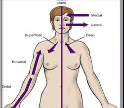
proximal
area closer to median plane
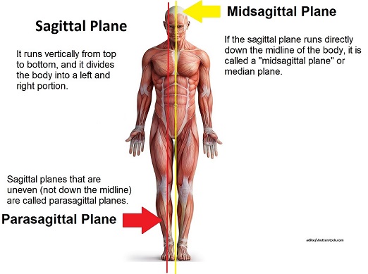
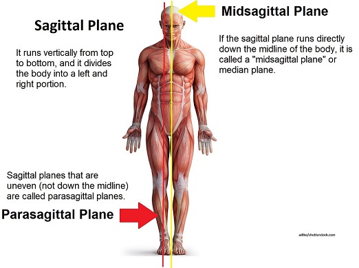
sagittal plane
planes of body related to any imaginary plane parallel to medial plane
superficial
structure located toward surface
superior
area facing toward head and away from feet
transverse section
section through any horizontal plane
splits the body “hamburger style”
ventral
front of area
opposite of dorsal
anatomic variation
there are often variation of head and neck structures
number of bones and muscles is usually constant but some specific details may vary
bones may have different sizes, muscles may differ in size and attachment details
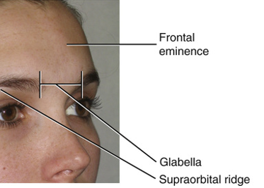
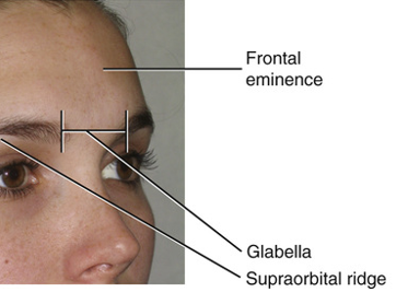
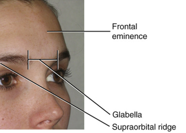
frontal region of face
includes forehead and the area superior to the eyes and is defined by the deeper skull bone
frontal eminence
supraorbital ridge
glabella
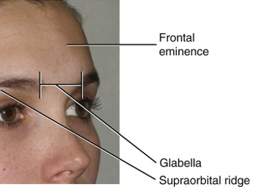
frontal eminence
prominence of the forehead
is usually more pronounced in children and adult females
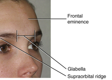
supraorbital region
directly inferior to each eyebrow
more pronounced in males
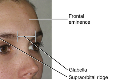
glabella
smooth and elevated area between the eyebrows
tends to be flat in children and adult females
forms a rounded prominence in adult males
parietal and occipital region
both covered by the scalp and defined by the deeper skull bones
consists of soft layers of tissue overlying the bones of the braincase
large areas of scalp may additionally be covered by hair
temporal region
within the temporal region is the temple, the superficial side of the head posterior to each eye, which is defined by the deeper skull bone
temple
posterior to eye
auricular region
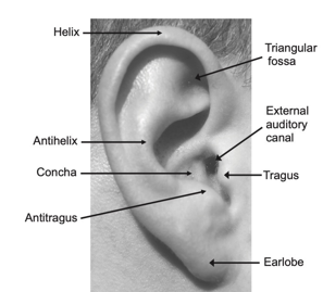
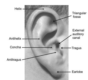
auricular region of each side of the head has the external ear as a prominent feature
external ear
auricle
helix
lobule
traugus
anti tragus
intertragic notch
external acoustic meatus
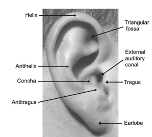
helix
superior and posterior free margin of the auricle
ends inferiorly at the lobule, the fleshy protuberance of the earlobe
upper helix level with eyebrow and glabella
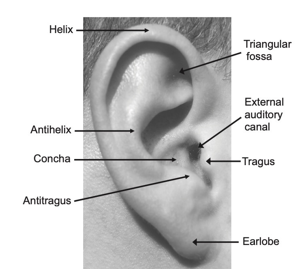
lobule
the fleshy protuberance of the earlobe
approximately at the level of the apex of the nose
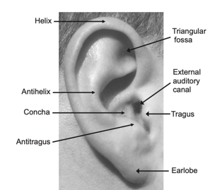
tragus
smaller flap of tissue of the auricles anterior to the external acoustic meatus
flexible when palpated due to cartilage
antitragus
the other flap of tissue opposite the tragus
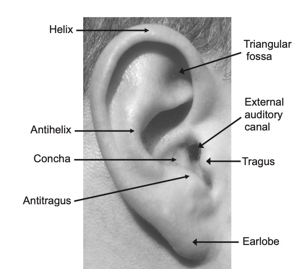
intertragic notch
between the tragus and antitragus
small groove
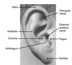
the _ _ _ and _are important landmarks when taking certain radiographs and administering certain local anesthesia blocks
external aucustic meatus
tragus
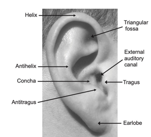
external acoustic meatus
tube through which sound waves are transmitted to the middle ear within the skull
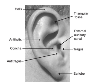
auricle
collects sound waves
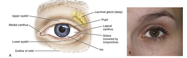
orbital region
orbit
eye
lacrimal glands
medial canthus
lateral canthus
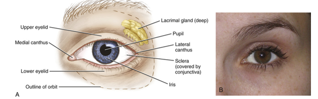
orbit
bony socket that contains the eyeball and its supporting structures
formed by various skull bones
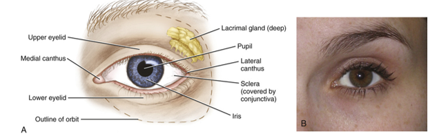
eye
eyes usually meet near the midpoint of the vertical height of the head
width of each eye is usually the same as the distance between the eyes
scelera (white)
iris (color)
pupil (center of iris, appears black and changes size due to light)
conjunctiva
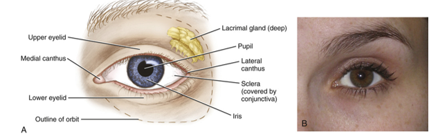
lacrimal glands
produce lacrimal fluid (tears)
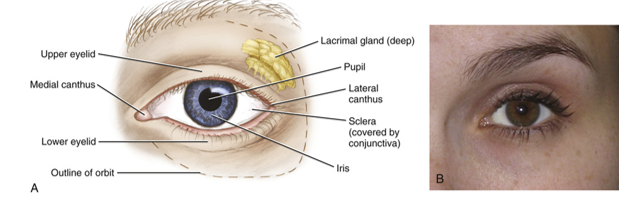
conjunctiva
delicate and thin membrane lining the inside of the eyelids and the front of the eyeball
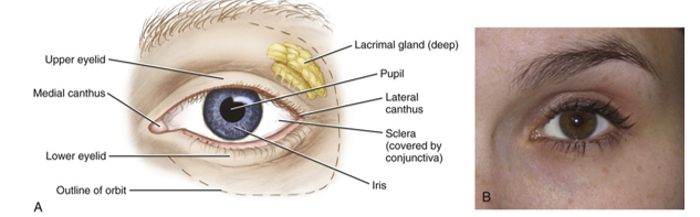
lateral canthus
canthi for plural
the outer corners where the upper and lower eyelids meet
also known as outer canthus
important landmark for taking extraoral radiographs
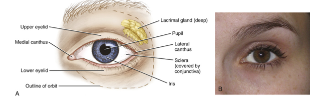
medial canthus
inner canthus
inner angles of eye located lateral to root of nose
important landmark when taking radiographs
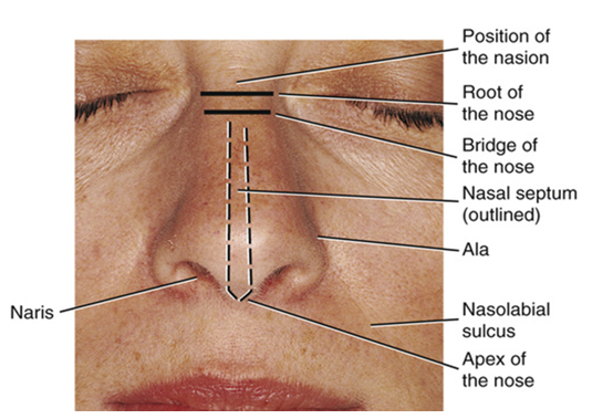
nasal region
contains the external nose
root of nose
nasion
bridge of nose
nares
nasal septum
alae
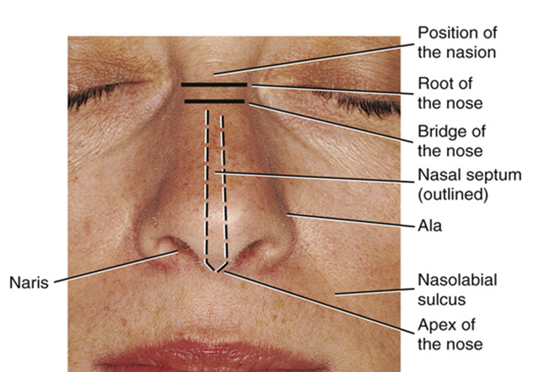
root of nose
located between eyes
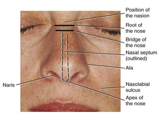
nasion
inferior to glabella is a midpoint cephalometric landmark of the nasal region that corresponds with the junction between the underlying bones
landmark for external radiograph
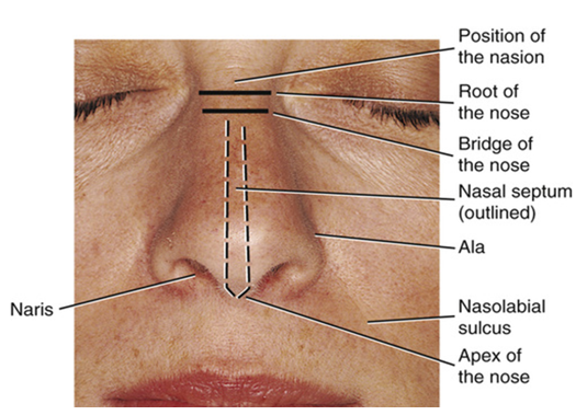
bridge of nose
inferior to nasion is the bony structure of the skull that forms the __ of the nose
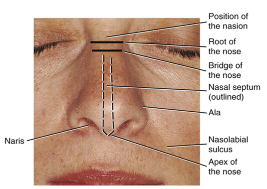
apex of nose
formed by cartilage
is flexible when palpated
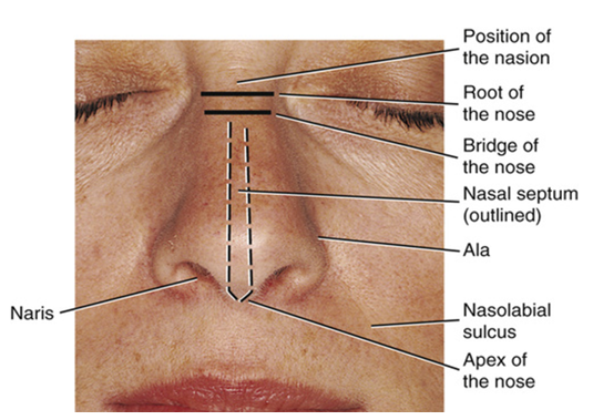
nares
nostrils
inferior to the apex on each side of the nose
the opening
separated by the midline nasal septum
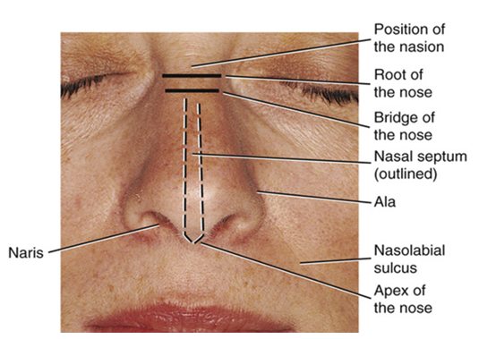
nasal septum
formed by various skull bones and the adjoining nasal septal cartilage
separates nares
midline of nose
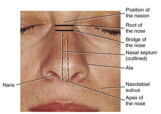
ala
tissue surrounding nostrils
winglike cartilaginous structures
the width between the ale should be approximately the same width as one eye or the space between eyes
landmark for external radiograph
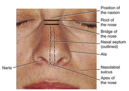
infraorbital region
located inferior to orbital region and lateral to nasal region
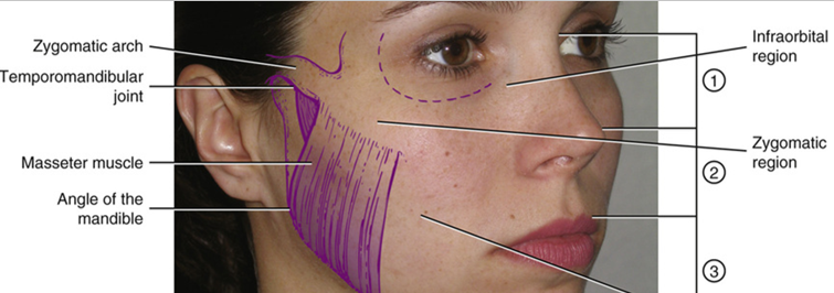
zygomatic region
overlies cheekbone, the zygomatic arch
extends from just inferior to the lateral margin of eye toward middle part of ear
zygomatic arch is formed from various skull bones
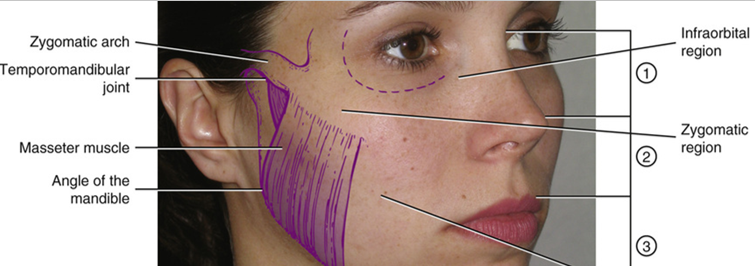
temporomandibular joint (TMJ)
inferior to zygomatic arch and anterior to middle ear
upper skull forms joint with lower jaw
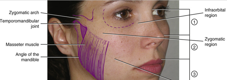
buccal region
soft tissue of cheek
includes the masseter muscle
felt when patient clenches teeth
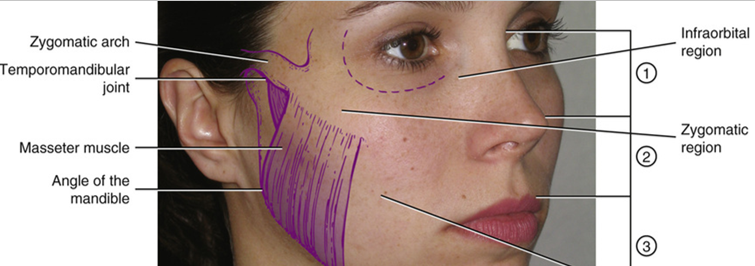
oral region!
includes
lips
oral cavity
palate
tongue
floor of mouth
part of throat
pharynx
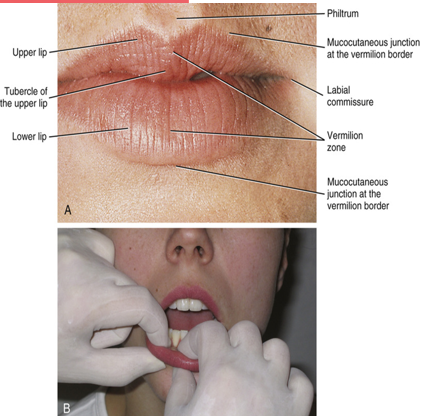
vermillion zone
has a darker appearance than the surrounding skin
darker shade of lips
width of lips should be approximately the same distance as between the irises of the eyes
mucocutaneous junction
outline of lips at vermillion border
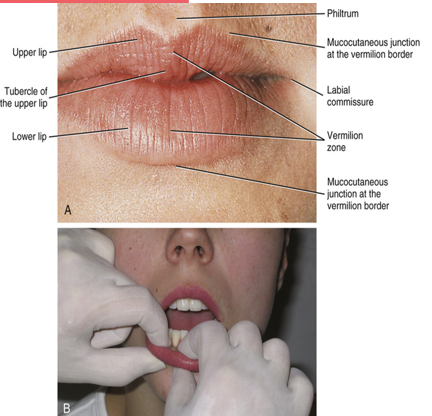
vermillion border
the lips are outlined from the surrounding skin by the transition of the mucocutaneous junction at the __ __
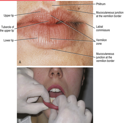
philtrum
superior to the midline of upper lip, extending downward from the nasal septum
vertical groove of upper lip
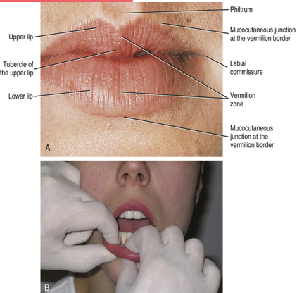
tubercle
inferior to philtrum
midline of upper lip
the midline of the upper lip terminates in a thicker area, or __ of the upper lip
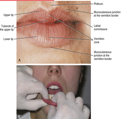
labial commissures
corners of mouth where upper and lower lips meet
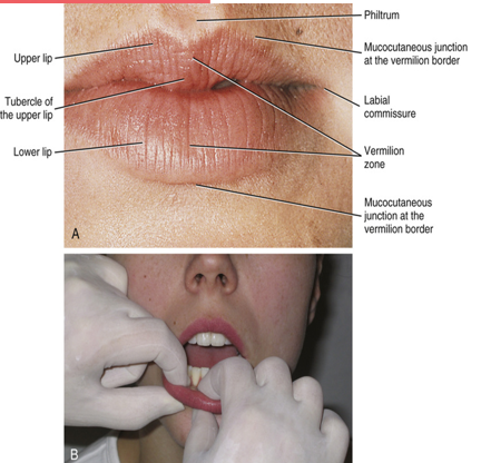
nasolabial sulcus
the groove running upward between each labial commissure and each ala of the nose
labiomental groove
lower lip extends to the horizontally placed __
separates the lower lip from the chin in the mental region
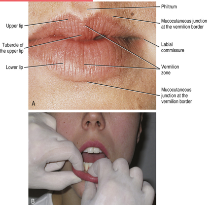
facial
structure located closest to face
labial
structure located closest to lips
buccal
structures closest to inner cheek
lingual
structures located closest to tongue
palatal
structures that are closest to palate
vertical dimensions of face
division of face into thirds
when we age, or even lose teeth, there can be a loss of “vertical dimension” resulting in a sinking appearance
used to compare portions of face for functional and esthetic purposes
golden proportions
used in vertical dimension as a set of guidelines
used to compare portions of face for functional and esthetic purposes
oral cavity is lined by __ mucosa
the oral cavity is lined by oral mucosa
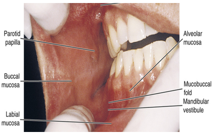
lips are lined with __ mucosa
the inner parts of the lips are lined by a pink and thick labial mucosa
is continuous with the equally pink and thick buccal mucosa
can vary in color and pigmentation while still being healthy
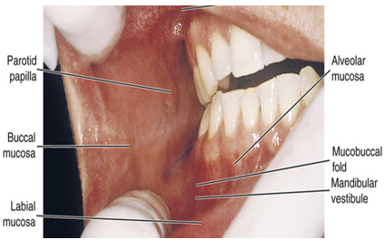
cheeks are lined with __ mucosa
buccal mucosa
covers the buccal fat bad which is a dense pad of inner tissue
can vary in color and pigmentation while still being healthy
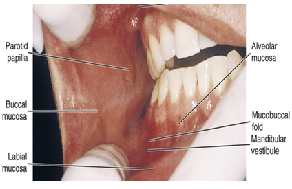
parotid papilla
located opposite to maxillary second molar
a small elevation of tissue that protects the ductal opening of parotid salivary gland
this is why buccal maxillary molars always should be checked for calculus
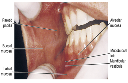
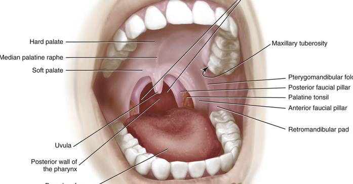
maxillary tuberosity
tissue covered elevation on the posterior aspects of the maxilla just posterior to the most distal maxillary molar
retromolar pad
dense pad of tissue just posterior to most distal mandibular molar
gingiva
surrounds maxillary and mandibular teeth
composed of a firm pink oral mucosa
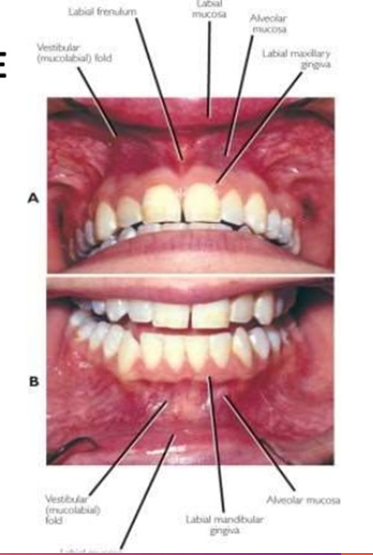
attached gingiva
the gingiva that tightly adheres to the bone around the roots of the teeth
may also have localized areas of pigmentation
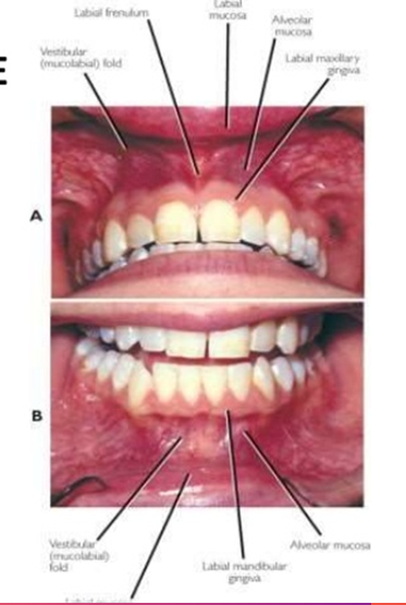
alveolar mucosa
moveable, red mucosa that makes up the oral vestibules
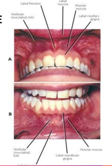
mucogingival junction
the line of demarcation between the firmer and pinker attached gingiva and the moveable and redder alveolar mucosa
scallop-shaped
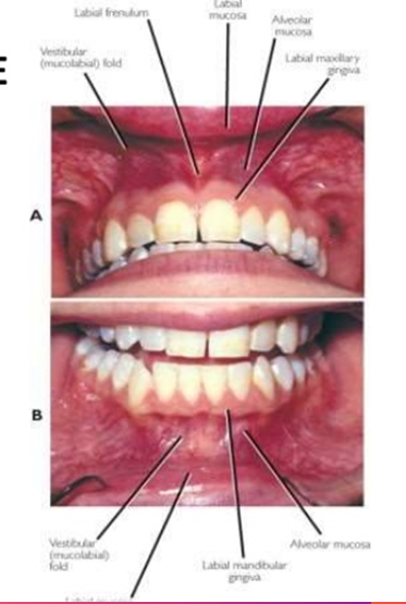
vestibules
upper and lower horseshoe shaped spaced between lips and cheek anteriorly and laterally as well as the teeth and their soft tissues medially and posteriorly
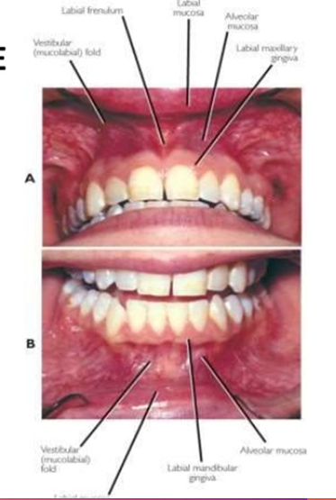
vestibular fornix
where the pink and thick labial or buccal musoca meets the redder and thinner alveolar mucosa at the mucobuccal fold
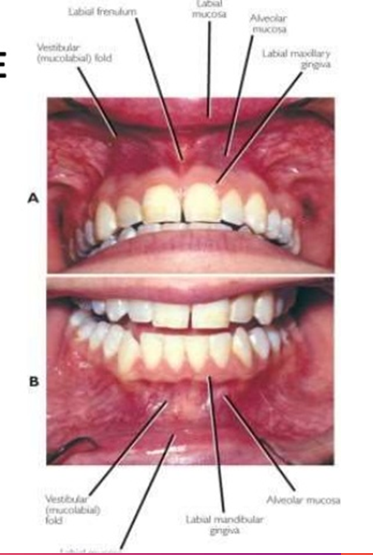
labial frenum
fold of tissue located as the midline between the labial mucosa and the alveolar mucosa on both the maxilla and mandible
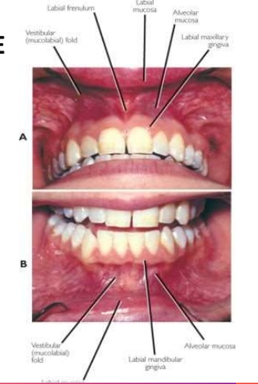
boundaries of oral cavity
anteriorly - lips
laterally - cheeks
posteriorly and medially - teeth and gums
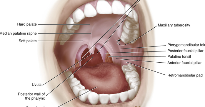
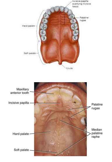
soft palate
yellower and looser posterior part of the palate
only compromises 15% of palate
uvula
median palatine raphe
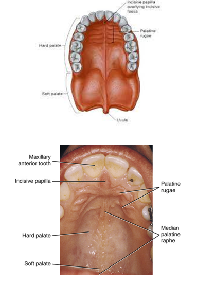
uvula
midline muscular structure
hands from the posterior margin of soft palate
median palate raphe
midline ridge of tissue on the palate
runs from incisive papilla to the uvula
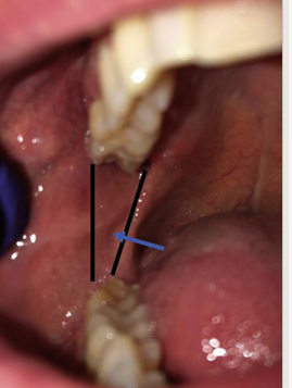
pterygomandibular fold
extends from junction of hard and soft palates down to the mandible on each side just posterior to most distal mandibular molar
stretches when patient opens mouth wider
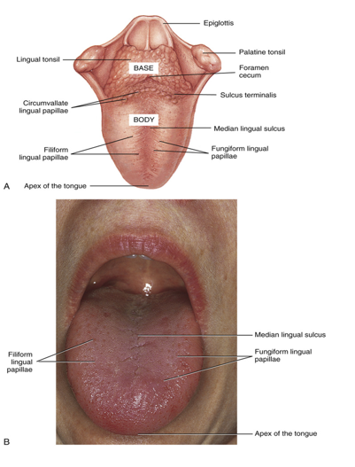
pharyngeal part of tongue
also known as the base of the tongue
the posterior 1/3
attaches to the floor of the mouth
does not lie withing the oral cavity but within the oral part of the throat or pharynx
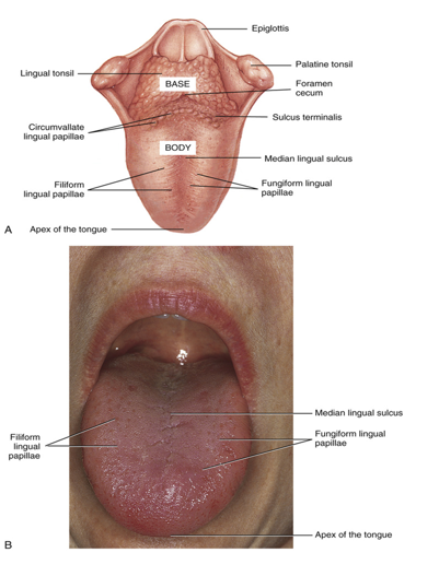
body of tongue
anterior 2/3rds of the tongue
lies within the oral cavity
covered in lingual papillae on dorsal surface
filliform
fungiform
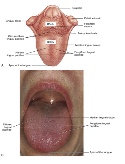
apex of tongue
tip of the tongue
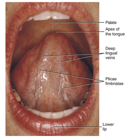
filliform tastebuds
slender, threadlike papillae making tongue velvety