Parasitology: Protozoa
 Call Kai
 Learn
Learn Practice Test
 Spaced Repetition
 Match
Match Flashcards
 Knowt Play
1/45

There's no tags or description
Looks like no tags are added yet.
Name | Mastery | Learn | Test | Matching | Spaced | Call with Kai |
|---|
No analytics yet
Send a link to your students to track their progress
46 Terms
What types of protozoa are covered in this unit?
intestinal amoeba
free living amoeba
flagellates
intestinal ciliates
intestinal coccidia
Most intestinal amoeba are ___
non-pathogenic
What do intestinal amoeba use for motility?
pseudopods (cytoplasmic extensions, “false feet”)
How are amoeba identified?
by their:
size
shape
number of nuclei
granularity of cytoplasm
vacuoles
ingested materials
Protozoa cyst form
found in formed stool
rigid, non-motile, infectious
resistant to environmental changes
found in asymptomatic hosts or carriers
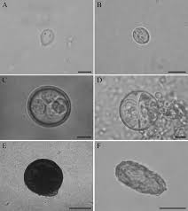
Protozoa trophozoite form
found in liquid stool
pleomorphic (appears in various forms)
seen in active disease
sensitive to environmental changes
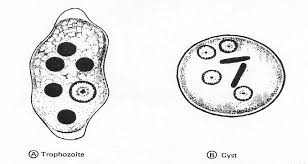
What is it called when a protozoan in cyst form converts to its trophozoite form?
excystation
occurs when the host ingests cysts
Entamoeba histolytica
causes amoebic dysentery
symptoms
cramping, fever, bloody/mucusy diarrhea, characteristic lesions in the intestines (flask-shaped ulcers)
can cause hepatic abscess
organism can erode intestinal mucosa, enter circulation, and colonize the liver (causes increased WBCs and liver enzymes)
picture: trophozoite form
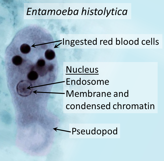
Entamoeba histolytica cyst and trophozoite forms
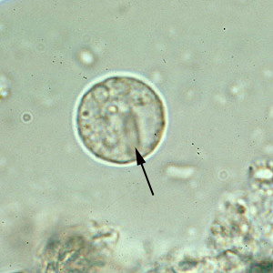
cyst form (picture)
contains 1 to 4 nuclei with a central karyosome, cytoplasm may have bars
trophozoite form
“bullseye” nucleus
seen in wet mounts and trichrome stains
Blastocystis hominis
most common intestinal protozoa, but not considered a common cause of diarrheal disease
associated with traveling abroad
Toxoplasma gondii accidental host and definitive host
accidental: humans
definitive: cats (oocysts are shed from cat feces)
Where do the sexual and asexual life cycles of Toxoplasma gondii occur?
sexual (enteric cycle)
cats’ intestinal tract, caused by ingesting reservoir meat
asexual (extraintestinal cycle)
humans, ingested oocysts or trophozoites
can multiply in the brain and other host cells
What infections does Toxoplasma gondii cause? What happens to pregnant women who are infected?
encephalitis, myocarditis, pneumonitis, and dissemination
in pregnant women, the infection can be spread congenitally, causing CNS involvement in the fetus, severity depends on when the exposure occurs during the pregnancy
Toxoplasma gondii diagnosis
extremely difficult
oocysts are pear or crescent-shaped and are called tachyzoites (visualized by hematoxylin stain)
IgM specific tests can diagnose acute infections in neonates
IFA and EIA tests are used routinely
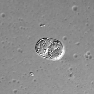
What does Naegleria fowleri cause, and what are the stages?
Primary Amoebic Meningoencephalitis (PAM)
early stage: vague upper resp distress, headache, lethargy
acute stage: sore throat, nasal congestion, severe headache, fever, vomiting, stiff neck, coma, and death
How does Naegleria fowleri cause infection?
by penetrating nasal mucosa and traveling by the olfactory nerve to the brain
causes infection of the brain and meninges
rapidly fatal
What is the 3 stage life cycle of Naegleria fowleri?
cyst
trophozoite
flagellated
Infections caused by Acanthamoeba spp. (free living amoeba)
granulomatous amoebic encephalitis (GAE)
brain lesions, acute fever, headache, and neck pain
amoebic keratitis
occurs in those wearing soft or extended-wear contacts
trichrome stains and giemsa stains visualize trophozoites in specimens
flagellates
more rigid than amoeba
causes infections of the GI tract, blood, and urogenital tract
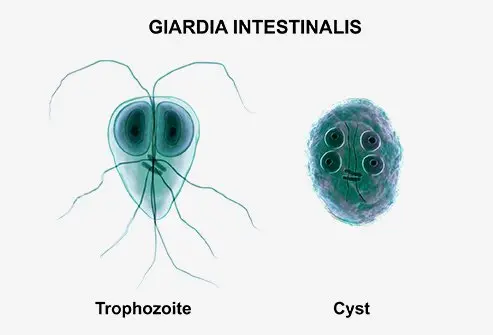
Giardia duodenalis
Giardia duodenalis trophozoite and cyst forms
Giardiasis
low infectious dose
trophozoites
pear or teardrop shaped, face-like, bilateral symmetry
falling leaf motility and a large ventral sucking disk
cysts
oval, up to 4 nuclei, retracted flagella, “cluttered” internal appearance
How does Giardia duodenalis cause infection and what are some common symptoms?
organism adheres to intestinal mucosa after ingestion and interferes with nutrient absorption
symptoms
acute diarrhea, cramps, nausea, malaise
12 to 14 days after: severe diarrhea with no blood
Giardia duodenalis treatment
metronidazole or albendazole
Dientamoeba fragilis
only binucleate flagellate that infects humans
sluggish motility
diarrhea
lacks a cyst stage
trophozoite (picture)
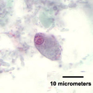
Which intestinal flagellate is this?
Chilomastix mesnili
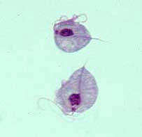
Trichomonas vaginalis
STD
associated with adverse pregnancy outcomes
transmission to newborn, cervical neoplasia, pelvic inflammatory disease
increased risk of HIV due to compromised mucosal barriers
infections: vaginalis and urethritis
Trichomonas vaginalis in the lab
lacks a cyst stage!
trophozoite
pear-shaped with an undulating membrane, 4 flagella, jerky motility
wet mounts used for observation
giemsa stain shows a prominent axostyle (midline) and a single nucleus
Balantidium coli
only pathogenic intestinal ciliate
lives in the large intestine and does not migrate
cyst (infective form)
trophozoite
oval shaped, slightly pointed end
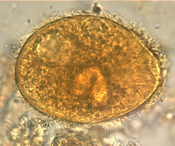
Where can Cystoisospora belli (intestinal coccidia) be found? What infection does it cause? What stains are used to identify?
location
the cytoplasm of the epithelial cells of the small intestine
infection
cystoisosporiasis
identification
modified acid fast stain or iodine prep
oocysts are not infective when passed in the stool
but once ingested, they excyst, releasing sporozoites that invade the epithelial cells
How are the Cryptosporidium infections transmissed and how are they diagnosed?
transmission: contact with contaminated water (including chlorinated pools)
diagnosis: modified acid fast stain with carbolfuschin
Which species of Cryptosporidium causes apicomplexa?
C. parvum
What is the vector of Plasmodium? How is it transmissed? What type of host are humans?
vector: mosquitoes
transmission: bites from female mosquitoes, blood transfusions, infected needles, or transplacental transmission
humans are intermediate hosts
What types of patients are immune from Plasmodium infections?
patients with sickle cell anemia, hemoglobin C, or G6PD deficiency
schizogony
The asexual reproduction process of Plasmodium
exoerythrocytic schizogony of Plasmodium
occurs after a mosquito bite
sporozoites from the mosquito travel from the blood to liver cells where the first asexual cycle occurs
merozoites (daughter cells) are released and enter the host’s RBCs
erythrocytic cycle of Plasmodium
in the RBCs, the merozoites (daughter cells) develop into ring forms and mature into trophozoites
hemozoin pigment can be seen as the merozoites use Hgb for metabolism
followed by the formation of schizonts (asexual forms), which then rupture and release new merozoites
symptoms of Malaria
prodromal period: headache, anorexia, and low grade fever
paroxysms (shaking chills) occur as RBCs rupture
includes fever, limb and back pain, disorientation, night sweats, and fatigue
anemia, splenomegaly, and black water fever (hemozoin pigment released into urine)
Plasmodium vivax
usually invades reticulocytes
enlarged, infected RBCs
Schuffner’s stippling may be present (fine pink dots)
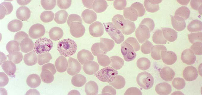
Plasmodium malariae
invades older RBCs
may have a band appearance with dark, course, brown pigment
mature schizont has 8 merozoites
Plasmodium ovale
least common
resembles vivax
enlarged RBCs, may have an oval shape with fringed edges
mature schizont has 6-12 merozoites
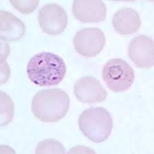
Plasmodium falciparum
infects any and all RBCs
cresent-shaped gametocyte (sexual stage) (upper left of picture)
frequently shows multiple rings in one RBC
20-24 merozoites in schizonts
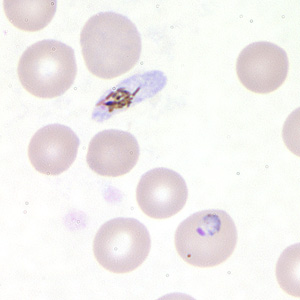
Babesia (malaria-like parasite)
tickborne parasite (humans are accidental hosts)
infection resembles malaria, involves hemolytic anemia, splenomegaly, and hepatomegaly
Babesia microti
vector: ixodes species of tick (same tick that spread lyme disease)
symptoms generally only appear in the young, elderly, and immunocompromised
diagnosis
Wright or giemsa stains show pleomorphic ring forms
trophozoites form tetrad arrangments of 4 merozoites, “Maltese cross”
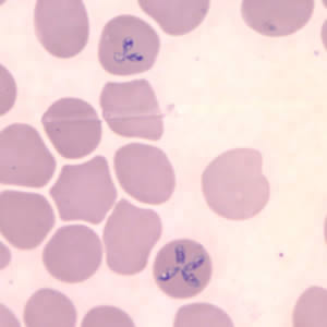
What does the Trypanosomes species of hemoflagellates cause?
African trypanosomiasis (sleeping sickness) and Chagas disease
African trypanosomiasis
caused by T. brucei subsp. gambiense (West African sleeping sickness) and T. brucei subsp. rhodesiense (East African sleeping sickness)
infects the blood and lymph nodes
vector: tsetse flies
T. cruzi (Chagas Disease)
vector: kissing bugs
transmission can also occur through blood transfusion, transplant, or congenitally
diagnosis: C-shaped trypomastigotes found in thick and thin blood smears and body fluids
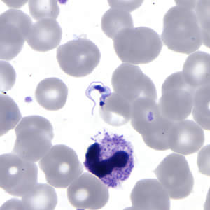
What infections does Leishmania spp. cause?
cutaneous Leishmaniasis
skin ulcers
visceral Leishmaniasis
organism spread through the lymphatics and invades the liver (increased liver enzymes), spleen, BM, and lymph nodes