oral radiology ch 8, 18, and 19 quiz
 Call Kai
 Learn
Learn Practice Test
 Spaced Repetition
 Match
Match Flashcards
 Knowt Play
1/19

There's no tags or description
Looks like no tags are added yet.
Name | Mastery | Learn | Test | Matching | Spaced | Call with Kai |
|---|
No analytics yet
Send a link to your students to track their progress
20 Terms
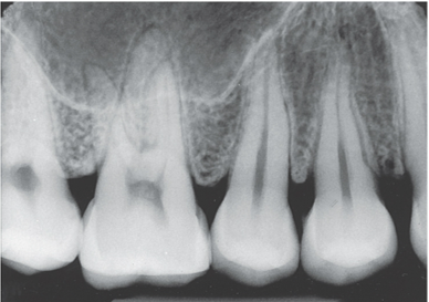
Why do we take periapical radiographs?
used to examine the entire tooth and supporting bone, examine “around apical”
uses __ receptor
utilized paralleling and bisecting technique*
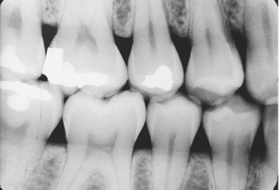
Why do we take interproximal (BWX’s) radiographs?
to examine the crowns and contacts of both the maxillary and mandibular teeth on a single film
used for interproximal examination, therefore no overlapping can occur
uses bite-wing receptor
uses bite-wing technique
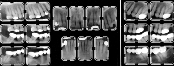
What is covered on a FMX?:
also known as CMS or FMS
a full-mouth series or complete series that consists of tooth-bearing areas (dentulous or edentulous areas)
includes periapical and bitewings
14 to 20 films may be taken
What makes a x-ray image diagnostic?
must show the entire crowns and roots of teeth being examined, as well as 2 to 3 mm beyond root apices
BWX images must show open contacts, optimum density, contrast, definition, and detail with the least amount of distortion possible
What are the basic principles of paralleling?:
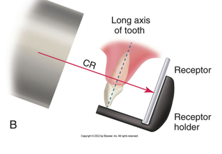
receptor is placed in the mouth parallel to long axis of tooth
central ray of x-ray beam is directed perpendicular to the film and the long axis of the tooth
beam alignment device must be used to keep the receptor parallel with the long axis of tooth
recePtor → Parallel
Central Ray → perpendiCulaR to sensor and tooth
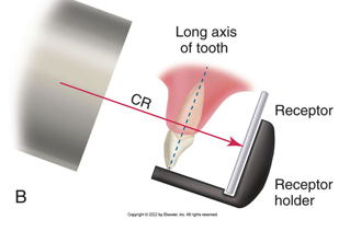
How do we achieve parallelism between the receptor and the tooth
increase object-receptor distance to insure receptor is parallel with long axis of tooth
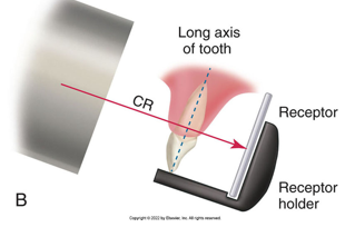
basic rules of paralleling
Receptor placement: Position the image receptor so that it will cover the correct teeth to be examined
Receptor position: the image receptor must be positioned parallel to the long axis of the tooth
Vertical angulation: the central ray of the x-ray beam must be directed perpendicular to the image receptor nd the long axis of the tooth
Horizontal angulation: the central ray of the x-ray beam through the contact areas between the teeth
Film receptor exposure: The X-ray beam must be centered on the receptor to ensure that all areas are exposed. Failure to do so will result in “cone cut.”
Digital image
an image composed of pixels that can be stored in a computer
commonly used to detect lesions, disease, and conditions of teeth and supporting structures
use LESS radiation exposure than film (50-90% less exposure time)
Analog image
produced from conventional film
Bit-depth image
number of possible gray-scale combinations for each pixel
254 shades of gray possible
Digital imaging:
filmless imaging system
a method of capturing an image using sensor, breaking it into electronic pieces, and presenting and storing image in computer software
digital subtraction
one feature of digital imaging
method of reversing the gray scale as image is viewed
radiolucent appear white
radiopaque appear black
direct digital imaging
a sensor is placed into the mouth of the patient and exposed
the sensor captures the image and transmits it to the computer monitor
software is used to enhance and store image
indirect digital imaging
method of obtaining a digital image
an exsisting x-ray film is digitized using a CCD camera
the image is displayed on a computer monitor
line pairs/ millimeter (lp/mm)
measurement used to evaluate the ability of the computer to capture the resolution (or detail) of an image
pixel
a discrete unit of information
picture element
digital information is contained in, and presented as, discrete units of information
sensor
a receptor that is used to capture an intraoral or extraoral image
we will be using a size 2 receptor
storage phosphor imaging
method of obtaining a digital image in which the image is recorded on a phosphor coated plate and then placed into an electronic processor where a laser scans the plate and produces an image on a computer monitor
charge coupled device (CCD)
solid state silicone chip detector that converts lights or x-ray photons into an electrical charge or signal
found in sensor
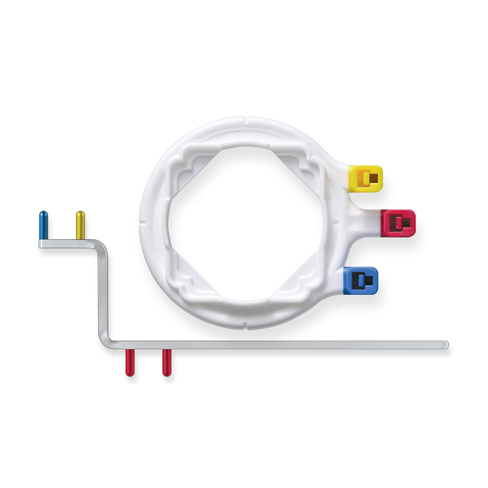
Rinn XCP-ORA one ring and arm positioning system
one ring
one arm
is a beam alignment device that is used to position receptor in the mouth and retain receptor in position during exposure