NCM 109: Fetal Circulation and Congenital Heart Defects
1/79
There's no tags or description
Looks like no tags are added yet.
Name | Mastery | Learn | Test | Matching | Spaced | Call with Kai |
|---|
No analytics yet
Send a link to your students to track their progress
80 Terms
Difference between Adult and Fetal Circulation
Fetal Circulation
- Fluid-filled lungs: The placenta performs gas exchange, not the lungs
*High-pressure pulmonary system
*Mixing of oxygenated and deoxygenated blood
*No mixing of fetal and maternal blood
- Fetal Shunts
- R > L side pressure
Adult Circulation
- Gas filled lungs
- L > R side pressure
- No mixing of oxygenated and deoxygenated blood
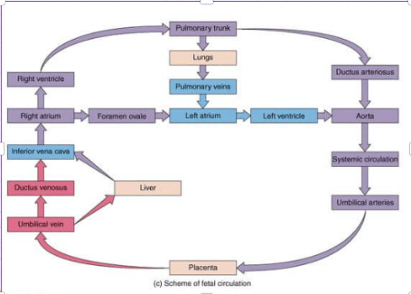
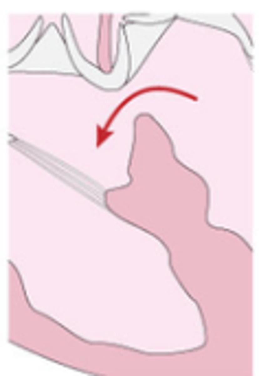
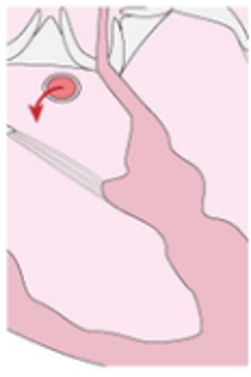
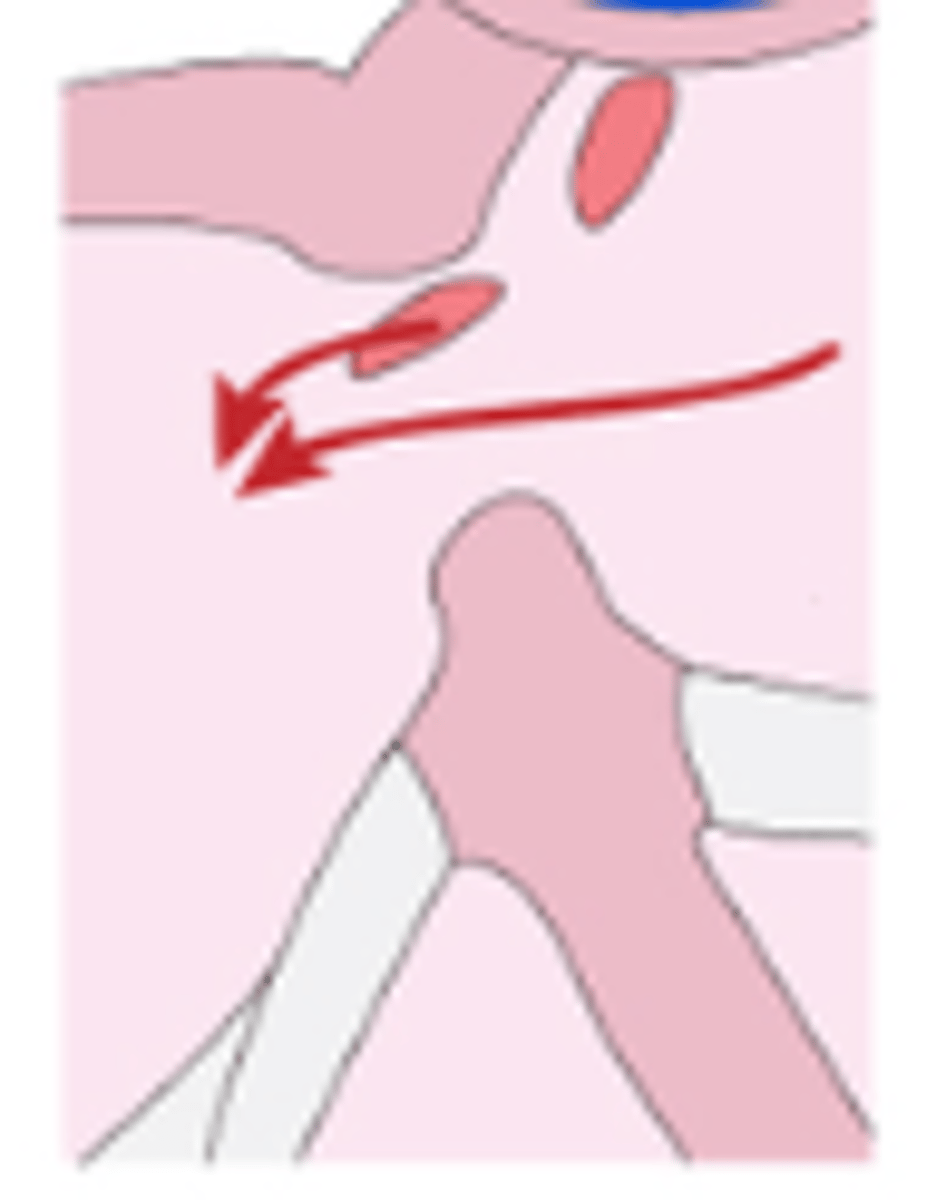
Foramen Ovale
- Leads from right atrium directly to left atrium,
- Blood skips the lungs
- 40% of oxygenated blood passes here
- The remaining IVC blood mixes in the right atrium with the deoxygenated blood that has returned from the head and upper extremities via the SVC.
Ductus arteriosus
- Leads from the Pulmonary Trunk directly to the Aorta
- 90% of blood from the right ventricle passes here
- 10% goes to the lungs for growth and development
Ductus venosus
- Leads newly oxygenated blood from the umbilical vein directly to the inferior vena cava
- As blood returns from the placenta through the umbilical vein, some go into the hepatic veins and slightly more than half passes through the ductus venosus
Fetal Circulation (Flow)
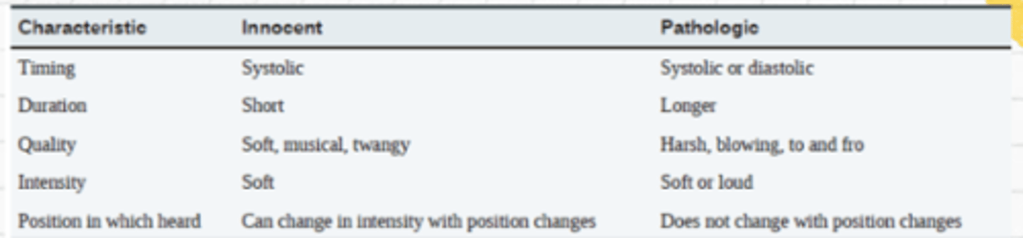
Murmurs
sounds created by abnormal, turbulent flow of blood in the heart
- May be innocent or may indicate congenital heart defects
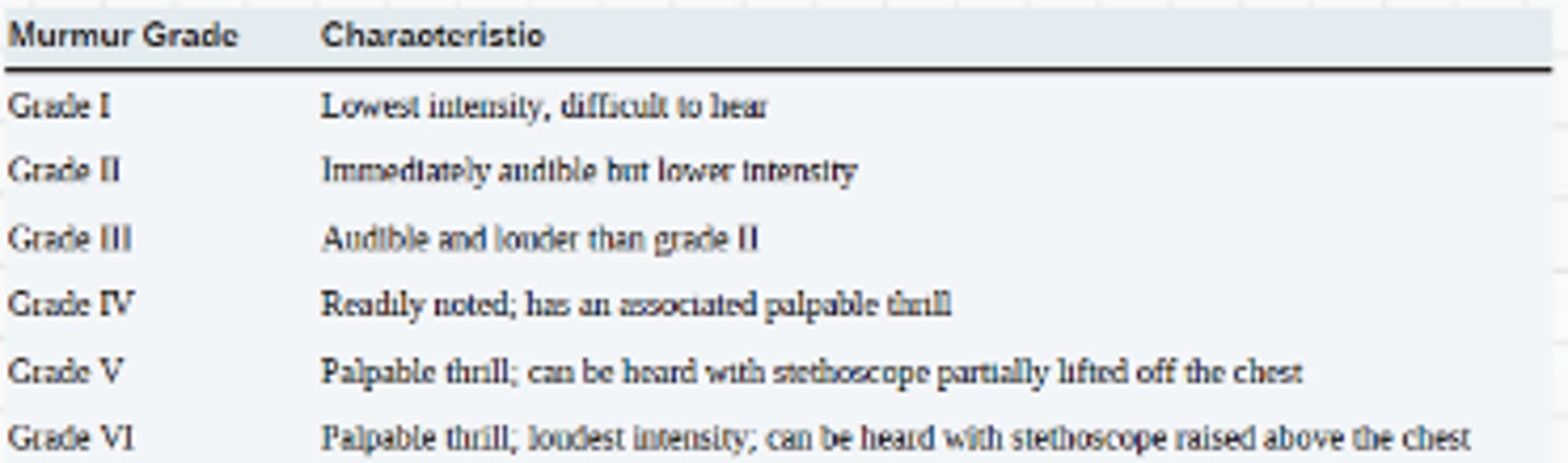
Levine Scale
grading of murmurs:
1/6- faint, not heard in all positions
2/6- quiet, but not difficult to hear
3/6- moderately loud
4/6- moderate w/ thrills
5/6- heard with stethoscope barely on chest, loud w/ thrills
6/6- heard with stethoscope off chest, very loud w/ thrills
Systolic = I to VI
Diastolic = I to IV
Thrills
Palpabrle vibration felt secondary to significant cardiac murmurs
Lift
Forceful cardiac contraction that causes the hand to move up
Heave
Very forceful; causes the hand to move up and laterally
Innocent vs. Pathologic Murmur
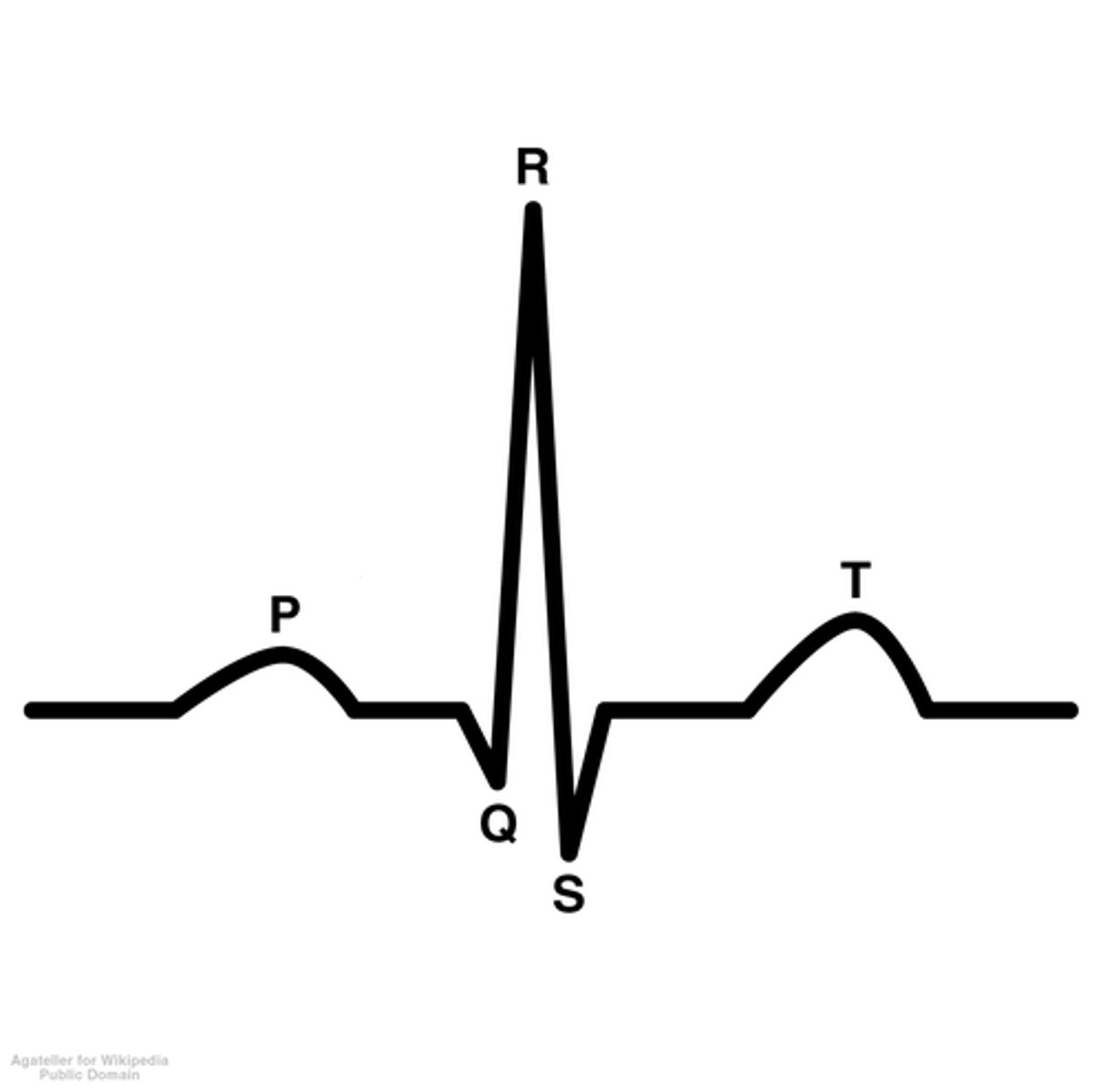
ECG
electrocardiogram
- a written record of the electrical activity generated by the heart and provides information about heart rate, rhythm, state of the myocardium, presence or absence of hypertrophy (thickening of the heart walls), ischemia or necrosis
- Changes in the size or shape of the various waveforms may indicate chamber enlargement or poor function.
parts of ECG wave
P wave, QRS complex, T wave
P wave = atrial depolarization (contraction)
QRS = ventricular depolarization
T wave = atrial repolarization (relaxation)
S1 sound meaning
closing of AV valve
S2 sound meaning
closing of seminlunar valves (aortic/pulmonic)
S3 Heart sound
↑ventricular filling pressure (e.g., mitral regurgitation, HF), common in dilated ventricles
S4 Heart sound
Stiff/hypertrophic ventricle (aortic stenosis, restrictive cardiomyopathy)
low frequency
Pericardial Friction rub
scraping or grating noise heard on auscultation of the heart; suggestive of pericarditis
Holter Monitor
- worn for 24 to 72 hours and gives a complete account of every heart beat the child experiences.
- allows for evaluation of average rate, rhythm, and frequency of ectopic beats.
Transthoracic Echocardiogram
noninvasive ultrasound of the heart that gives detailed information about heart structure and function.
Exercise Stress Testing
used to evaluate a child's clinical condition during periods of increased myocardial demand, such as with exerciseaccomplished with a treadmill, bicycle, or a 6-minute walk to increase the child's heart rate.
Cardiac Catheterization
invasive; catheters are inserted through a large vein and artery and floated into the heart.allows for direct measurements of pressure and oxygen saturations as well as visualization of the heart and all blood vessels with the aid of a contrast medium
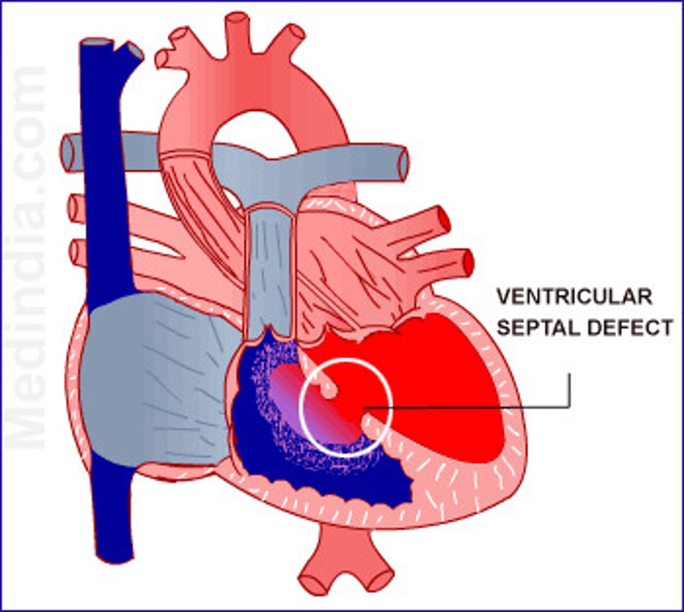
Ventricular Septal Defect (Definition)
- It occurs when there is an opening in the ventricular septum, or dividing wall between the 2 lower chambers of the heart known as the right and left ventricles.
- A VSD occurs when a portion of the ventricular septum does not completely close.
- VSDs may be single or multiple and are defined based on their anatomic location within the septum
Ventricular Septal Defect (Membranous)
a hole is found at the upper section of the ventricular septum and is very close to the tricuspid and aortic valves.
Ventricular Septal Defect (Muscular)
found at the lower part of the septum in the muscle layer. Small muscular VSDs have a high chance of closing on their own.
Ventricular Septal Defect (Outlet (conal or subarterial)
found at structures where blood is leaving the heart around the pulmonic and aortic valve locations.
Ventricular Septal Defect (Inlet (atrioventricular canal))
found at structures where blood is entering the heart around the tricuspid and bicuspid valve (mitral).
Ventricular Septal Defect (Assessment)
An acyanotic defect allows oxygenated blood to flow from the left side of the heart to the right side of the heart.
Left heart dilatation
- Due to increasing pulmonary blood flow that ultimately increases volume of blood returning to the left heart.
Harsh and Holosystolic/ Pansystolic murmur
- Noted at the left lower sternal border.
- May not be heard at birth because the pulmonary vascular resistance is still high, which limits blood flow across the defect.4-6 weeks of life:
Small defect:
- May have a murmur
- No clinical symptomsFirst 2 weeks of life:
- Most small VSD close spontaneously.
Large Defect:
- increased pulmonary blood flow manifests as symptoms of HF:
- Tachypnea
- Poor feeding/failure to thrive
Ventricular Septal Defect (S/S HOLE)
○ Heart Failure and pulmonary hypertension: Dyspnea, fatigue, swelling extremities, crackles, sweating, clammy with activity
○ Often experiences lung infection: With the narrowed arteries, it can cause possible secondary bacterial infection.
○ Low growth rate and loss of weight: It ties back to the heart and breathing problems burning a lot of energy to maintain the life of the patient. The heart has to pump harder.
○ Extra heart sounds: Murmurs heard at the left sternal border, at the 3rd or 4th interspace.
Ventricular Septal Defect (Management)
- Furosemide:
* Management if the child does exhibit signs of pulmonary overload, such as tachypnea, retractions, or rales.
- Increase caloric density of the child’s formula or human milk
* If there are concerns for poor weight gain
* Breastfeeding/chestfeeeding demands a significant amount of energy from the child.
* Parents may need to manually express the human milk and feed the infant through a bottle or nasogastric tube.
Referred for closure If closure does not occur overtime.Child experiences persistent failure to thrive or dilatation of the left heart.
* Cardiac Catheterization (transcatheter closure of a VSD/VSD transcatheter repair)
* Median Sternotomy
* VSD repairs can be at a low risk for heart block or other arrhythmias depending on the location of the VSD and the size of the child.
Atrial Septal Defect (Definition)
- An ASD occurs when a portion of the atrial septal tissue does not completely form.
- R-> L shunting
- Due to the lower overall pressure in the atria than the ventricles, the amount of blood flow through this defect is limited.
- ASD may not have a murmur associated with it; therefore, it may go unnoticed if no clinical symptoms exist.
- Regardless of the presence of a murmur, an ASD may have a fixed split-S2 heart sound.
Atrial Septal Defect (Primum Defect)
found low in the atrial septum near the IVC.
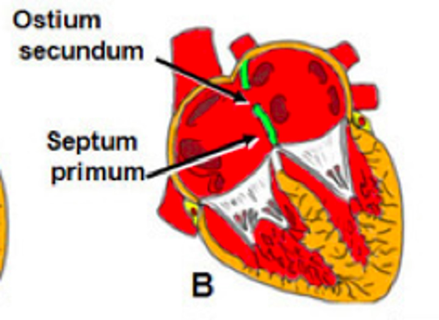
Atrial Septal Defect (Secundum defect)
located in the center of the atrial septum.
Atrial Septal Defect (Sinus venosus defect)
found high in the septum where the pulmonary veins enter the left atrium.allow communication of one or more of the pulmonary veins with the right atrium.
Atrial Septal Defect (Assessment)
Small defect
- Undetected or if noted may cause no clinical concern and require no intervention.
- Many small ASDs will close spontaneously in the first few years of life.
o Large defect:
- Child may show symptoms of ASD such as:
- Pulmonary overcirculation
- Rales
- Congestion
- Tiring with activity
- Frequent respiratory infections
- Poor weight gain
- Right heart may also dilateIncreased volume
- Harsh Systolic murmurs
- split heart sound heard at 2nd or 3rd intercostal space
.
- Turbulence
- Increased pulmonary blood flow that produces systolic murmur heard best at the left upper sternal border
Atrial Septal Defect (Management)
Will not require closure
- If ASD is noted to be small and is producing no clinical symptoms.
Diuretics
- May be attempted first in an effort to allow the defect to close spontaneously and to manage symptoms of ASD.
Referred for closure
- Defects that fail to close with persistent shunting after an observation period to decrease the incidence of supraventricular dysrhythmias and prevent pulmonary vascular disease. If the defect is larger with evidence of increased pulmonary blood flow.
Cardiac catheterization (transcatheter ASD closure)
- For secundum ASDs
Median Sternotomy incision and Cardiopulmonary bypass
- For sinus venosus
- For primum defects
- And very large secundum ASDs
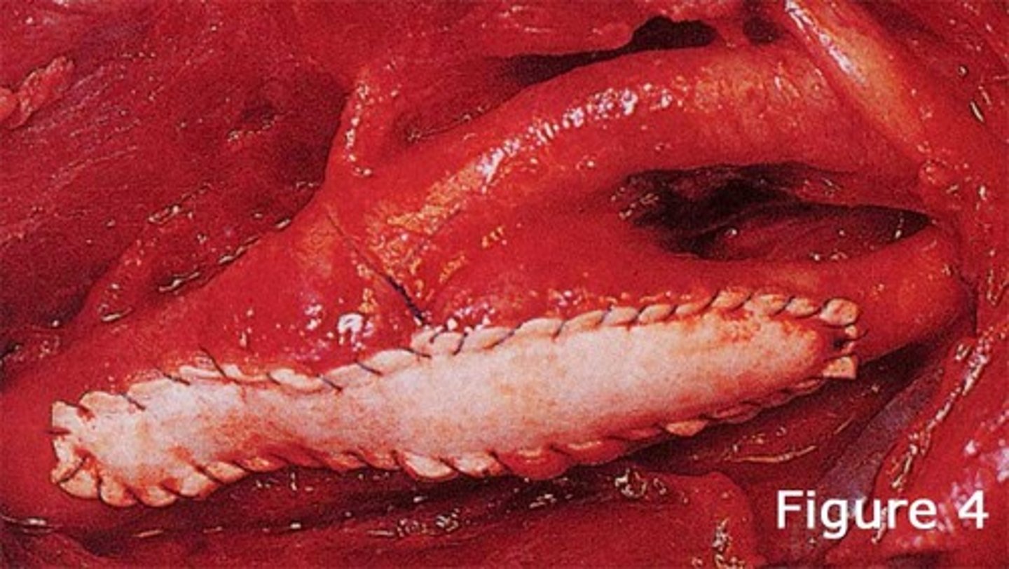
Dacron patch
used to repair atrial, ventricular septal defects in pediatric patients
Atrioventricular Canal Defect (Definition)
- Blood freely mixes between the right and left sides.
- Blood flow in AVSD is predominately left to right, resulting in increased pulmonary blood flow and HF as pulmonary vascular resistance falls in the first 6 to 8 weeks of life.
- only one large malformed AV valve
- The atria can become dilated through regurgitation of blood through the incompetent valves.
- AVSD is the most common type of CHD in children with trisomy 21.
Atrioventricular Canal Defect (S/S)
▪ The same symptoms of ASDs
▪ Right ventricular hypertrophy
▪ Increased pulmonary blood flow
▪ Fixed S2 splitting (fixed second heart sound splitting)
▪ Caused by closure of the aortic and pulmonary valve are not synchronized during inspiration
Atrioventricular Canal Defect (Management)
Pharmacological Management:
Furosemide
Digoxin
ACE inhibitors like captopril/enalapril
Referred for surgery: Children without Down Syndrome
- Surgical correction by 3 months age or earlier if the transthoracic echocardiogram demonstrates signs of increased pulmonary pressures.
Children with Down Syndrome
- referred for repair by 5 or 6 months of age or as clinically necessary.
Pulmonary Artery Band
- If surgical repair needs to be delayed, a temporary measure to decrease the amount of pulmonary blood flow can be performed.
- Which is a band surgically placed around the pulmonary artery that constricts it to decrease some of the overall pulmonary blood flow
After surgery:
- Risk for heart block
- May have persistent mitral valve dysfunction (stenosis or regurgitation)
Further surgical interventions
- If it progresses to severe dysfunction.
Artificial Valve placement
- If the goal to repair the mitral valve cannot be achieved.
Patent Ductus Arteriosus (Definition)
It is a persistent vascular connection between the pulmonary artery and the aorta that persist after birth.
= Patent occurs more frequently in children born prematurely.
Patent Ductus Arteriosus (S/S CALL)
Cardiac
- Continuous "machine-like" murmur: Hallmark sign; The heart sound that is unique. Continuous meaning you're gonna hear in both diastole and systole and you are gonna hear it in the left upper sternal border.
- Risk for endocarditis due to inc. pulmonary blood flow
Activity Intolerance
- Diaphoresis
- Fatigue due
Lungs
- Pulmonary Hypertension
Loss of Weight
- Difficulty feeding
Patent Ductus Arteriosus (Management)
-IV indomethacin 1st line treatment (closes the PDA) VERY SLOWLY
-surgical correction if indomethacin fails (best if done before 1-3 years of age)
Pulmonary Stenosis (Definition)
- a narrowing of the pulmonary valve or pulmonary artery that results in obstruction of blood flow from the ventricles
- Inability of the right ventricle to evacuate blood by way of the pulmonary artery because of the obstruction
- can be noted at several sites; most typical is subpulmonary valve stenosis with anomalies of the actual pulmonary valve also seen.
Pulmonary Stenosis (Complications)
Mild: infants with pulmonary stenosis may be asymptomatic or mild right sided heart failure
- right ventricular hypertrophy
Severe: cyanosis because of the inability of adequate blood to reach the lungs for oxygenation or right to left shunting across the foramen ovale because of the increased right sided heart pressure
systolic ejection murmur (grade 5, crescendo decrescendo murmur) heard at upper left sternal border
Pulmonary Stenosis (Management)
- ECG will reveal right ventricular hypertrophy
- Cardiac catheterization: Rarely necessary for diagnosis but is used for interventional enlargement of the stenosis valve
- Balloon angioplasty is a catheter with an un-inflated balloon at its tip inserted and passed through the heart into the stenosed valve. As the balloon is inflated, it breaks valve adhesions and releases the stenosis. Following the procedure, although children may have residual heart murmur, you can expect a normal life span.
Aortic Stenosis (Definition)
narrowing of the aortic valve
subvalvar, valvar, or supravalvar.
The narrowing prevents blood from passing freely from the left ventricle of the heart into the aorta causing the heart to work harder to pass blood through, resulting to increased pressure and hypertrophy in the left ventricle
Aortic Stenosis (Complications)
left ventricular hypertrophy
- possible back pressure causing pulmonary edema
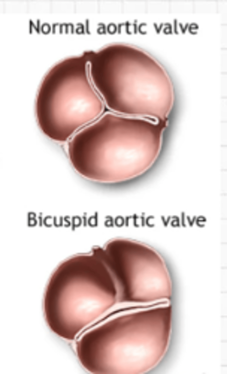
Aortic Stenosis (Valvar)
Most common location
- bicuspid aortic valve instead of tricuspid
Aortic Stenosis (Assessment)
- Present with systolic heart murmur
- diastolic murmur may also be noted
- Thrill or palpable purring sensation in the suprasternal notch
- left ventricular hypertrophy
In severe cases, there will be decreased cardiac output as evidenced by:
- Faint pulses
- Hypotension
- Tachycardia
Aortic Stenosis (Management)
Beta-blocker - Metoprolol, may be necessary to reduce or prevent further ventricular hypertrophy.
Routine Echocardiogram and cardiac catheterization - to best quantify the pressure gradient across the valve.
Echocardiogram - known as cardiac ultrasound, is the use of ultrasound to examine the heart. It is a type of medical imaging, using standard ultrasound or Doppler ultrasound.
Balloon valvuloplasty is a surgical treatment of choice. It is a surgery that involves dividing the stenotic valve or dilating constrictive aortic ring for severe defects. Such repair may lead to aortic valve insufficiency in later life at which time further surgery may be needed.
Coarctation of the Aorta (Definition)
narrowing of the aorta
- This defect is typically located at the level of the ductus arteriosus insertion;
- the mechanism is thought to be migration or extension of ductal tissue into the wall of the fetal thoracic aorta
- causing the tissue to constrict following birth.
- It occurs more commonly in males than in females
Coarctation of the Aorta (Assessment)
Frequently, a child is not diagnosed with a coarctation until they grow and the narrowed area does not grow along with them. The narrowed area is most frequently distal to the right subclavian artery = elevated blood pressure in the right arm.
This narrowing increases the resistance to the left ventricle and can lead to left ventricular hypertension and hypertrophy over time.
- a systolic murmur heard along the left sternal border and the left midscapular area..
- unequal upper and lower extremity pulses
- obtain blood pressures in the right arm and either leg.
- Coarctation noted in infancy is usually more critical and can produce HF within days to weeks of birth
Coarctation of the Aorta (Management)
Balloon or Stent Angioplasty
- With this procedure, a catheter with an inflated balloon at its tip is inserted and passed through the heart and into the aorta. As the balloon is inflated it breaks the adhesions and reveals the stenosis.
Echocardiogram, MRI, X-ray Exam
- Reveals left sided heart enlargement from back pressure and also notching the ribs for large collateral vessels
Left Thoracotomy Incision
- resection of the narrowed tissue and the remaining ends of the aorta sutured together (end-to-end anastomosis)
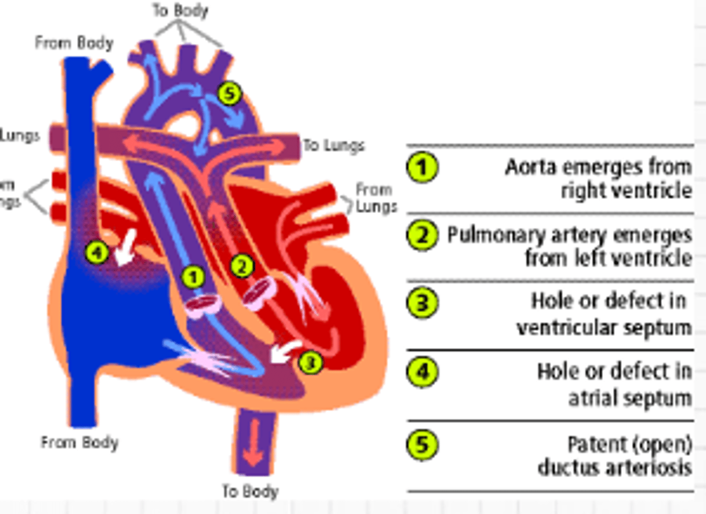
Transposition of the Great Arteries (Definition)
This is a heart defect that occurs when the two main blood vessels leaving the heart are in abnormal positions. It is a life-threatening congenital condition
Aorta emerges from right ventricle
Pulmonary artery emerges from left ventricle
Hole or defect in ventricular septum
Hole in atrial septum
Patent Ductus arteriosus
Each side of the heart has its own circulation without communication.
Mixing of blood seen in ASD, VSD, PDA
Transposition of the Great Arteries (SWAP)
S: Severe cyanosis
W: Watch heart rate, rhythm, and O2 saturation levels
A: Alprostadil (Prostaglandin E)
P: Procedures to correct:
Transposition of the Great Arteries (S: Severe Cyanosis)
- Will not resolve without treatment.
- Degree vary if CHD is present (worse as structures close normally).
- Low oxygen: Increased HR & RR (body's way of trying to compensate to pump more oxygen but it does not happen because there is no connection between right and left side of heart).
- Poor feeding lead to decreased growth rate.
- Cool extremities.
Transposition of the Great Arteries (A: Alprostadil (Prostaglandin E))
Keeps the connection between aorta and pulmonary artery (PDA); Keep ductus arteriosus open; Buys us some time until surgery.
Transposition of the Great Arteries (P: Procedures to correct:)
Balloon atrial septal pull-through
- Enlarged Septal Opening
- Temporary
- First few days of infants
- Done by cardiac catheterization wherein balloon is passed from foramen ovale through right atrium
- Creates artificial ASD
Arterial Switch Procedure
- Permanent
- Done to 1 week to 3 months of age major vessels are switched in position
- Survival heart of 95%
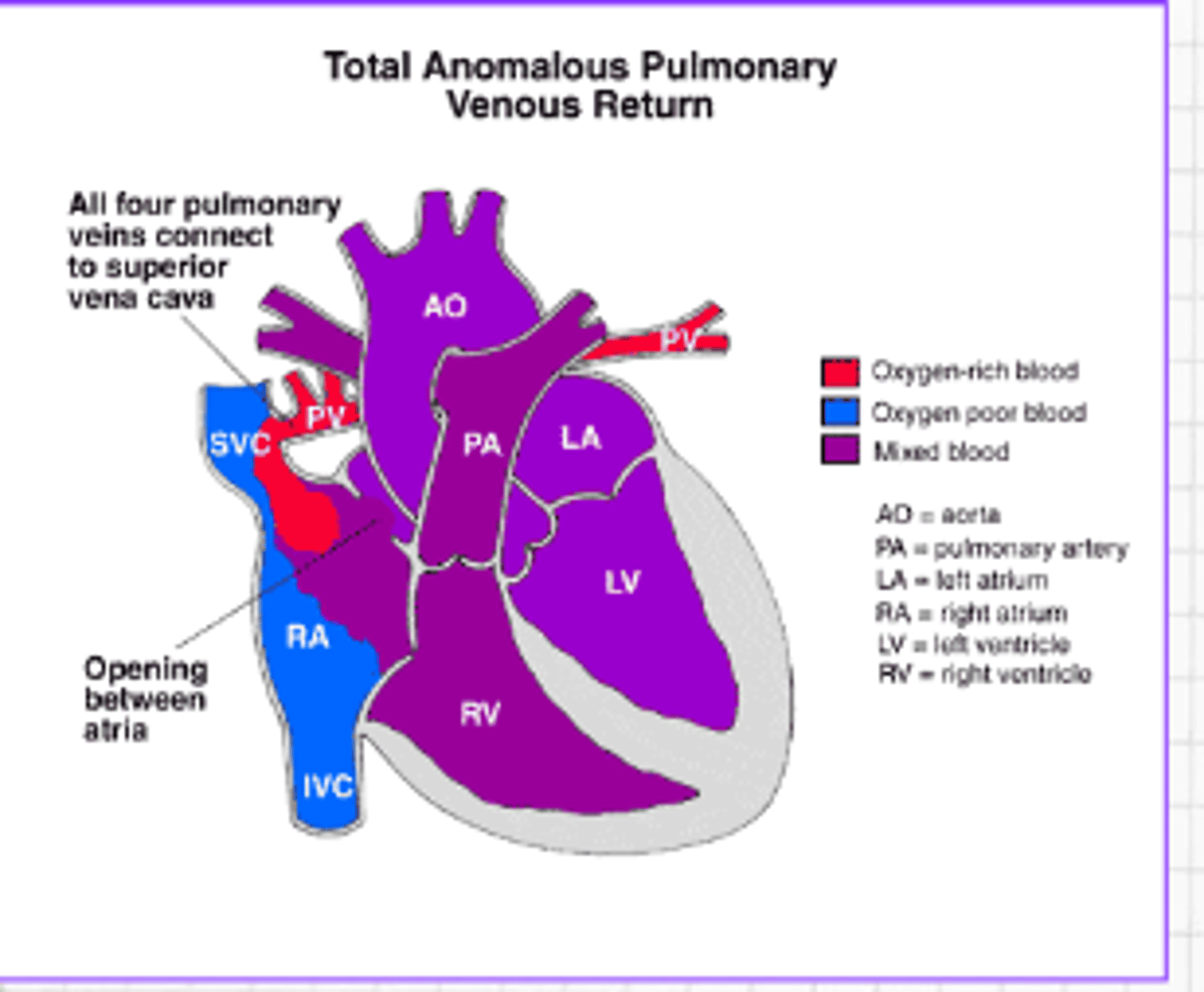
Total Anomalous Pulmonary Venous Return (Definition)
Oxygen-rich blood does not return from the lungs to the left atrium or to a vein flowing to the right atrium or SVC. Instead, the oxygen-rich blood returns to the right side of the heart via the superior vena cava at the right atrium. Thus, oxygen-rich blood mixes with oxygen-poor blood.
Total Anomalous Pulmonary Venous Return (S/S)
- Tire easily
- Trouble breathing
- Mildly cyanotic
- If the ductus arteriosus closes or the septal defect is small, cyanosis increases.
- Right sided heart failure develops as a complication
Total Anomalous Pulmonary Venous Return (Management)
- Continuous infusion of prostaglandin to help keep the ductus arteriosus open
- Balloon atrial septal pull-through procedure to enlarge a small foramen ovale
- Continuous IV infusion with prostaglandin to help keep ductus arteriosus open
- Surgery: Reimplanting the pulmonary veins into the left atrium (permanent correction)
Medication to keep open PDA
Prostaglandin E
Medication to close PDA
Indomethacin
Truncus Arteriosus (Definition)
A rare type of heart disease in which a single blood vessel (trunk) comes out of the right & left ventricles, instead of the normal 2 vessels (pulmonary artery & aorta).
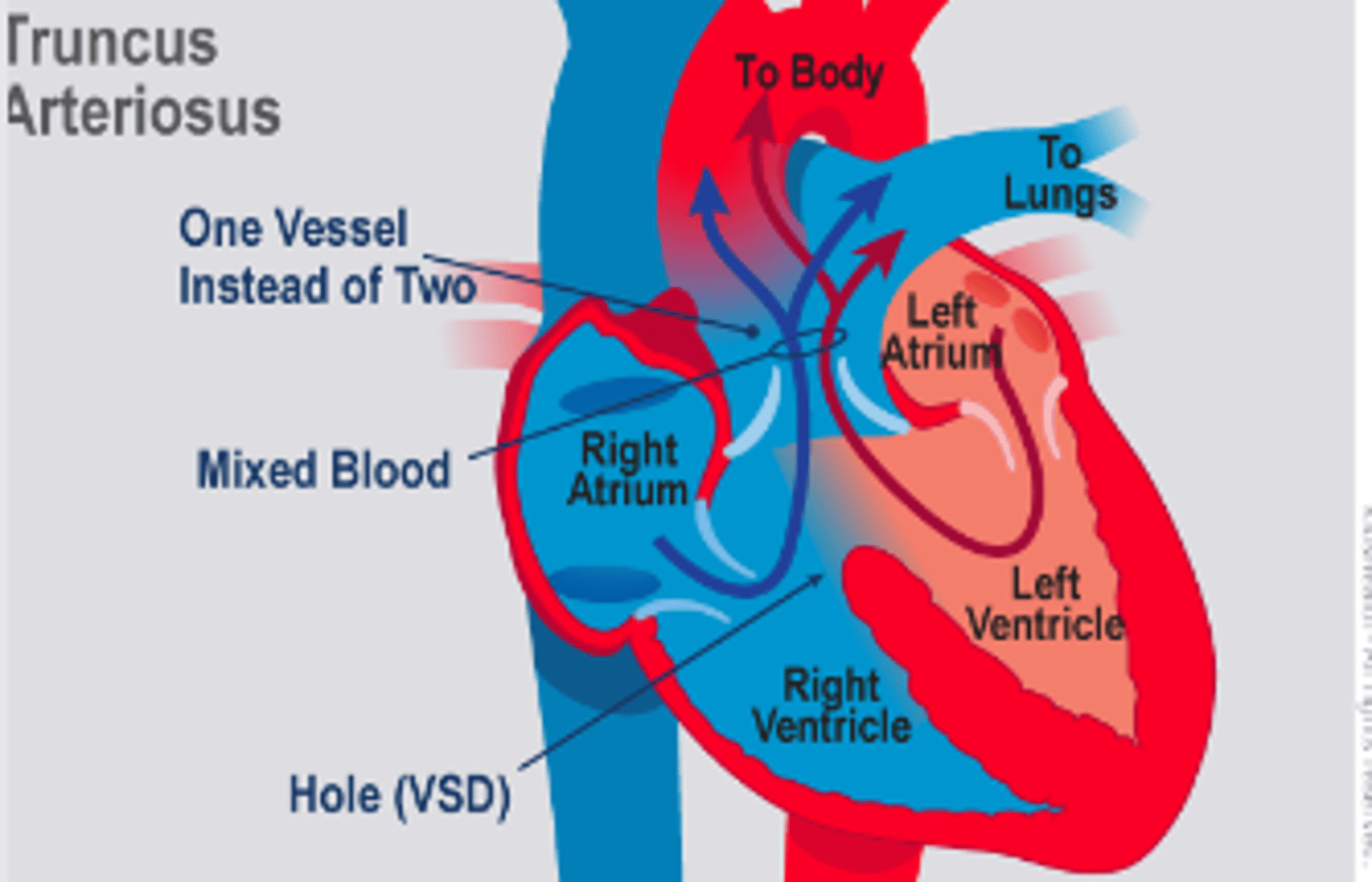
Truncus Arteriosus (S/S)
- Cyanotic: Decreased oxygenated blood in the system.
- Increased RR.
- Extreme fatigue
- Poor feeding
- Decreased cardiac intolerance, cold, clammy skin
- May have a typical VSD murmur:
* Usually heard at the upper sternal border
Truncus Arteriosus (Management)
- Some children need a 2nd surgical procedure during school age as the graft inserted to separate the aorta & pulmonary artery may be large (permanent correction).
- Done for the first 2 weeks
- Digoxin, diuretics and ACE inhibitors decrease pressure in the pulmonary arteries: Given before surgery; Goal is to decrease the stress of the heart while increasing the contraction.
Hypoplastic Left Heart Syndrome (Definition)
left side of the heart is underdeveloped.
- Left ventricles not functioning or is too small
- Absence of mitral and aortic valve.
- Aorta (main artery leaving the heart) is smaller than normal.
- The left side of the heart can’t effectively pump blood to the body.
- Instead, the right side of the heart must pump blood to the lungs and to the rest of the body.
Hypoplastic Left Heart Syndrome (S/S)
Blue or purple tint to lips, skin and nails (cyanosis)
Difficulty breathing
Difficulty feeding
Lethargy (sleepy or unresponsive)
Hypoplastic Left Heart Syndrome (Management)
Prostaglandin therapy: To maintain PDA
Inhaled nitrogen combined with oxygen: To decrease PO2
Surgery (restructuring of the heart)
- There is limited success in this syndrome
- Norwood procedure
Heart transplantation: Ultimate answer for prolonging the child’s life; donor hearts for newborns are limited.
Norwood Procedure
-Treats hypoplastic left heart
-3 stages
-done shortly after birth
-Right ventricle is converted: connects aorta to right ventricle
-Between step 1&2 is highest risk for death
-bi-directional Glenn operation (stage 2, under 6 mo. old)
-superior vena cava redirected to lungs
-Fontan operation (1.5-3 yrs old)
-inferior vena cava redirected to lungs
Heart transplant is another option
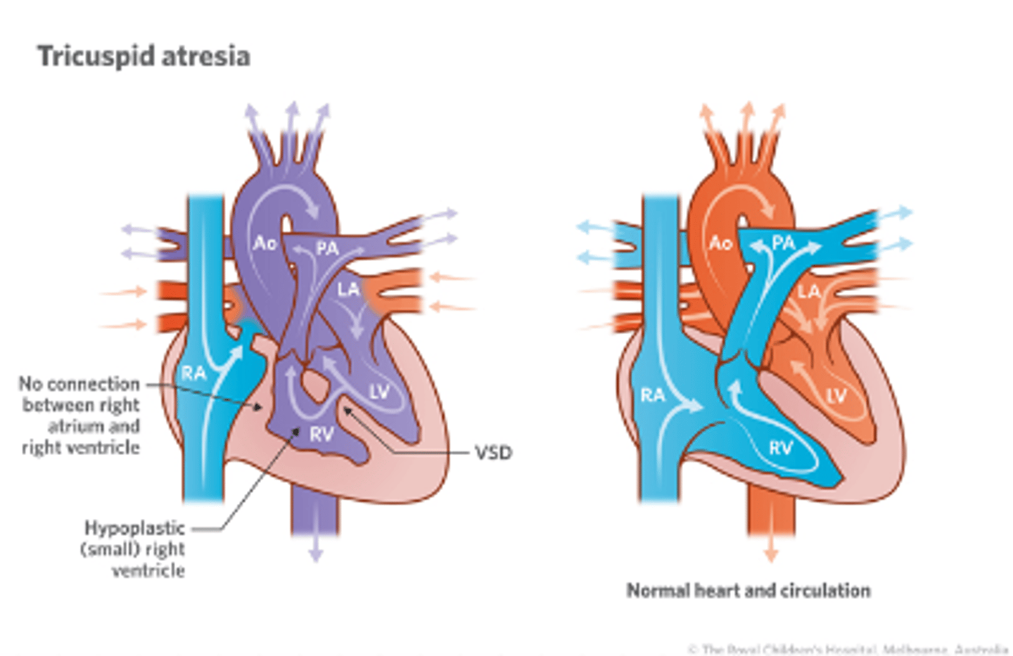
Tricuspid Atresia (Definition)
- Is a birth defect of the tricuspid valve, which is the valve that controls blood flow from the right atrium (upper right chamber of the heart) to the right ventral (lower right chamber of the heart.)
- Occurs when the tricuspid valve doesn’t form at all , and no blood can go from the right atrium through the right ventricle to the lungs for oxygen.
- Blood will flow from the heart's right upper chamber directly to the left upper chamber through a hole in the wall between them. (PFO/ASD)
Tricuspid Atresia (S/S)
- Bluish looking skin color or cyanosis due to low blood oxygen levels
- Difficulty breathing
- Poor feeding
- Extreme sleepiness, especially during feedings
- Slow growth and poor weight gain.
Tricuspid Atresia (Management)
Medicines:
IV infusion of Prostaglandin helps keep the ductus arteriosus open. Keeping the ductus open in babies with tricuspid atresia improves the flow of blood to the lungs.
Surgery:
1. Glenn procedure
2. Fontan Procedure
3. Cardiac Catheterization:
Can make or enlarge openings in the wall between the two atria and between the two ventricles. It also can be used to place a stent (mesh tube) in the ductus to keep open.
Tricuspid Atresia (Glenn Procedure)
- This usually is performed when an infant is 4 to 6 months of age.
- This procedure creates a direct connection between the main pulmonary artery and the superior vena cava, the vessel returning oxygen-poor blood from the upper part of the body to the heart.
- This allows blood returning from the body to flow directly to the lungs and bypass the heart.
Tricuspid Atresia (Fontan Procedure)
- This procedure usually is done sometime around 2 years of age.
- Doctors connect the main pulmonary artery and the inferior vena cava, the vessel returning oxygen-poor blood from the lower part of the body to the heart, allowing the rest of the blood coming back from the body to go to the lungs.
- Once this procedure is complete, oxygen-rich and oxygen-poor blood no longer mix in the heart and an infant’s skin will no longer look bluish.
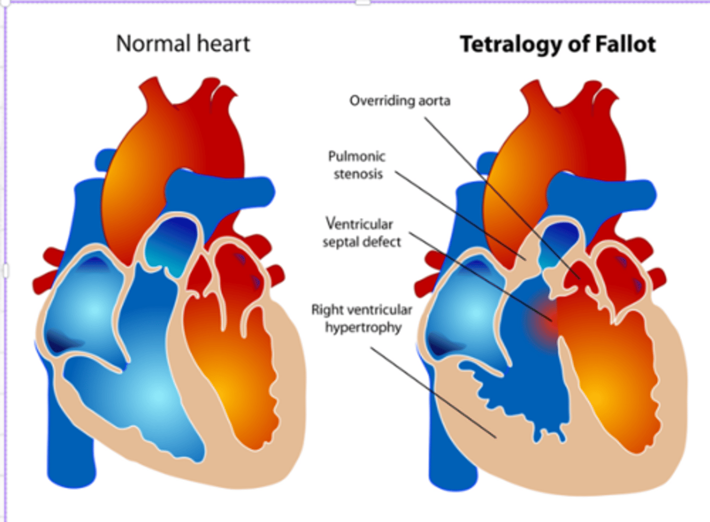
Tetralogy of Fallot (Definition)_
Four Heart Defects that occur at once
- poor pulmonary flow
RAPS
1. Right ventricle Hypertrophy: The right lower chamber of the heart is bigger or thicker than normal, making it harder for blood to go through the pulmonary valve
2. Aorta Displacement:
- The aorta, which is the artery that carries oxygen-rich blood to the body, is out of place and rises above both ventricles. As a result, the body gets too much oxygen-poor blood.
1. Pulmonary stenosis:
- It is a narrowing of the pulmonary valve or pulmonary artery just distal to the valve. pulmonary valve does not open properly
2. Septal defect (VSD):
- VSD is a hole between the two lower chambers (ventricles) of the heart. The hole allows oxygen-rich blood and oxygen-poor blood to mix, so the body does not receive enough oxygen-rich blood.
Tetralogy of Fallot (S/S)
Cyanosis
Tet spells:
Difficult or rapid breathing
Fatigue
Getting tired easily during play or exercise
Heart murmur
Trouble feeding or gaining weight
Clubbing
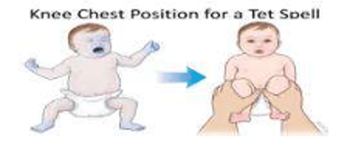
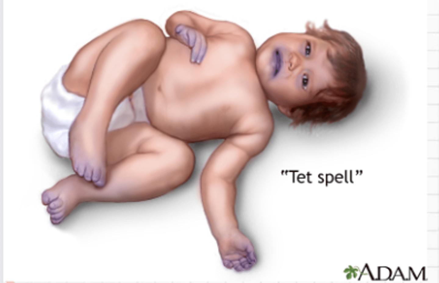
Tetralogy of Fallot (Tet Spells)
- Some babies with ToF suddenly develops blue or gray, skin, nails and lips.
- This happens when the baby cries, eats or is upset
- Due to rapid drop in the amount of oxygen in the blood.
Tetralogy of Fallot (Management)
If baby is experiencing tet spells:
- Lift knee to chest or squats (for older children): Bringing the infant’s knees tightly to the chest or Squatting increases the systemic vascular resistance, it decreases the right to left shunting which is going to improve the blood flow and help increase the oxygen level.
Temporary Shunt Operation:
- Babies who are not strong enough for a full repair or have other underlying health problems may have a shunt operation or a palliative surgery. A shunt is a tube that is sewn into place between the aorta and the pulmonary artery. It helps blood flow to the lungs. A shunt operation is not permanent; babies with a shunt still need a full repair surgery later.
Open-Heart surgery or Complete/Full repair:
- This procedure is usually done in the first year of life. The surgeon patches the hole between the lower heart chambers and repairs or replaces the pulmonary valve. The surgeon may remove thickened muscle below the pulmonary valve or widen the smaller lung arteries.