MB 251 Exam 1
 Call Kai
 Learn
Learn Practice Test
 Spaced Repetition
 Match
Match Flashcards
 Knowt Play
1/119

There's no tags or description
Looks like no tags are added yet.
Name | Mastery | Learn | Test | Matching | Spaced | Call with Kai |
|---|
No analytics yet
Send a link to your students to track their progress
120 Terms
Microbiology
Study of small Biological Specimens
Bacteriology
Study of bacteria
Virology
Study of viruses
Immunology
Study of immune cells/systems
Microorganism
Organism that can’t be seen by naked eye; Bacteria, Archaea, Viruses, Yeast
All Bacteria = Microorganisms
Not all Microorganisms = Bacteria
Colony
Pure Culture made from 1 species
Robert Hooke
1st to describe, observe, and visualize microbes/microorganisms. Drew fruiting structures of molds and invented modern microscope
Antoni Van Leeuwenhoek
First to describe Bacteria. Called them Animalcules. Used magnifiers to see 500x
Louis Pasteur
Found living organisms discriminate between optical isomers. Disproved Theory of Spontaneous Generation. Created vaccines for Anthrax, Fowl Cholera, and Rabies. Invented process of pasteurization. Create Swan Neck flask
Spontaneous Generation
Microorganisms arose spontaneously and Microorganisms developed from seeds/germs from air
John Needham (1745) and Lazzaro Spallanzani
Boiled broth to see if microorganisms would grow or be killed off
Edward Jenner
Pioneer of Vaccionology. Created 1st documented Vaccine for Smallpox
Ignaz Semmelweis
Puerperal Fever (Childbed Fever). Proposed washed hands and deaths plummeted
Joseph Lister
Hypothesized infections were from contamination of wounds by living creatures. Used phenol to reduce infections. Treating infections/tools eliminated infections
Robert Koch
Linked Microbes and Infectious Diseases. Germs cause disease. Developed techniques for obtaining pure cultures of microbes
What is the first postulate of Robert Koch's postulates?
All diseased hosts must have the pathogen associated with them, and all healthy animals must be absent of the pathogen.
What is the second postulate of Robert Koch's postulates?
The pathogen must be one species of bacteria.
What is the third postulate of Robert Koch's postulates?
Cells from a pure culture of the pathogen must cause disease in a healthy animal.
What is the fourth postulate of Robert Koch's postulates?
The suspected pathogen must be reisolated and shown to be the same as the original.
Fanny Hesse (Koch Associate)
Suggested use of Agar in place of gelatin that was stable at higher temperature and not degradable by bacteria
Julius Petri (Koch Associate)
Used bell-shaped glass as a cover. Created the Petri dish
Strict Anaerobes
Anaerobic Microorganisms that do not use Oxygen (O2) for any metabolic properties. Microbes expel oxygen (toxic to them)
Largest Known Bacteria
Thiomargarita Namibiensis (750 microns)
Advantages of being a small Bacteria
More surface area relative to cell volume, grow faster, greater nutrient exchange per unit cell volume
Obligate Intracellular Bacteria
Too small to survive in the environment by themselves die to lack of needed organelles. Survive in host cells to carry out metabolic processes. Does not have necessary protein molecules
Smallest Known Bacteria
Part of Mycoplasma Genus (0.2 microns)
Magnification
Making an object larger for visualization
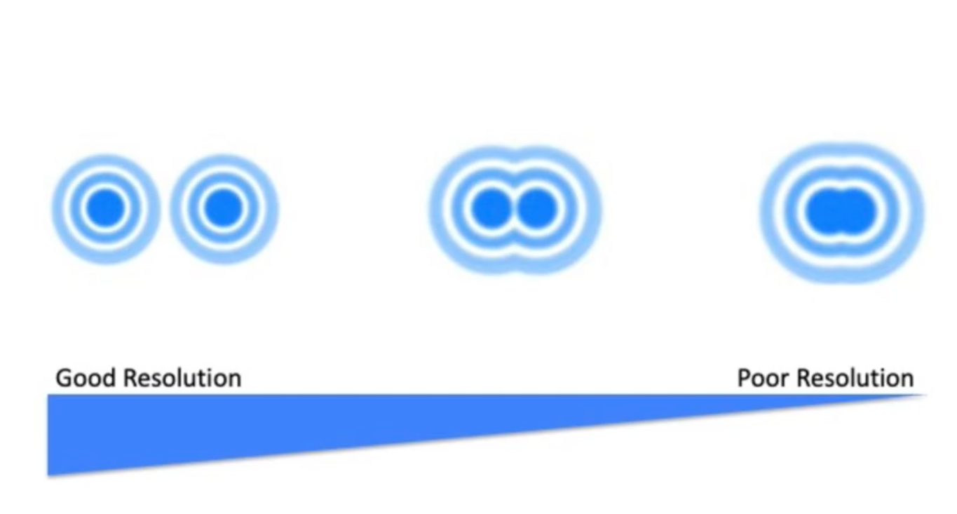
Resolution
Distinguish between two objects (Clarity).
Resolving Power of Light Microscope
Must be at least 0.2 Microns to see separation
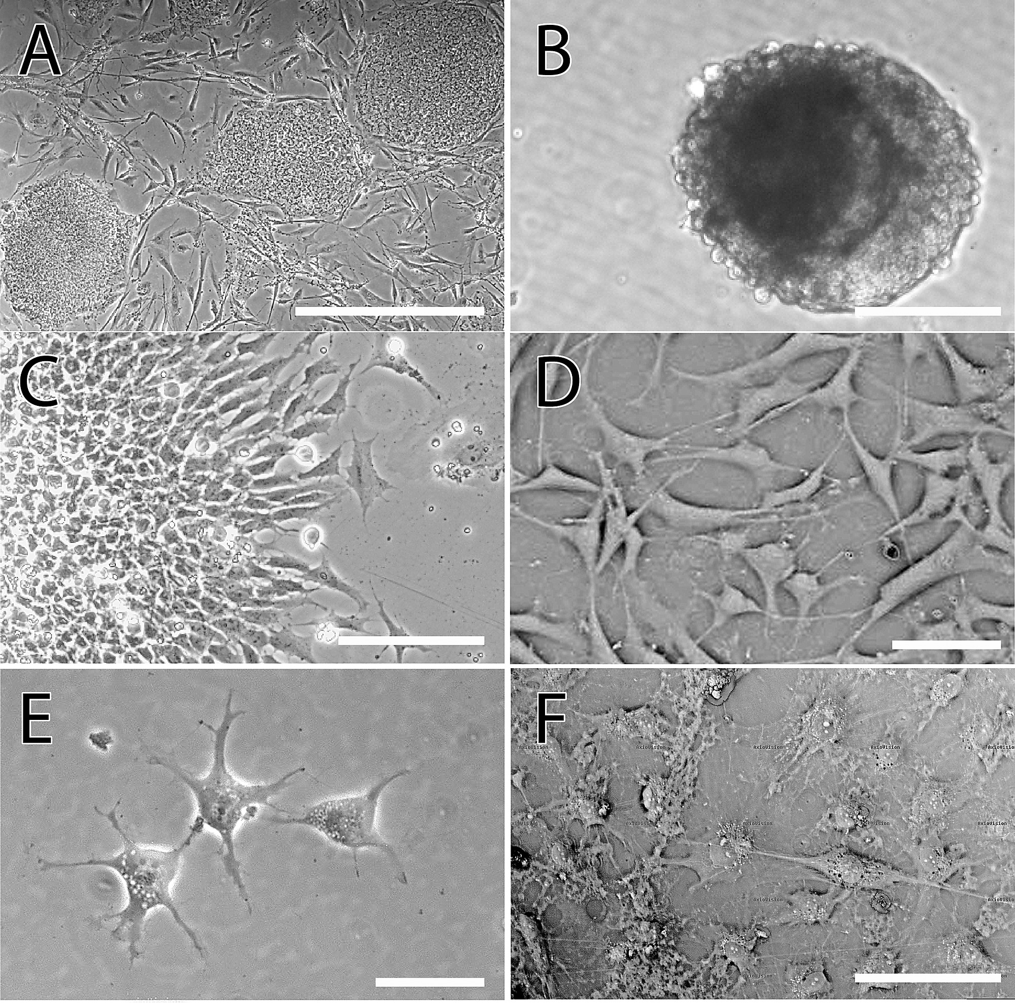
Bright-Field Microscopy
Visualize differences in density/contrast between specimens/surroundings
Pros: Easy/Live cells
Cons: Need 0.2 microns, stain sample to see structures clearly, stained samples = dead
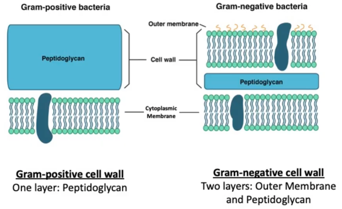
Gram Negative
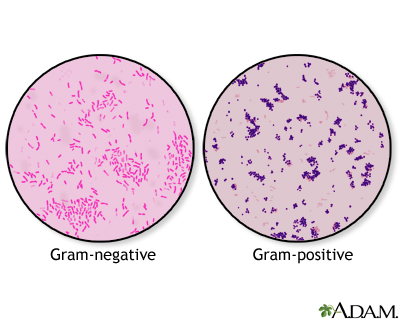
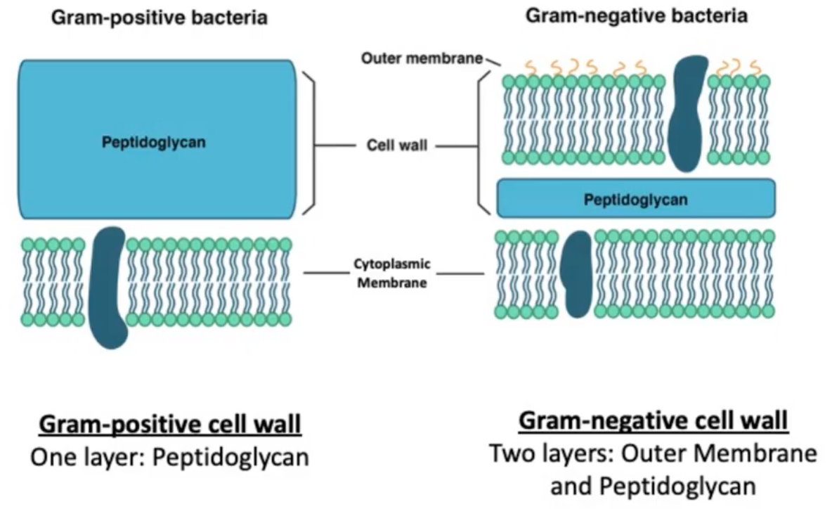
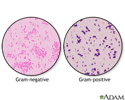
Two Layers: Outer Membrane and Thin Peptidoglycan. Outer Membrane is less selective than Cytoplasmic Membrane. Stain Red/Pink due to thin cell wall (pink color = Safrinin)
LPS/Out Membrane, Single Amino Acid Crosslinks, and Think Peptidoglycan Layer
Gram Positive
One Layer: Thick Peptidoglycan. Teichoic acids in cell wall that support/withstand pressure in cell wall. Stain Purple due to think cell wall
Amino Acid Interbridges, Teichoic Acids, and Thick Peptidoglycan Layer
Fluorescence
Emitting light of 1 wavelength after absorbing light of a different wavelength (Can not see with naked eye)
Autofluorescence
Natural fluorescence Chlorophyll or Pyoverdine (Siderophore)
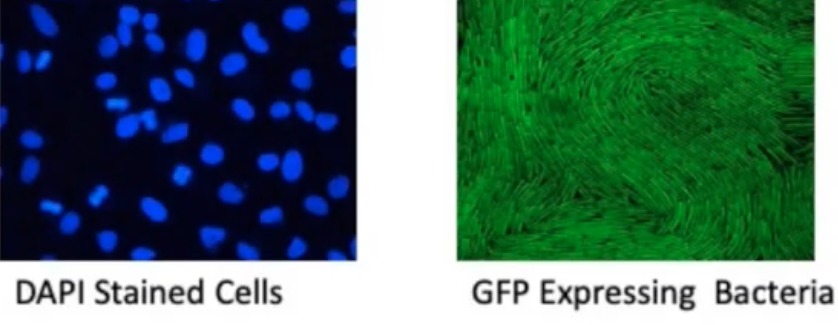
(DAPI) and Tagged Proteins (GFP)
Artificial stains made to have color. Not natural
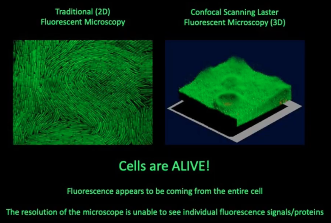
Fluorescent Microscopy
Traditional (2D/Flat) Fluorescent Microscopy and Confocal Scanning Laster (3D). Cells are Alive!! Fluorescence comes from cell resolution can not see individual signals/proteins
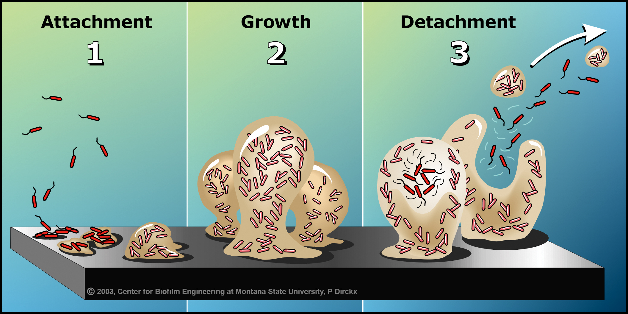
Bacteria Biofilm
Bacteria come together, secrete extracellular components (DNA/RNA/Proteins) to form mass to prevent external forces from killing them. Community of bacteria embedded in a matrix to stick to a surface. Form on tissues inside of host and non living thins
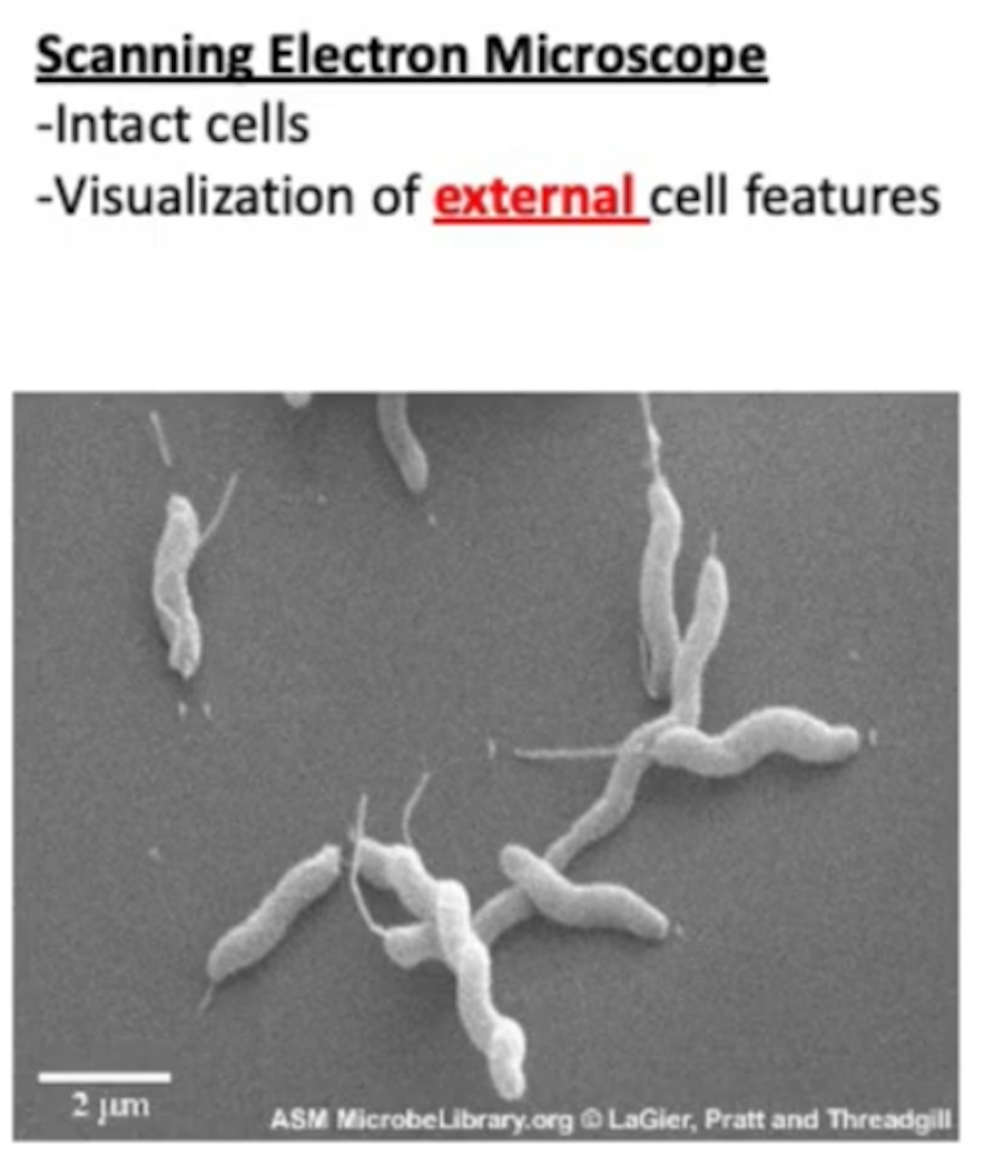
Scanning Electron Microscope (Electron Microscopy)
Uses electrons instead of photons/visible light. Can see 0.0002 microns. Samples are stained for better contrast/cover them in heavy metals; Lead Salt, Uranium, Lanthanum, gold. Cells are dead
Intact Cells; Visualization of External cell features
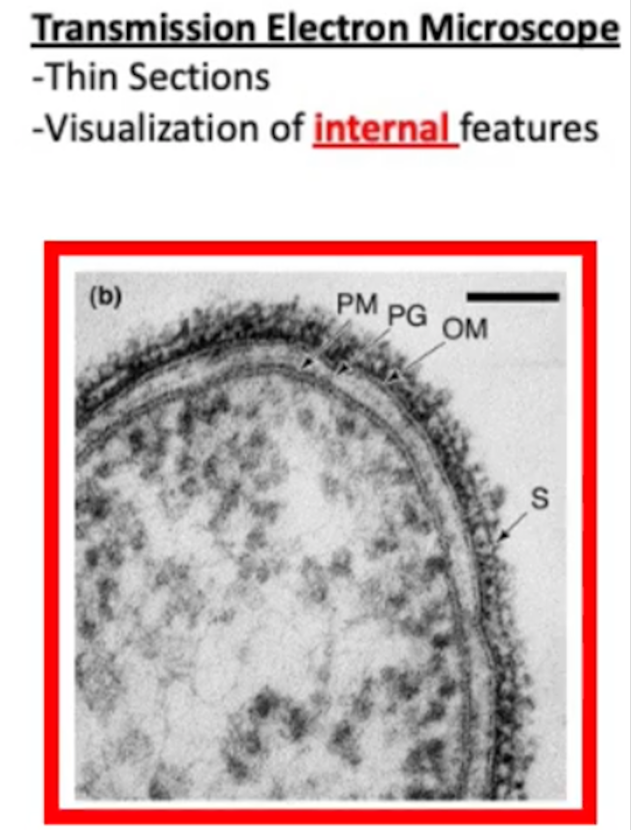
Transmission Electron Microscope (Electron Microscopy)
Uses electrons instead of photons/visible light. Can see 0.0002 microns. Samples are stained for better contrast/cover them in heavy metals; Lead Salt, Uranium, Lanthanum, gold. Cells are dead
Thin sections with visualization of Internal cell features
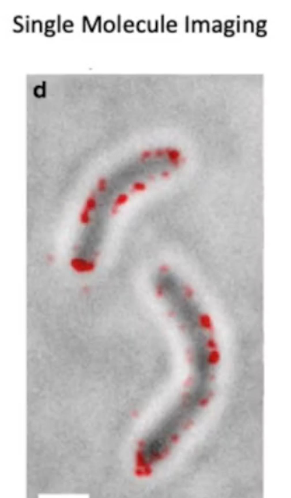
Single Molecule Imaging
Resolves fluorescent proteins. Allows us to track protein movement in cells
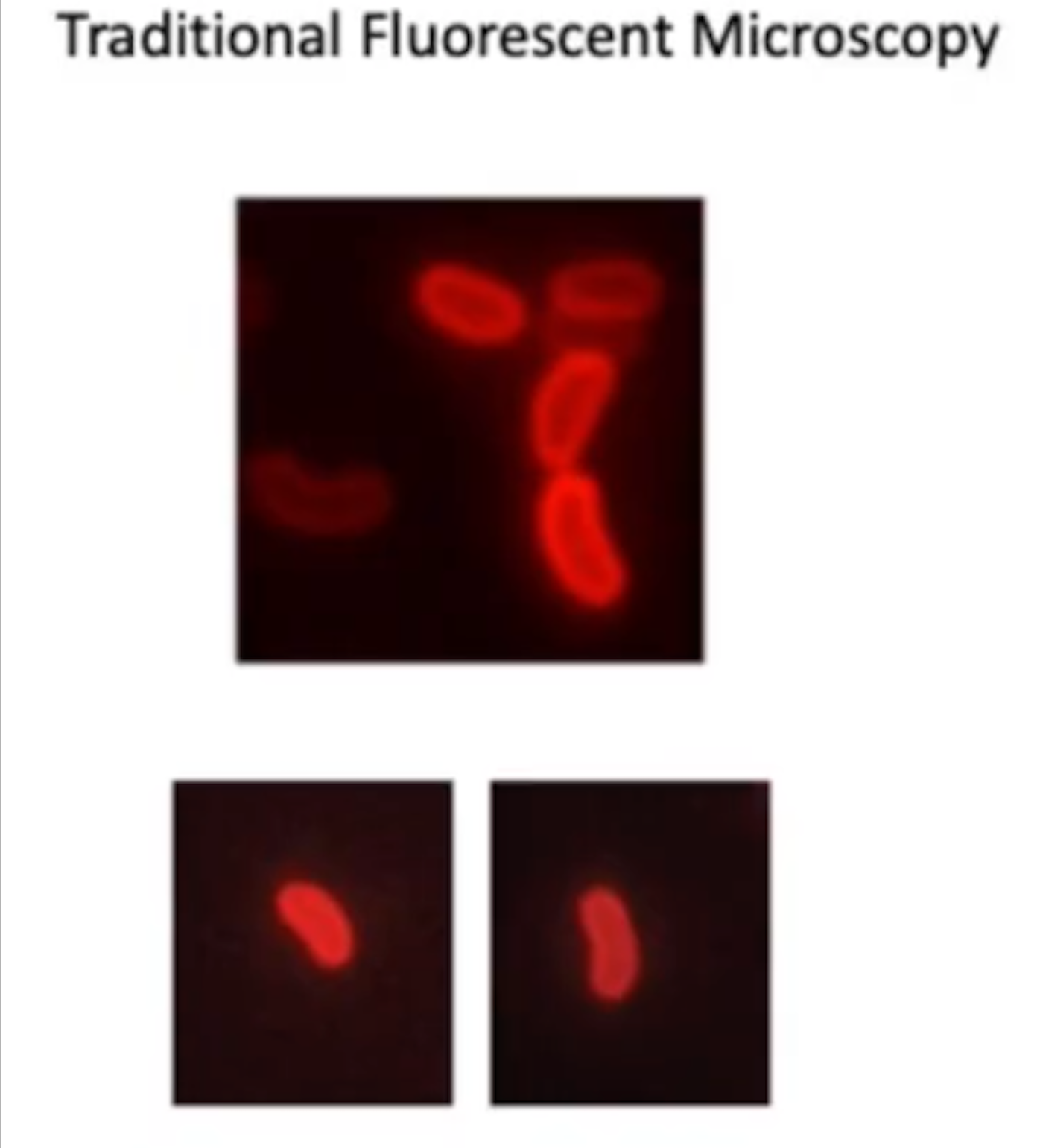
Traditional Fluorescent Microscopy
Can not see individual proteins (highlights whole cell)
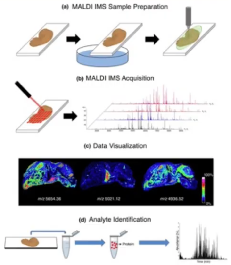
MALDI-IMS (Matrix-Assisted, Laser Desorption, Mass Spectrometry) Microscopy
Tracks individual molecules Lipids, Proteins, Metabolites
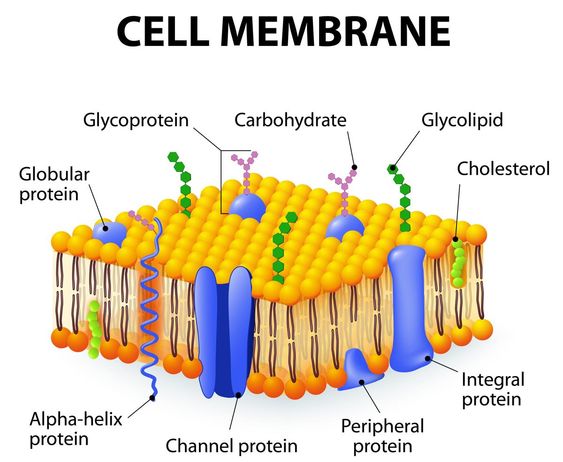
Cytoplasmic Membrane
Highly selective permeability barrier; enables acquisition of specific metabolites and excretion of waste. So strict it does not allow protons to pass (Must have specific protein)
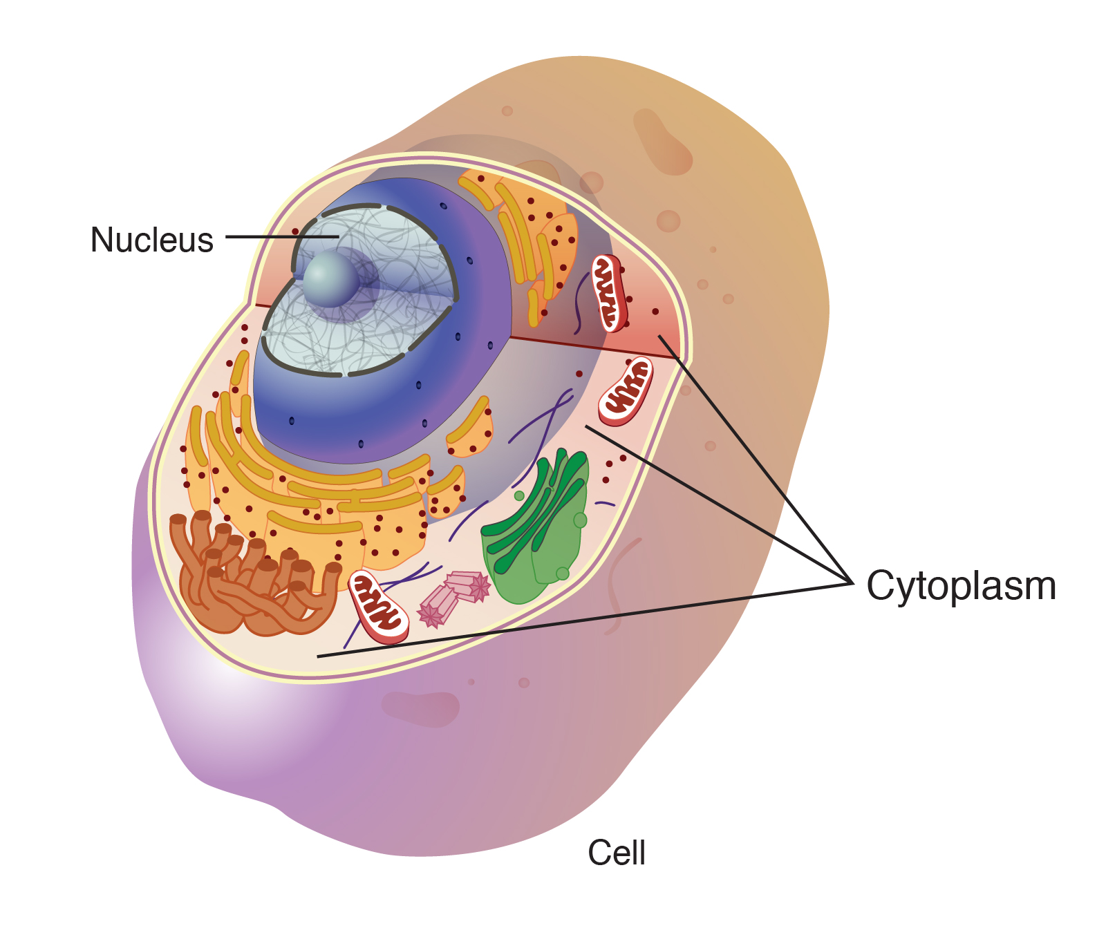
Cytoplasm
Aqueous mixture of macromolecules, ions, and ribosome
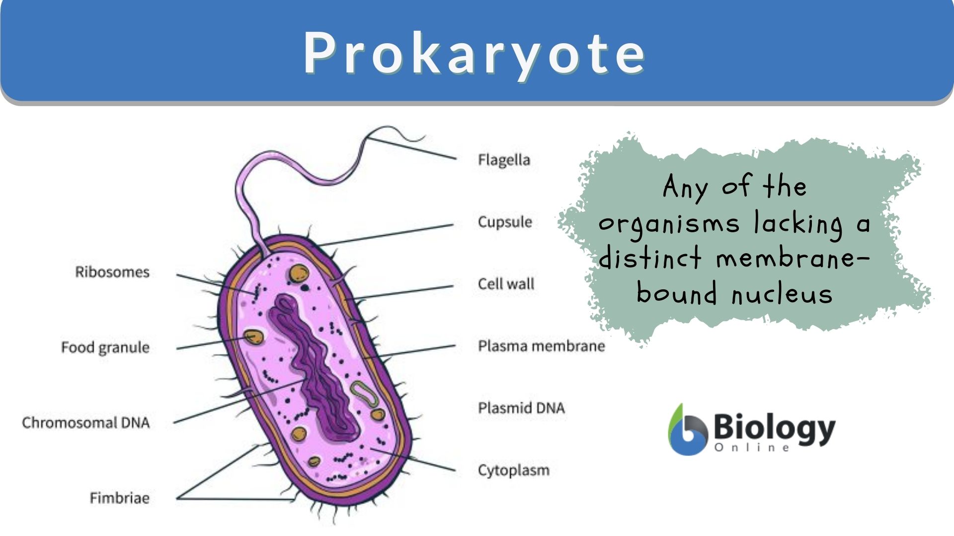
Prokaryote
No membrane bound organelles; no nucleus, singular chromosomes, plasma membrane (can lose DNA and survive), Non essential functions
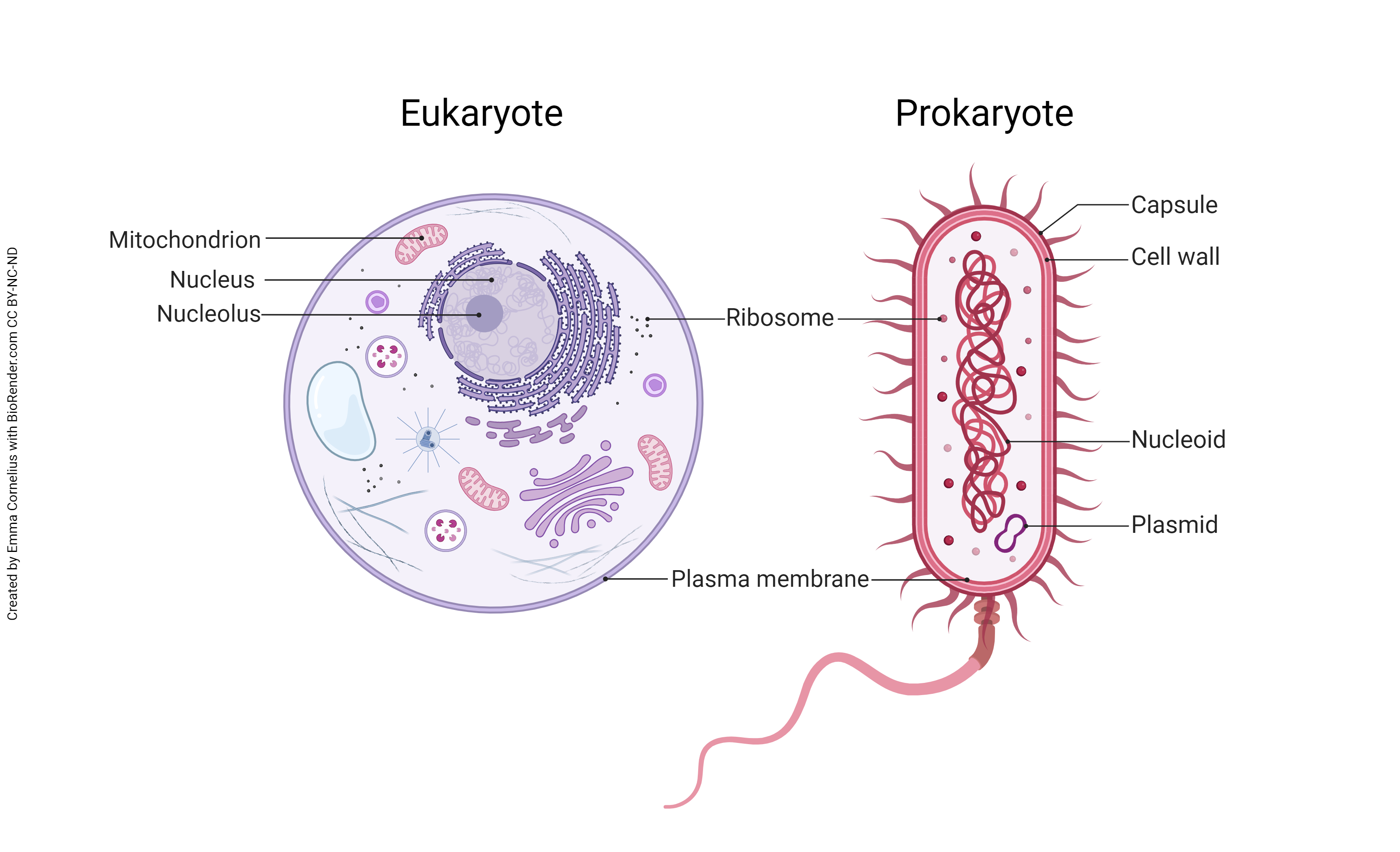
Eukaryotes
DNA in membrane bound organelles, large, linear chromosomes,
Cytoplasm is where functions happen
Protein Anchors
Proteins that participate in transport, bioenergetics, and chemotaxis
Energy Conservation
Site of generation and dissipation of proton motive force
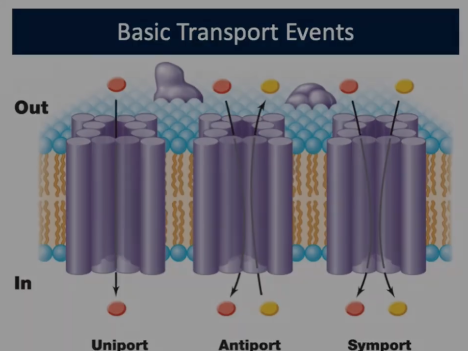
Basic Transport Events (Uniport)
1 molecule going in or out
Group Translocation (Glucose)
Chemical modification of transported substance driven by phosphoenolpyruvate
ABC Transporter (Zinc)
Periplasmic binding proteins involved and energy comes from ATP
Driven by ATP hydrolysis to power transport
Ester/Ether linkages
Stability for higher temperatures. Ester is not at stable
Archaeal Membrane Monolayers
Covalently linked membranes with no hydrophobic interactions
Important for stability at higher temperatures
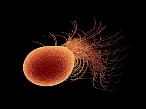
Pyrococcus Furiosus: Nerve Agent Detoxification
Breaks down Proline-Proline Amino Acid Linkages
Periplasmic Space
Small molecules can pass. Large molecules or charged molecules can not pass. Space between Outer Membrane and Cytoplasmic Membrane. Peptidoglycoglycan of Gram-Negative bacteria located in Periplasmic Space
Embedded Proteins
ABC transporters, enzymes, and chemoreceptors
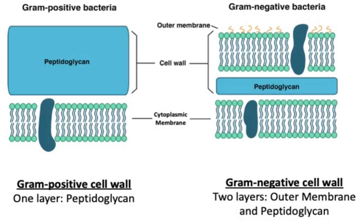
Peptidoglycan Layer
Structure that provides strength and rigidity for blueprint for cell shape. Antibiotics target this structure to kill bacteria. Layers of Polysaccharides (NAG-NAM)
Lysis
When a cell bursts open.
Transglycosylases
Enzymes involved in linking NAG-NAM sugars in the peptidoglycan backbone. Target for Antibiotics that disrupt cell wall. Insert precursors into growing cell wall and catalyze the glycosidic bond formation between NAG-NAM
Transpeptidases
Enzymes that form amino acid links between NAM sugars of different layers. Target for Antibiotics that disrupt cell wall
B-Lactamase Enzymes
Degrade B-Lactam ring making antibiotics ineffective
Lysozyme
Destroys peptidoglycan. Cleaves bonds between NAG-NAM beta 1,4 linkages
Lipid A
Allows imbedding into phospholipid bilayer
Gram-Stain Process
Safranin (Heat Fixing): Kill cell and sticks to glass side
Crystal Violet stain: Stain both types of cells Purple
Iodine Treatment: Mordant making it harder to wash out
Destain: Acid/Alco hol mixture to wash out Crystal Violet from cell wall (7-10 seconds)
Hans Christian Gram
Developed staining method that could differentiate the two major groups of bacteria.
Capsule
Made of Polysaccharides/Amino Acids
Attaches to Peptidoglycan
Works in attachment, immune evasion/cell protection (desiccation)
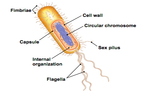
Fimbriae
Short Hair like appendages Visible with electron microscope. Used for attachment
EndoSpore
Seed that recognizes threats (heat, low food) and turns into Spore
ExoSporium
Outermost Layer that encloses spores like Bacillus (Protein)
Spore Coat
Layers of spore proteins. Acts as a barrier to large molecules. Enzymes for germination
Dipicolonic Acid
Only in the spore. Binds to water and used to dehydrate spore
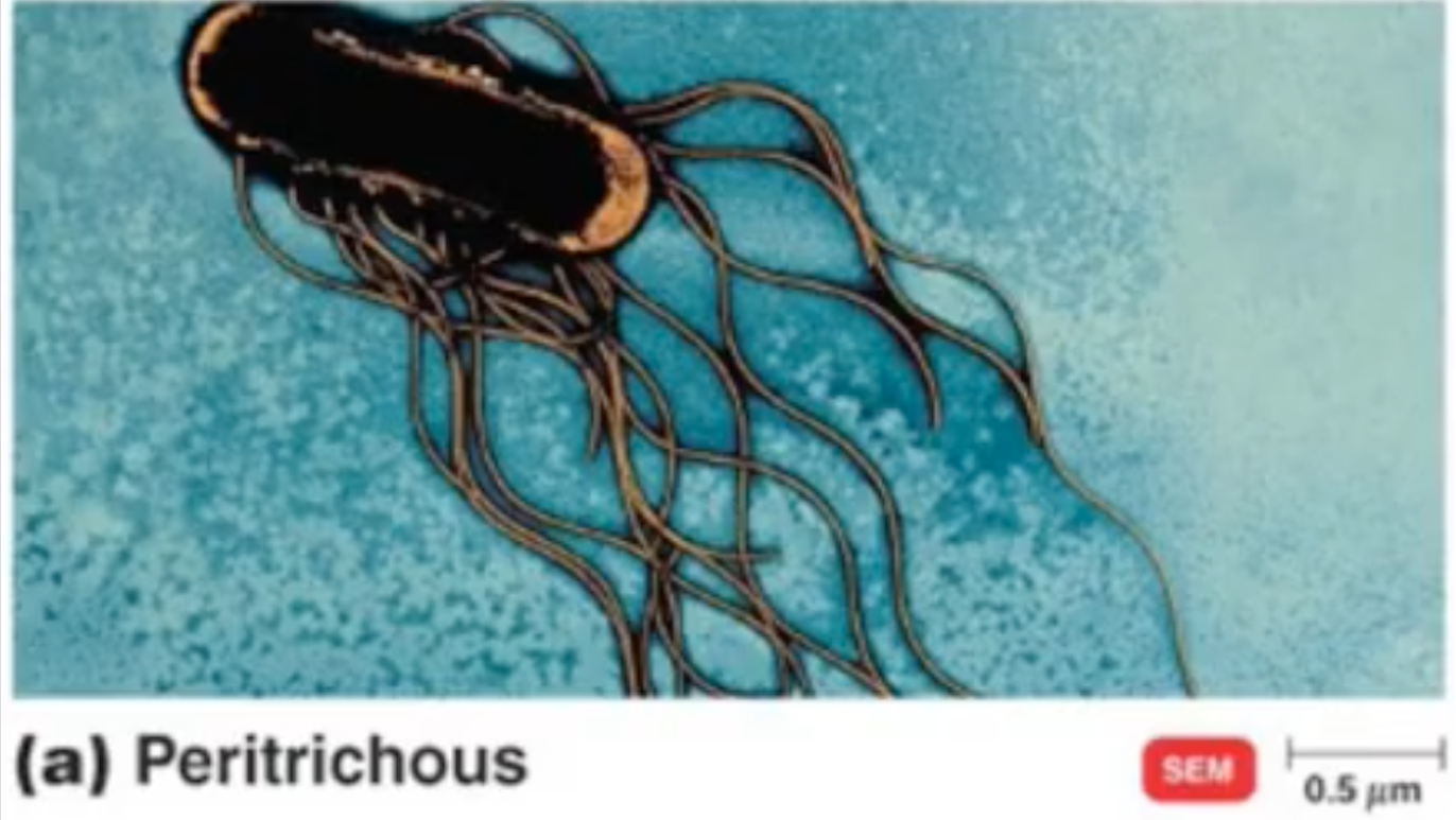
Peritrichous
Gliding Motility
Bacterial movement across solid surface
Does not use Flagella
Slower and smoother
Happens through excretion of polysaccharides
Twitching Motility
Allows movement on surface but uses Pilus/Pili that pulls cells across a surface
Flagellum/Flagella
Allows movement in liquid environments
Rotate up to 1,000 revolutions per minute
10-50 movements per second
Not every bacteria produces this
Motor powered by Proton Motive Force (PMF)
Chemotaxis
Chemical Movement. Movement of bacteria through chemical signals
Use to direct Bacteria to more favorable environments
Attractant
Positive; Directs movement towards positive stimulus
Repellant
Negative; less Bacteria in tub because they move away
Chemoreceptors
Methyl-Accepting Chemotaxis Proteins (MCPs)
Proteins that sense chemical signals in environment (Uses Flagella)
Cell encodes many sensors
Each sensor has substrate/class they can sense
Aerotaxis
Movement in response to oxygen
Phototaxis
Movement in response to light
Microbial Growth
Increase in the number of cells (Not size of cell)
Binary Fission
Asexual cell division, parent cell splits into two
Each daughter cell gets chromosode and copies of all other cells needs to exist by itself (Balanced Growth)
If a parent cell does not equally divide, possibility that one/both cells may not survive
Generation Time
Time needed to double in number
Bacterial Growth Requirements
Media (Nutrients/Food to grow), Temperature, and Oxygen
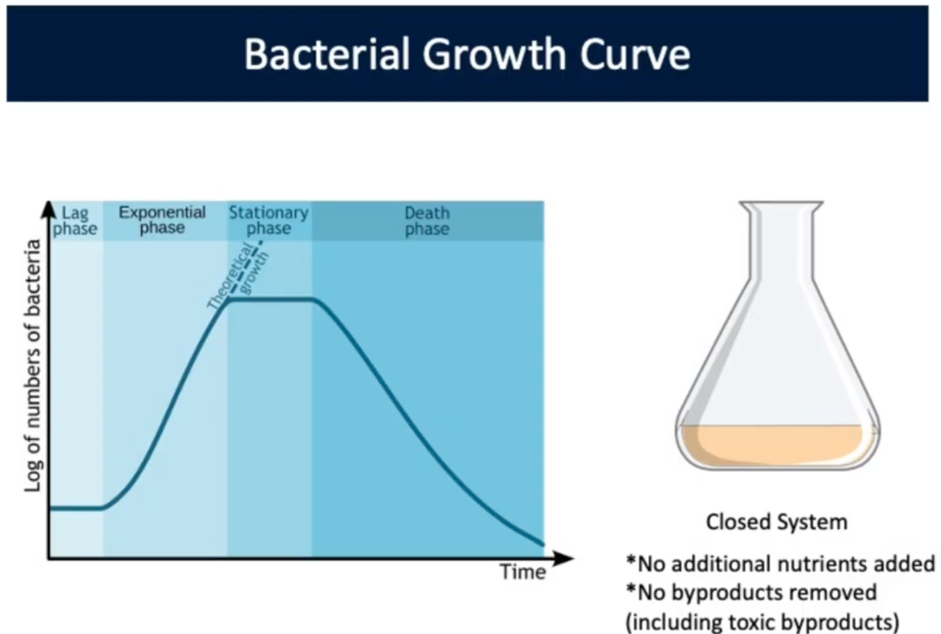
Bacterial Growth Curve (Lag Phase)
Cells getting accustomed to environment, producing all materials to divide
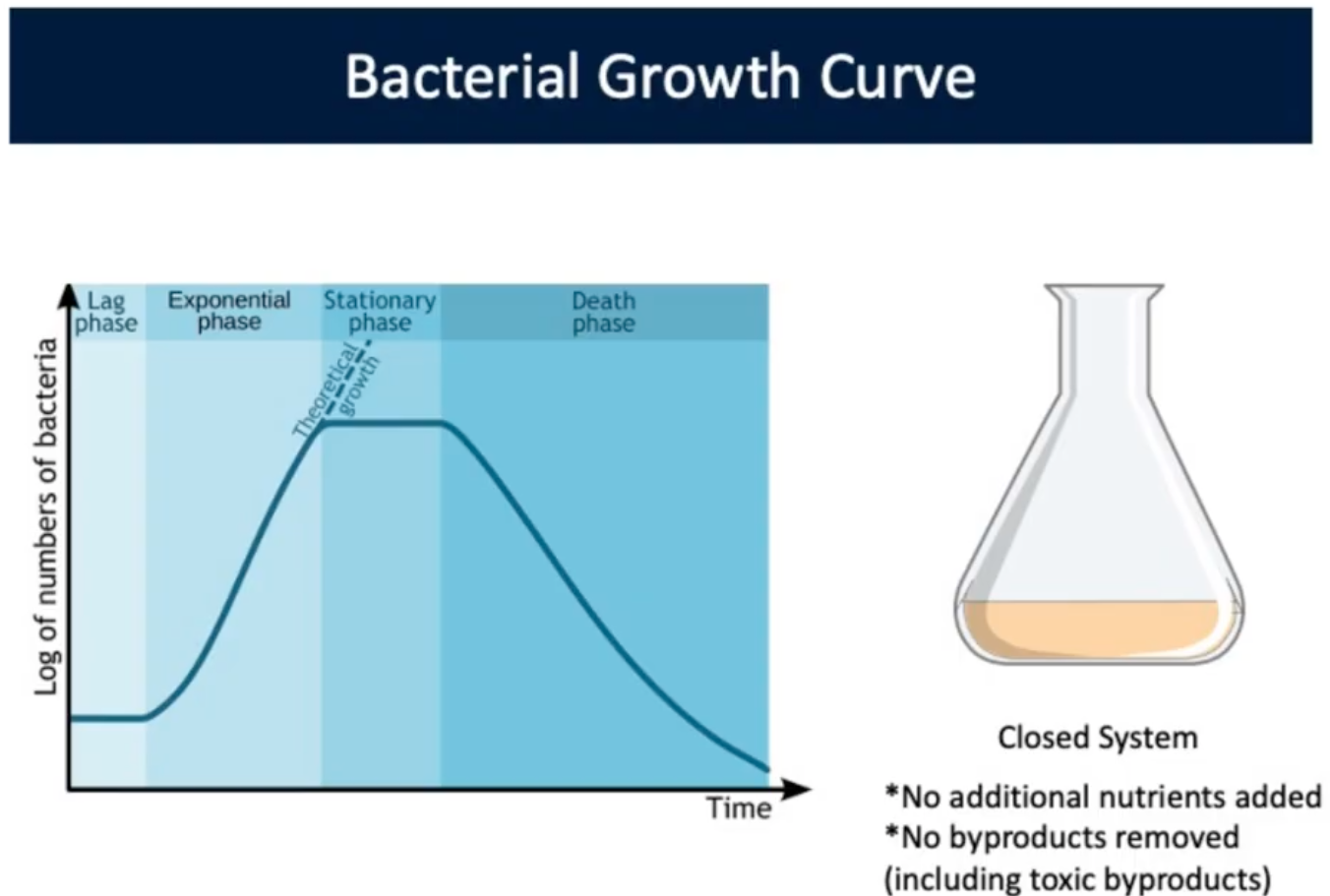
Bacterial Growth Curve (Exponential Phase)
Cells actively dividing, and growing at their max rate in the specified environment (Balanced Growth)
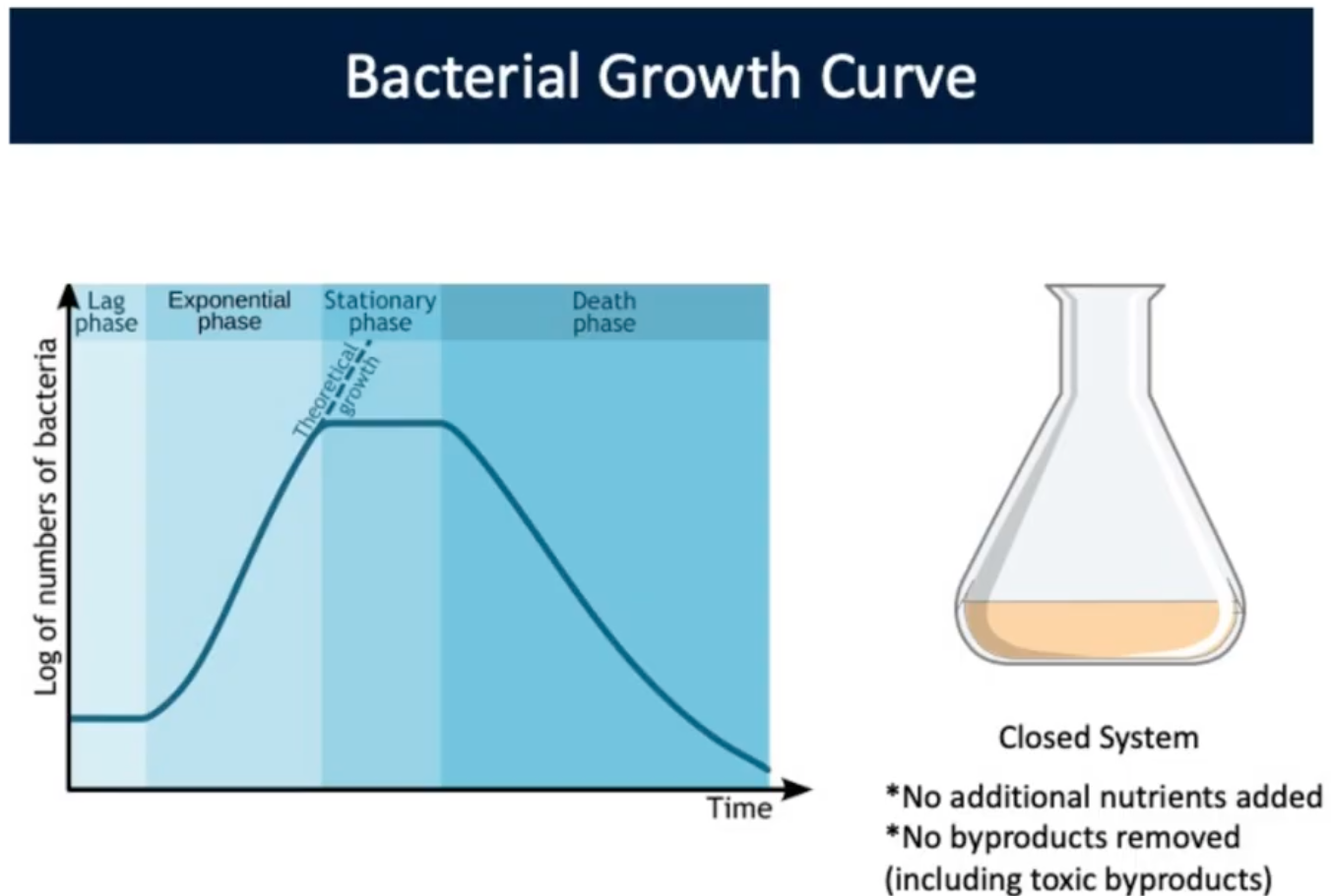
Bacterial Growth Curve (Stationary Phase)
Growth rate and death rate are equal. The number of cells stays constant. Nutrient limitation/accumulation of toxic metabolic byproducts
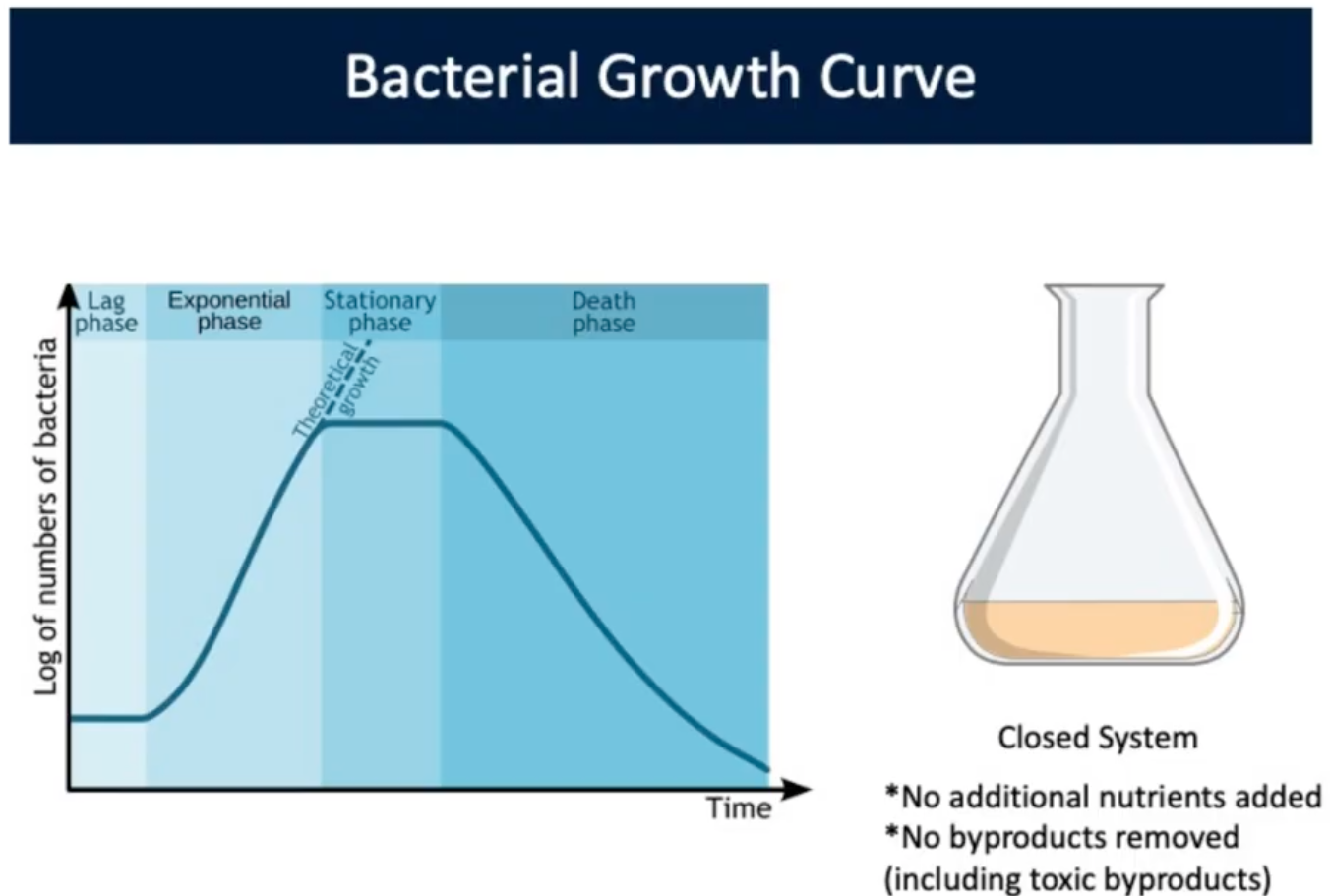
Bacterial Growth Curve (Death Phase)
No nutrients remain and waste products remain. Population dies out
Theoretical Growth
Increase in population continues if there is a constant supply of new nutrients/removal of waste byproducts
Fts Proteins: Filamentous temperature sensitive proteins
Needed for cell division in all Bacteria (most Archaea)
Proteins interact to form divisome (cell division apparatus)
FtsZ: Forms ring around center of cell; related to tubulin
Min System (C,D, & E)
Ensure cell division occurs in the middle of a cell.
C: Direct inhibitor that drives cell Division
D: ATPase that binds to inner cell membrane recruiting MinC to block cell division
E: Oscillates from pole to pole. Clears C and D allowing FtsZ to assemble in the center of the ring
FtsZ
Responsible for cell dividing, forming a ring at the center of the cell
Bacteria Cell Morphology
Morphology = Cell Shape that is genetically coded
MreB
Rod Shaped Morphology determining factor in Prokaryotes
Forms cytoskeleton in Bacteria
Forms spiral shaped bands around inside of cell, underneath cytoplasmic membrane
Not found in coccus shaped bacteria
Localizes synthesis of new peptidoglycan and other cell wall components to locations in cylinder of rod shaped cell during growth
Localize peptidoglycan to elongate a cell. Does not have MreB protein would be coccus
Autolysins
Small openings in the wall that begin at the FtsZ ring. Linkages in preexisting peptidoglycan
Bactoprenol
Carrier molecule involved in transport of NAG-NAM peptidoglycan precursors across cytoplasmic membranes. Makes precursors hydrophobic to cross cytoplasmic membrane
Transpeptidation
Final step in cell wall synthesis (Ftsl). Forms peptide cross links between muramic acid residues in adjacent glycine chains
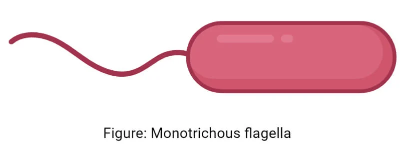
Monotrichous
1 tail