oral radiology final

0.0(0)
Studied by 0 people
call kaiCall Kai
learnLearn
examPractice Test
spaced repetitionSpaced Repetition
heart puzzleMatch
flashcardsFlashcards
GameKnowt Play
Card Sorting

1/89

encourage image

There's no tags or description

Looks like no tags are added yet.

Last updated 8:07 PM on 5/29/26
Name
Mastery
Learn
Test
Matching
Spaced
Call with Kai

No analytics yet

Send a link to your students to track their progress

90 Terms

1
New cards

tomography

  • Is an imaging technique that allows the imaging of one layer, or section, of the body while blurring the images of structure in other planes

  • panoramic imaging process in which the movement of the receptor and tubehead produces an image

2
New cards

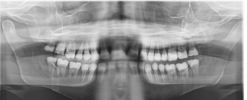
what is a panoramic image?

  • shows a wide view of the maxilla and the mandible and surrounding structures

  • Allows for the visualization of the patients’ oral and facial structures spread out across a flat surface

  • structures in a panoramic image are in a 2D curved zone

3
New cards

how is a pan taken?

·       Is an extraoral method of radiography

·       Extraoral receptor is placed and positioned outside the mouth during x-ray exposure

·       Patient is positioned in correct manner

·       Receptor and tube head rotated around patient taking a series of x-ray images that will combine into one
<p><span><span>·</span></span><span style="font-family: &quot;Times New Roman&quot;; line-height: normal; font-size: 7pt;"><span>&nbsp;&nbsp;&nbsp;&nbsp;&nbsp;&nbsp; </span></span><span><span>Is an extraoral method of radiography</span></span></p><p class="MsoListParagraphCxSpMiddle"><span><span>·</span></span><span style="font-family: &quot;Times New Roman&quot;; line-height: normal; font-size: 7pt;"><span>&nbsp;&nbsp;&nbsp;&nbsp;&nbsp;&nbsp; </span></span><span><span>Extraoral receptor is placed and positioned outside the mouth during x-ray exposure</span></span></p><p class="MsoListParagraphCxSpMiddle"><span><span>·</span></span><span style="font-family: &quot;Times New Roman&quot;; line-height: normal; font-size: 7pt;"><span>&nbsp;&nbsp;&nbsp;&nbsp;&nbsp;&nbsp; </span></span><span><span>Patient is positioned in correct manner</span></span></p><p class="MsoListParagraphCxSpLast"><span><span>·</span></span><span style="font-family: &quot;Times New Roman&quot;; line-height: normal; font-size: 7pt;"><span>&nbsp;&nbsp;&nbsp;&nbsp;&nbsp;&nbsp; </span></span><span><span>Receptor and tube head rotated around patient taking a series of x-ray images that will combine into one</span></span></p>
4
New cards

 What rotation occurs when taking panoramic images

·       Both the receptor and tube head rotate around the patient in opposing directions, producing a series of individual images

·       When the images are combined, an overall view of the maxilla and mandible are created

·       tomography

  • tubehead rotates BEHIND patient, receptor rotates IN FRONT of patient

5
New cards

Why do we use Pans

·       To evaluate the dentition and supporting structures

·       To evaluate impacted teeth

·       To evaluate eruption patterns, growth, and development

·       To detect diseases, lesions, and conditions of the jaws

·       To examine the extent of large lesion

·       To evaluate trauma

6
New cards

disadvantages of taking panoramic images

  • image quality

  • focal trough limitations

  • distortion

  • equipment cost

7
New cards

advantages of taking panoramic images

  • field size

  • simplicity

  • patient cooperation

  • minimal exposure

  • can aid in detection of

    • jaw fractures, impacted 3rd molars, large lesions in mandible

8
New cards

focal trough

·       Theoretical concept used to determine where the dental arches must be positioned to obtain the sharpest image

·       Image layer

·       Three dimensional curved zone in which structures located within the focal trough appear reasonably well defined on the resulting panoramic image

9
New cards

collimation in pano machine vs collimation in x-ray tubehead

·       In intraoral imaging it is a lead plate with a small round or rectangular opening in the middle while the one used in panoramic imaging is a lead plate with a small opening in the shape of a narrow vertical slit

·       Both are used to restrict the size and shape of the x-ray beam

10
New cards

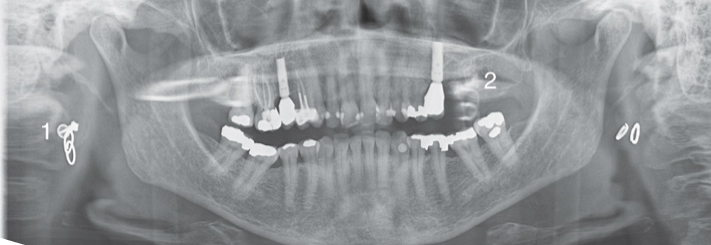
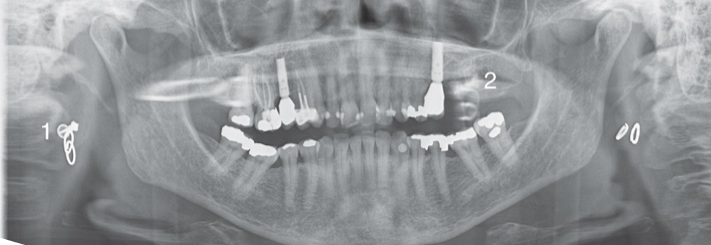
what are ghost images

·       An artifact seen on a panoramic image that is produced when a metallic or dense object is penetrated twice by the x-ray beam

·       Could obscure diagnostic information

  • often appear higher than the counterpart
<p><span>·</span><span style="font-family: &quot;Times New Roman&quot;; line-height: normal; font-size: 7pt;"><span>&nbsp;&nbsp;&nbsp;&nbsp;&nbsp;&nbsp; </span></span><span>An artifact seen on a panoramic image that is produced when a metallic or dense object is penetrated twice by the x-ray beam</span></p><p><span>·&nbsp;&nbsp;&nbsp;&nbsp;&nbsp;&nbsp; Could obscure diagnostic information</span></p><ul><li><p>often appear higher than the counterpart</p></li></ul><p></p>
11
New cards

appearance of ghost images

·       Found on the opposite side of the receptor

·       Appears indistinct, larger, and higher than its actual counterpart

·       Could obscure diagnostic information
<p><span><span>·</span></span><span style="font-family: &quot;Times New Roman&quot;; line-height: normal; font-size: 7pt;"><span>&nbsp;&nbsp;&nbsp;&nbsp;&nbsp;&nbsp; </span></span><span><span>Found on the opposite side of the receptor</span></span></p><p class="MsoListParagraphCxSpMiddle"><span><span>·</span></span><span style="font-family: &quot;Times New Roman&quot;; line-height: normal; font-size: 7pt;"><span>&nbsp;&nbsp;&nbsp;&nbsp;&nbsp;&nbsp; </span></span><span><span>Appears indistinct, larger, and higher than its actual counterpart</span></span></p><p class="MsoListParagraphCxSpLast"><span><span>·</span></span><span style="font-family: &quot;Times New Roman&quot;; line-height: normal; font-size: 7pt;"><span>&nbsp;&nbsp;&nbsp;&nbsp;&nbsp;&nbsp; </span></span><span><span>Could obscure diagnostic information</span></span></p>
12
New cards

What happens if a patient does not close their lips around the bite block when taking a Pan?

A dark radiolucent shadow results that obscures the anterior teeth

13
New cards

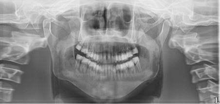
What happens on the Pan image if the patients chin is too low?

·       An “exaggerated smile line” is apparent on the image

·       Smaller mandible, teeth, and a high arch can be seen
<p><span><span>·</span></span><span style="font-family: &quot;Times New Roman&quot;; line-height: normal; font-size: 7pt;"><span>&nbsp;&nbsp;&nbsp;&nbsp;&nbsp;&nbsp; </span></span><span><span>An “exaggerated smile line” is apparent on the image</span></span></p><p class="MsoListParagraphCxSpLast"><span><span>·</span></span><span style="font-family: &quot;Times New Roman&quot;; line-height: normal; font-size: 7pt;"><span>&nbsp;&nbsp;&nbsp;&nbsp;&nbsp;&nbsp; </span></span><span><span>Smaller mandible, teeth, and a high arch can be seen</span></span></p>
14
New cards

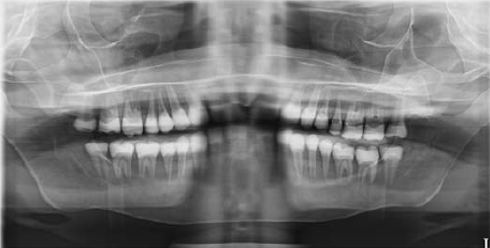
The patient's chin is too high?

·       A “reverse smile line” is apparent on the image
<p><span><span>·</span></span><span style="font-family: &quot;Times New Roman&quot;; line-height: normal; font-size: 7pt;"><span>&nbsp;&nbsp;&nbsp;&nbsp;&nbsp;&nbsp; </span></span><span><span>A “reverse smile line” is apparent on the image</span></span></p>
15
New cards

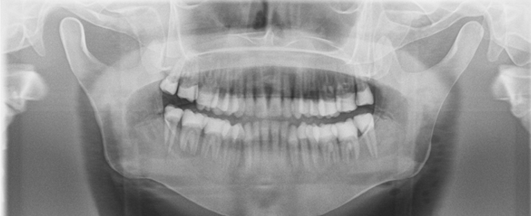
The patient's teeth are too far forward on the bite block?

·       Anterior teeth appear skinny and out of focus
<p><span><span>·</span></span><span style="font-family: &quot;Times New Roman&quot;; line-height: normal; font-size: 7pt;"><span>&nbsp;&nbsp;&nbsp;&nbsp;&nbsp;&nbsp; </span></span><span><span>Anterior teeth appear skinny and out of focus</span></span></p>
16
New cards

The patient’s teeth are too far back on the bite block?

Anterior teeth appear fat and out of focus

  • big back

<p><span style="font-family: Aptos, sans-serif; line-height: 115%;"><span>Anterior teeth appear fat and out of focus</span></span></p><ul><li><p>big back</p></li></ul><p></p>
17
New cards

When learning about CBCT scans what is multiplanar reconstruction?

·       The sagittal plane is a vertical plane that divides the anatomic features within the FOV into right and left slices

·       When viewed together, axial, coronal, and sagittal images are referred to as this

·       MPR images

18
New cards

What does DICOM stand for?

·       Digital  imaging and communications in medicine

19
New cards

What are common uses of 3-dimensional imaging

  • Provides accurate and detailed information regarding the evaluation of pathology

  • distances to critical anatomic landmarks

  • locations of impacted teeth

  • eruption patterns

  • other concerns of the oral and maxillofacial complex

20
New cards

What areas of dentistry use 3-dimensional imaging

·       Every single specialty!

o   Public health

o   Orthodontics

o   Oral medicine

o   Endodontics

o   Oral surgery

o   Etc..

21
New cards

What happens on a CBCT scan if the patient has multiple metallic restorations?

·       Beam hardening

·       X-ray beam scatter reduces image quality

22
New cards

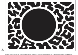
Is a uniocular lesion with corticated borders usually benign or malignant?  Fast-growing or slow-growing?

·       This lesion exhibits a thin, well-demarcated radiopaque rim of bone at the periphery

  • one compartment, non expansible

·       Usually indicative of a benign, slow-growing process
<p><span>·</span><span style="font-family: &quot;Times New Roman&quot;; line-height: normal; font-size: 7pt;"><span>&nbsp;&nbsp;&nbsp;&nbsp;&nbsp;&nbsp; </span></span><span>This lesion exhibits a thin, well-demarcated radiopaque rim of bone at the periphery</span></p><ul><li><p><strong>one compartment, non expansible</strong></p></li></ul><p class="MsoListParagraphCxSpLast"><span>·</span><span style="font-family: &quot;Times New Roman&quot;; line-height: normal; font-size: 7pt;"><span>&nbsp;&nbsp;&nbsp;&nbsp;&nbsp;&nbsp; </span></span><span>Usually indicative of a benign, slow-growing process</span></p>
23
New cards

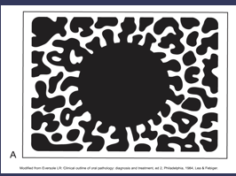
Unilocular lesions with no corticated borders

·       This lesion does not exhibit a thin radiopaque rim of bone at the periphery

·       The periphery appears fuzzy or poorly defined

·       May represent either a benign or malignant process
<p><span><span>·</span></span><span style="font-family: &quot;Times New Roman&quot;; line-height: normal; font-size: 7pt;"><span>&nbsp;&nbsp;&nbsp;&nbsp;&nbsp;&nbsp; </span></span><span><span>This lesion does not exhibit a thin radiopaque rim of bone at the periphery</span></span></p><p class="MsoListParagraphCxSpMiddle"><span><span>·</span></span><span style="font-family: &quot;Times New Roman&quot;; line-height: normal; font-size: 7pt;"><span>&nbsp;&nbsp;&nbsp;&nbsp;&nbsp;&nbsp; </span></span><span><span>The periphery appears fuzzy or poorly defined</span></span></p><p class="MsoListParagraphCxSpLast"><span><span>·</span></span><span style="font-family: &quot;Times New Roman&quot;; line-height: normal; font-size: 7pt;"><span>&nbsp;&nbsp;&nbsp;&nbsp;&nbsp;&nbsp; </span></span><span><span>May represent either a benign or malignant process</span></span></p>
24
New cards

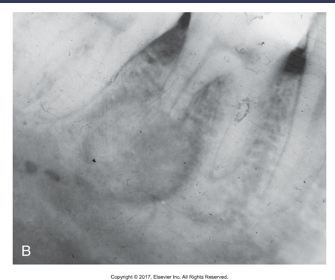
What is a Target-lesion?  What do they look like?

·       A well-defined, localized radiopaque area surrounded by a uniform radiolucent halo
<p><span><span>·</span></span><span style="font-family: &quot;Times New Roman&quot;; line-height: normal; font-size: 7pt;"><span>&nbsp;&nbsp;&nbsp;&nbsp;&nbsp;&nbsp; </span></span><span><span>A well-defined, localized radiopaque area surrounded by a uniform radiolucent halo</span></span></p>
25
New cards

What is the definition of Radiation?

·       A form of energy carried by waves or a stream of particles

26
New cards

What is the definition of a Radiograph?

·       An image or picture produced on a receptor by exposure to ionizing radiation; a two dimensional representation of a three dimensional object

27
New cards

Density is defined as?

·       The overall darkness or blackness of an image

28
New cards

What does millamperage control?

·       Controls the quantity of electrons produced

·       KLMN

·       Regulates the temperature of the cathode filament, the higher the setting the more electrons that are produced

29
New cards

What does kilovoltage control?

·       Controls quality of dental x-ray images

·       Regulates the speed and energy of electrons and determines the penetrating ability of the x-ray beam

·       KLMN

30
New cards

When do we use a thyroid collar on a patient?

  •   When taking x-rays, radiations is exposed to critical organs of the neck

  • A critical organ is an organ that, if damaged, diminished the quality of a persons life

    • Thyroid gland

    • Bone marrow

    • Skin

    • Lens of eye

    • Reproductive organs

    • Lymphocytes

31
New cards

What are the basic principles of the Paralleling technique?

  1. receptor placement

  2. receptor position

  3. vertical angulation

  4. horizontal angulation

  5. receptor exposure

32
New cards

receptor placement

  • the receptor must be positioned to cover the prescribed area of teeth to be examined

33
New cards

receptor position

o   The receptor must be positioned parallel to the long axis of the tooth

o   The receptor and beam alignment device must be placed away from the teeth and toward the middle of the oral cavity

34
New cards

vertical angulation

o   The central ray of the x-ray beam must be directed perpendicular (at a right angle) to the receptor and the long axis of the tooth

35
New cards

horizontal angulation

o   The central ray if the x-ray beam must be directed through the contact areas between the teeth

36
New cards

receptor exposure

o   The x-ray beam must be centered on the receptor to ensure that all areas are exposed

o   failure to center the x-ray beam results in a partial image on the receptor or a cone cut

o   cone cuts can be produced with either a round PID or a rectangular

37
New cards

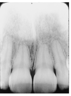
What is Foreshortening?  How is it corrected?

·       Caused by excessive vertical angulation

·       Appear as short teeth with blunted roots

·       Can be corrected by reducing amount of vertical angulation
<p><span><span>·</span></span><span style="font-family: &quot;Times New Roman&quot;; line-height: normal; font-size: 7pt;"><span>&nbsp;&nbsp;&nbsp;&nbsp;&nbsp;&nbsp; </span></span><span><span>Caused by </span><strong><span>excessive</span></strong><span> vertical angulation</span></span></p><p class="MsoListParagraphCxSpMiddle"><span><span>·</span></span><span style="font-family: &quot;Times New Roman&quot;; line-height: normal; font-size: 7pt;"><span>&nbsp;&nbsp;&nbsp;&nbsp;&nbsp;&nbsp; </span></span><span><span>Appear as short teeth with blunted roots</span></span></p><p class="MsoListParagraphCxSpLast"><span><span>·</span></span><span style="font-family: &quot;Times New Roman&quot;; line-height: normal; font-size: 7pt;"><span>&nbsp;&nbsp;&nbsp;&nbsp;&nbsp;&nbsp; </span></span><span><span>Can be corrected by reducing amount of vertical angulation</span></span></p>
38
New cards

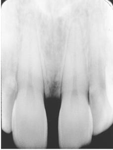
What is Elongation?  How is it corrected?

·       Caused by insufficient vertical angulation

·       Appear as long, distorted teeth

·       Can be corrected by increasing the amount of vertical angulation
<p><span><span>·</span></span><span style="font-family: &quot;Times New Roman&quot;; line-height: normal; font-size: 7pt;"><span>&nbsp;&nbsp;&nbsp;&nbsp;&nbsp;&nbsp; </span></span><span><span>Caused by </span><strong><span>insufficient</span></strong><span> vertical angulation</span></span></p><p class="MsoListParagraphCxSpMiddle"><span><span>·</span></span><span style="font-family: &quot;Times New Roman&quot;; line-height: normal; font-size: 7pt;"><span>&nbsp;&nbsp;&nbsp;&nbsp;&nbsp;&nbsp; </span></span><span><span>Appear as long, distorted teeth</span></span></p><p class="MsoListParagraphCxSpLast"><span><span>·</span></span><span style="font-family: &quot;Times New Roman&quot;; line-height: normal; font-size: 7pt;"><span>&nbsp;&nbsp;&nbsp;&nbsp;&nbsp;&nbsp; </span></span><span><span>Can be corrected by increasing the amount of vertical angulation</span></span></p>
39
New cards

Contrast

·       Refers to how sharply dark and light areas are differentiated or separated on an image

40
New cards

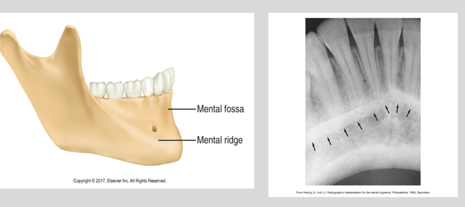
Mental Ridge

·       A linear prominence of cortical bone located on the external surface of the anterior portion of the mandible

·       Often appears superimposed over the mandibular anterior teeth

·       A thick radiopaque band that extends from the premolar region to the incisor region
<p><span><span>·</span></span><span style="font-family: &quot;Times New Roman&quot;; line-height: normal; font-size: 7pt;"><span>&nbsp;&nbsp;&nbsp;&nbsp;&nbsp;&nbsp; </span></span><span><span>A linear prominence of cortical bone located on the external surface of the anterior portion of the mandible</span></span></p><p class="MsoListParagraphCxSpMiddle"><span><span>·</span></span><span style="font-family: &quot;Times New Roman&quot;; line-height: normal; font-size: 7pt;"><span>&nbsp;&nbsp;&nbsp;&nbsp;&nbsp;&nbsp; </span></span><span><span>Often appears superimposed over the mandibular anterior teeth</span></span></p><p class="MsoListParagraphCxSpLast"><span><span>·</span></span><span style="font-family: &quot;Times New Roman&quot;; line-height: normal; font-size: 7pt;"><span>&nbsp;&nbsp;&nbsp;&nbsp;&nbsp;&nbsp; </span></span><span><span>A thick radiopaque band that extends from the premolar region to the incisor region</span></span></p>
41
New cards

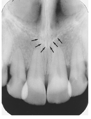
 Nasal Spine

·       A sharp projection of the maxilla located at the anterior and inferior portion of the nasal cavity

·       A v-shaped radiopaque area located at the intersection of the floor of the nasal cavity and nasal septum
<p><span><span>·</span></span><span style="font-family: &quot;Times New Roman&quot;; line-height: normal; font-size: 7pt;"><span>&nbsp;&nbsp;&nbsp;&nbsp;&nbsp;&nbsp; </span></span><span><span>A sharp projection of the maxilla located at the anterior and inferior portion of the nasal cavity</span></span></p><p class="MsoListParagraphCxSpLast"><span><span>·</span></span><span style="font-family: &quot;Times New Roman&quot;; line-height: normal; font-size: 7pt;"><span>&nbsp;&nbsp;&nbsp;&nbsp;&nbsp;&nbsp; </span></span><span><span>A v-shaped radiopaque area located at the intersection of the floor of the nasal cavity and nasal septum</span></span></p>
42
New cards

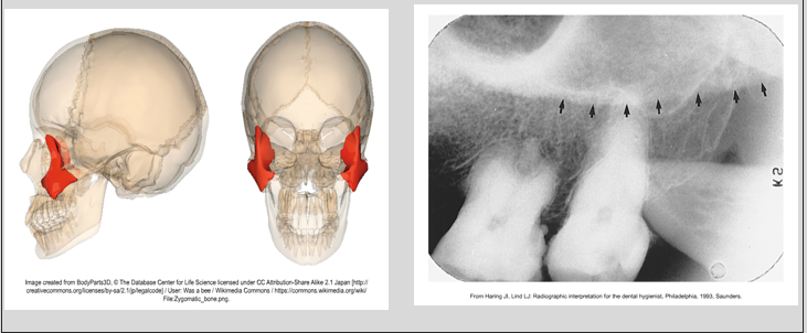
Zygoma

·       Articulates with the zygomatic process of maxilla

·       A diffuse, radiopaque band extending posteriorly from the zygomatic process of the maxilla

  • seen on the maxillary molar and premolar periapicals
<p><span>·</span><span style="font-family: &quot;Times New Roman&quot;; line-height: normal; font-size: 7pt;"><span>&nbsp;&nbsp;&nbsp;&nbsp;&nbsp;&nbsp; </span></span><span>Articulates with the zygomatic process of maxilla</span></p><p class="MsoListParagraphCxSpLast"><span>·</span><span style="font-family: &quot;Times New Roman&quot;; line-height: normal; font-size: 7pt;"><span>&nbsp;&nbsp;&nbsp;&nbsp;&nbsp;&nbsp; </span></span><span>A diffuse, radiopaque band extending posteriorly from the zygomatic process of the maxilla</span></p><ul><li><p class="MsoListParagraphCxSpLast">seen on the maxillary molar and premolar periapicals</p></li></ul><p></p>
43
New cards

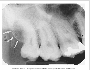
Maxillary tuberosity

·       Rounded prominence of bone that extends posterior to the third molar region

·       A radiopaque bulge distal to the third molar region
<p><span><span>·</span></span><span style="font-family: &quot;Times New Roman&quot;; line-height: normal; font-size: 7pt;"><span>&nbsp;&nbsp;&nbsp;&nbsp;&nbsp;&nbsp; </span></span><span><span>Rounded prominence of bone that extends posterior to the third molar region</span></span></p><p class="MsoListParagraphCxSpLast"><span><span>·</span></span><span style="font-family: &quot;Times New Roman&quot;; line-height: normal; font-size: 7pt;"><span>&nbsp;&nbsp;&nbsp;&nbsp;&nbsp;&nbsp; </span></span><span><span>A radiopaque bulge distal to the third molar region</span></span></p>
44
New cards

Mental Foramen

·       An opening or hole in bone located on the external surface of the mandible in the region of the mandibular premolars

·       A small ovoid or round radiolucent area located in the apical region of the mandibular premolars
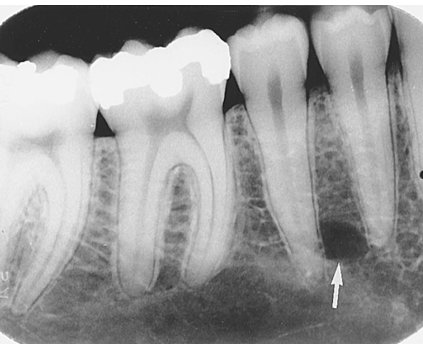
<p><span><span>·</span></span><span style="font-family: &quot;Times New Roman&quot;; line-height: normal; font-size: 7pt;"><span>&nbsp;&nbsp;&nbsp;&nbsp;&nbsp;&nbsp; </span></span><span><span>An opening or hole in bone located on the external surface of the mandible in the region of the mandibular premolars</span></span></p><p class="MsoListParagraphCxSpLast"><span><span>·</span></span><span style="font-family: &quot;Times New Roman&quot;; line-height: normal; font-size: 7pt;"><span>&nbsp;&nbsp;&nbsp;&nbsp;&nbsp;&nbsp; </span></span><span><span>A small ovoid or round radiolucent area located in the apical region of the mandibular premolars</span></span></p>
45
New cards

Maxillary sinuses

·       Paired cavities or compartments of bone located within the maxilla

·       Located above the maxillary premolar and molar teeth

·       A radiolucent area located above the apices of the maxillary premolars and molars
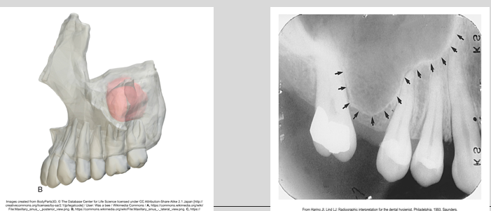
<p><span><span>·</span></span><span style="font-family: &quot;Times New Roman&quot;; line-height: normal; font-size: 7pt;"><span>&nbsp;&nbsp;&nbsp;&nbsp;&nbsp;&nbsp; </span></span><span><span>Paired cavities or compartments of bone located within the maxilla</span></span></p><p class="MsoListParagraphCxSpMiddle"><span><span>·</span></span><span style="font-family: &quot;Times New Roman&quot;; line-height: normal; font-size: 7pt;"><span>&nbsp;&nbsp;&nbsp;&nbsp;&nbsp;&nbsp; </span></span><span><span>Located above the maxillary premolar and molar teeth</span></span></p><p class="MsoListParagraphCxSpLast"><span><span>·</span></span><span style="font-family: &quot;Times New Roman&quot;; line-height: normal; font-size: 7pt;"><span>&nbsp;&nbsp;&nbsp;&nbsp;&nbsp;&nbsp; </span></span><span><span>A radiolucent area located above the apices of the maxillary premolars and molars</span></span></p>
46
New cards

where is the Inverted Y

·       The intersection of the maxillary sinus and the nasal cavity

·       A radiopaque upside-down Y formed by the intersection of the lateral walls of the nasal fossa and the anterior border of the maxillary sinus

·       Above maxillary canine
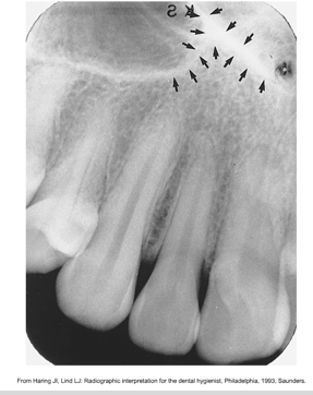
<p><span><span>·</span></span><span style="font-family: &quot;Times New Roman&quot;; line-height: normal; font-size: 7pt;"><span>&nbsp;&nbsp;&nbsp;&nbsp;&nbsp;&nbsp; </span></span><span><span>The intersection of the maxillary sinus and the nasal cavity</span></span></p><p class="MsoListParagraphCxSpMiddle"><span><span>·</span></span><span style="font-family: &quot;Times New Roman&quot;; line-height: normal; font-size: 7pt;"><span>&nbsp;&nbsp;&nbsp;&nbsp;&nbsp;&nbsp; </span></span><span><span>A radiopaque upside-down Y formed by the intersection of the lateral walls of the nasal fossa and the anterior border of the maxillary sinus</span></span></p><p class="MsoListParagraphCxSpLast"><span><span>·</span></span><span style="font-family: &quot;Times New Roman&quot;; line-height: normal; font-size: 7pt;"><span>&nbsp;&nbsp;&nbsp;&nbsp;&nbsp;&nbsp; </span></span><span><span>Above maxillary canine</span></span></p>
47
New cards

Where do we see Condensing Osteitis?

·       The most common periapical radiopacity, most commonly in the mandibular third molars

·       More common in women

·       Is seen below the apex of a tooth, varying in shape and size, with a history of pulpitis 

48
New cards

What is Luxation?

·       Is the abnormal displacement of teeth

·       Can be intrusion, the abnormal displacement of teeth into bone or

·       Extrusion, the abnormal displacement of teeth out of bone

49
New cards

Radiopaque

·       Refers to that portion of an image that appears light or white

·       Are dense and absorb or resist the passage of x-ray beam

50
New cards

Radiolucent

·       Refers to that portion of an image that is dark or black

·       A structure that lacks density and permits the passage of x-ray beam with little or no resistance

51
New cards

lamina dura

  • the wall of the tooth socket

  • made of dense cortical bone

  • a dense radiopaque line that surrounds the root of the tooth
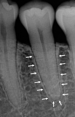
<ul><li><p>the wall of the tooth socket </p></li><li><p>made of dense cortical bone </p></li><li><p>a dense radiopaque line that surrounds the root of the tooth</p></li></ul><p></p>
52
New cards

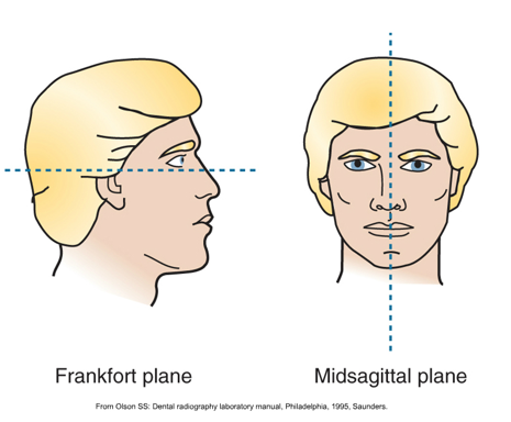
frankfort plane is

  • parallel with floor

53
New cards

midsagittal plane is

  • perpendicular with floor

54
New cards

In panoramic imaging the pivotal point or axis in which our machine rotates is the

  • rotation center

55
New cards

The rotation change in a panoramic machine allows the image layer to conform to which shape

  • elliptical shape of the dental arches

56
New cards

structures are clearly demonstrated on a panoramic image as a __

  • 2-Dimensional, curved image

57
New cards

Which component of a panromic machine tubehead is most different than intraoral tubehead

  • collimation- narrow vertical slit 

58
New cards

What is the angulation of a tubehead in a panoramic unit

  • slightly negative

59
New cards

Your patient has tooth letter A dislodged, which would be the acceptable x-ray technique for observing

  • periapical

60
New cards

why do ghost images appear

  • metallic accessories have not been properly removed

61
New cards

if a patients lips arent closed on a bite block, what shadow will appear

  • dark radiolucent shadow

62
New cards

What anatomy will appear on a panoramic image that is taken correctly

  • posterior teeth are the same size

  • anterior teeth should NOT be skinny

  • possibly see the hard palate

  • should be able to see the pulp chambers on visible teeth

63
New cards

multiplanar reconstruction

  • a technique used in medical imaging to create new 2D images from 3D volumetric data

    • axial

    • coronal

    • sagittal

    • oblique

64
New cards

What would a uniocular lesion with corticated borders be indicative of

  • benign, slow growing lesion

65
New cards

does most pathology on dental images appear with even thickness or different

  • everything will not look the same, they will appear with different varying levels of thickness, and as different shades of grey

66
New cards

radiation

  • energy carried by waves or streams of particles

67
New cards

radiography

  • the art and science of making radiographs by exposure of film to x-rays

68
New cards

who by law prescribes dental images

  • dentist

69
New cards

how often do we need to take a FMX

  • every 3 years

70
New cards

how often do we need to take bitewings

  • once a year on or past the last appointment date

71
New cards

How much of the apical area should we see on periapical images

  • 2-3 mm

72
New cards

angulation for bitewings

  • +10 degrees of positive vertical angulation

73
New cards

what do we do when we adjust horizontal angulation

  • side-to-side adjustment

74
New cards

why do we use vertical bitewings

  • loss of bone in patients with periodontal disease

75
New cards

in periodontal disease, dental images permit the evaluation of pocket depth, soft tissue inflammation, or bone

  • bone

76
New cards

where do we see condensing osteitis 

  • down in the apical area

  • frequently attributed to decay, tooth is nonvital

  • focal opacity

  • tooth is trying to save itself by condensing the bone around the apices 

77
New cards

PFM appearance

  • two components

    • a little or a lot of porcelin

    • solid, fluid metal structure that does not cover the full surface of the tooth

78
New cards

when do we interpret radiographs

  • when the patient is in the chair

79
New cards

where do we see post an core restorations

  • in endodontically treated teeth

80
New cards

where do we first begin to see decay in teeth

  • at contacts or just below

81
New cards

if the interproximal decay does not involve the dentin, but is halfway through the enamel, it is considered

  • moderate decay

82
New cards

who do we see rampant decay in

  • children and adults with minimal salivary flow (more often elders)

83
New cards

radiology

  • the science or study of radiation as used in medicine

84
New cards

what accessories need to be taken out for intraoral imaging

  • oral piercings

  • nose rings

  • glasses

85
New cards

what accessories need to be taken out for extraoral imaging

  • everything

    • earrings

    • glasses

    • facial jewelry

    • partial dentures

86
New cards

incipient

  • extends less than halfway through the enamel, cannot be found on a dental image

  • must be seen clinically

87
New cards

moderate

  • extends more than halfway through the enamel but does not involve the dentin

88
New cards

advanced

  • extends to or through the dentin but not more than halfway through the pulp

89
New cards

severe

  • extends through the enamel, dentin, and more than halfway through the pulp

90
New cards

multiplanar reconstruction

  • reconstruction of raw data into images when imported into viewing software to create 3 anatomic planes of the body