Week 6 Content 1
1/46
Earn XP
Description and Tags
Basal ganglia and cerebellum
Name | Mastery | Learn | Test | Matching | Spaced | Call with Kai |
|---|
No analytics yet
Send a link to your students to track their progress
47 Terms
Basal ganglia
Collection of five anatomical and functionally related grey matter structures = caudate, putamen, globus pallidus, subthalamic nuclei, substantia nigra
Globus pallidus
Consists of internus and externus sections - GPi and GPe
Substantia nigra
Consists of pars compacta SNpc and pars reticulata SNpr

Blue area
Striatum - caudate and putamen

Yellow area
Globus pallidus externus

Red area
Globus pallidus internus

Green area
Subthalamic nuclei STN
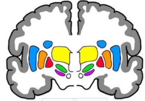
Purple area
Substantia nigra
Functions of BG
Circuits withing basal ganglia that provide insight into function
Four circuits of BG
Goal directed behaviour loops, social behaviour loop, emotion loop, motor loop - all non motor except for motor loop
BG movement control
Regulates desired movements and inhibits undesired movements by sending information to motor cortex via the thalamus
Motor loop
Cortico-basal ganglia-thalamic loop with three pathways = hyperdirect, direct and indirect
Direct pathway function
Allows movements by inhibiting globus pallidus internus and therefore allowing thalamus to activate
Direct pathway sequence
Dopamine from substantia nigra pars compacta and information from motor cortex to striatum, striatum releases GABA to inhibit globus pallidus internus, globus pallidus cannot inhibit thalamus and therefore allows movement
Indirect pathway function
Prevents undesired movements by inhibiting globus pallidus externus and therefore inhibits thalamus
Internal pathway sequence
Dopamine from substantia nigra pars compacta and information from motor cortex to striatum, striatum releases GABA to inhibit globus pallidus externus, allows subthalamic nuclei to release glutamate to globus pallidus internus to inhibit thalamus and therefore, inhibit movement
Dopamine
Fuel of basal ganglia
Dopaminergic producing neurons
Found in SNpc, enhanced action of direct pathway and inhibits action of indirect pathway with net effect of facilitation of movement
Hypokinetic movement disorder
Below normal amount of human movement due to overactive indirect pathway - parkinsons
Hyperkinetic movement disorder
Above normal amount of human movement due to overactive direct pathway - huntingtons
Cerebellar cortex
More regular than cerebral cortex, contains folds, fissures and gyri
First cortical layer of cerebellum
Molecular layer = few neurons, contains axons of granule cells and dendrites of purkinje cells
Second cortical layer of cerebellum
Purkinje cells = single row of huge cells, unique to cerebellum
Third cortical layer of cerebellum
Granular layer = numerous densely packed neurons
Structures of cerebellum
Two large cerebellar hemispheres with three lobes in each = anterior, posterior and flocculonodular
Cerebellum connection to rest of CNS
Connected by passing information via three cerebellar peduncles found on brainstem
First cerebellar peduncle
Superior cerebellar peduncle, located on midbrain, primarily cerebellar efferent fibres via thalamic nuclei to cortex
Second cerebellar peduncle
Middle cerebellar peduncle, located on pons, entirely afferent fibres, information to cerebellum from cerebru
Third cerebellar peduncle
Inferior cerebellar peduncle, located on medulla, afferent fibres from spinal cord and vestibular apparatus, efferent fibres to vestibular nuclei and reticular formation
Blood supply to cerebellum
Basilar artery gives rise to anterior inferior cerebellar artery, superior cerebellar artery and posterior inferior cerebellar artery
Functions of cerebellum
Coordinates human movement, depends on feedback for normal function, critical role in normal motor function, works as comparator
Roles of cerebellum
Maintaining posture and balance via input from vestibular receptors and proprioreceptors, coordination of timing, force, synchronisation of voluntary movement, motor learning
Functional areas of cerebellum 1
Spinocerebellum located in vermal and paravermal sections of cerebellum, has extensive connections with spinal cord
Spinocerebellum input
Movement commands from cortex, activity levels of spinal cord neurons
Spinocerebellum role
Role in making anticipatory, corrective and responsive adjustments or otherwise movement would be uncoordinated
Functional area of cerebellum 2
Vestibulocerebellum, located in flocculonodular lobe
Vestibulocerebellum input
Input from ipsilateral vestibular apparatus and ipsilateral vestibular nuclei in brainstem
Vestibulocerebellum output
Vestibular nuclei and reaches motor neurons via vestibulospinal tracts and tracts coordinating head and eye movement
Functional areas of cerebellum 3
Cerebrocerebellum, located in lateral cerebellar hemispheres, extensive connections with cerebral cortex
Cerebrocerebellum input
Input from cerebral cortex via pontine nucleus
Cerebrocerebellum output
Output from motor and premotor cortex via dentate and motor thalamus
Cerebrocerebellum role
Role in timing movements, planning movements, coordination of voluntary movements - influences corticospinal corticobrainstem and rubrospinal tracts
Cerebellum ataxia
Posture and balance impariments, dysmetria, intention tremor, nystagmus, dysarthria, dysdiadochokinesia, dyssynergia, decomposition of movement
Dyssynergia
Impairment of multijoint movements - movements are not properly sequenced or of proper range or direction
Cerebellar vs somatosensory ataxia
Movement coordination should be compared with eyes open vs eyes closed, cerebellar lesions cause ataxia regardless of use of vision
Vestibulocerebellum dysfunction signs
Unsteadiness, truncal ataxia, nystagmus
Spinocerebellum dysfunction signs
Intention tremor, ataxic gait, dysarthria, dysdiadochokinesia, dysmetria, movement decomposition